Single-session Coil Embolization of Multiple Intracranial Aneurysms
Keun Oh, Yong Cheol Lim
Department of Neurosurgery, Ajou University School of Medicine, Ajou University Hospital, Suwon, Korea
Objective : There is no clear treatment strategy for the management of multiple intracranial aneurysms because of variable anatomical distribution, difficult identification of the aneurysm ruptured, and poor overall outcomes.
The purpose of this study was to assess the efficacy and safety of sin- gle-session coil embolization for multiple intracranial aneurysms.
Methods : Between September 2008 and December 2012, 209 aneurysms in 117 patients were treated at our institute. Twenty eight among the 117 patients had multiple aneurysms with a total of 71, and 60 of the 71 aneurysms underwent coil embolization in a single-session.
Results : A total of 60 aneurysms were treated with a single-session coil embolization, of which the most frequent locations were in the posterior communicating artery, followed by the middle cerebral artery. Immediate post-embolization angiographies showed total occlusion in 49 (81.7%) aneur- ysms, remnant neck in 6 (10%), and body-filling in 5 (8.3%). Procedure-re- lated complications had developed in 2 (3.3%) of the 60 embolized aneur- ysms: an asymptomatic thromboembolic event, and a partial coil protrusion without a subsequent thromboembolic complication.
Conclusion : With careful evaluation of individual aneurysm characteristics and configuration, multiple intracranial aneurysms previously thought to require multimodality therapy can be safely treated in a single-session coil embolization.
J Cerebrovasc Endovasc Neurosurg.
2013 September;15(3):184-190 Received : 9 August 2013 Revised : 24 August 2013 Accepted : 28 August 2013 Correspondence to Yong Cheol Lim Department of Neurosurgery, Ajou University School of Medicine, 5 San, Woncheon-Dong, Yongtong-Ku, Suwon 442-721, Korea Tel : 82-31-219-5232
Fax : 82-31-219-5238 E-mail : [email protected]
This is an Open Access article distributed under the terms of the Creative Commons Attribution Non- Commercial License (http://creativecommons.org/li- censes/by-nc/3.0) which permits unrestricted non- commercial use, distribution, and reproduction in any medium, provided the original work is properly cited.
Keywords Multiple aneurysms, Coil embolization, Single-session
INTRODUCTION
Detection rate of multiple intracranial aneurysms has significantly increased with recent advances in imaging techniques. Approximately 15-35% of patients presenting with subarachnoid hemorrhage (SAH) were found to have multiple intracranial aneurysms.11)14) The management of patients with multiple intra- cranial aneurysms had been on discussion because of variations in anatomic distribution, difficulties in de- termination of the ruptured aneurysms, and poor
overall outcomes in the setting of SAH.15)
Although several groups have advocated micro- surgical clip ablation of all lesions in a single oper- ation or through multimodality approaches, no clear treatment strategy exists because many multiple aneurysms have bilateral distribution, and even lo- cated anterior-posteriorly in a subset of patients.2-4)12)16) In this study, we reviewed our experience of treat- ing patients with multiple intracranial aneurysms, and assessed the efficacy and safety of single-session coil embolization of multiple intracranial aneurysms.
PATIENTS AND METHODS
Studies
A review was performed for a consecutive 738 pa- tients whose intracranial aneurysms were treated in between September 2008 and December 2012. Patients excluded were those treated by microsurgical clip- ping, combination of microsurgical and endovascular treatment, and treatment of the aneurysms in 2 or more steps. Of the 738 patients, 117 (16%) had multi- ple intracranial aneurysms, a total of 209 aneurysms.
Eighty-nine of the patients were expected to require several treatment sessions for complete obliteration of all aneurysms, so they were excluded. Accordingly, 28 patients (5 men and 23 women, 31 to 77 years old) with 71 aneurysms were included for the analysis. All of these patients had multiple aneurysms and under- went coil embolization for more than 2 aneurysms in a single session. Out of the 71 aneurysms, 60 aneur- ysms were managed this way.
Initial clinical presentation was documented accord- ing to the Hunt and Hess grading system.8) Diagnosis of SAH was made using computed tomography (CT).
The amount of blood noted on the initial CT scan was classified according to the Fisher grading system.6) Immediate post-embolization angiography of each aneurysm was assigned to the 3 categories suggested by Raymond et al.;17) class 1= complete obliteration, class 2= residual neck; and class 3= residual aneurysm.
Endovascular treatment
All procedures were performed under general anesthesia. Patients with aneurysmal SAH underwent coil embolization irrespective of the Hunt and Hess grade (HHG). In patients with poor grades (HHG IV and V), external ventricular drainage was placed.
Biplane Intergris Allura angiography units (Phillips Healthcare, Amsterdam, Netherlands) were used. The size and neck of each aneurysm was evaluated in 3-dimensional (3D) angiographic images using the an- giographic unit software. The rupture site was de- termined by the following: 1) an aneurysm was con-
sidered to be ruptured if it was surrounded by more dense SAH or by focal accumulation of a clot on the 3DCT image, 2) the most irregular or the largest aneurysm was thought to be ruptured, if the CT im- age did not reveal a focal accumulation of blood clot or if there were multiple aneurysms near the focal clot; and, 3) irregularity (multilobulation, daughter sac, and noticeable elongation) was more important than size, and the existence of a daughter sac was the most important parameter.7)
Systemic heparinization was not given during coil embolization of the ruptured aneurysm. An initial bo- lus of 1000 U and intermittent boluses of 1000 U/h of heparin were administered during coil embolization of additional unruptured aneurysms. Basically, a con- ventional single or multiple-catheter technique was used to obliterate the presumably ruptured aneurysm in each patient. Additional aneurysms that could be approached in the same session were all also obli- terated, although any remaining unruptured lesions expected to require stent-assisted techniques were treated at a later stage for those patients who sig- nificantly recovered after the SAH.
In patients with unruptured aneurysms, antiplatelet medication was administrated before each procedure.
Systemic heparinization was performed during the procedure with a bolus of 2000 U of heparin after femoral sheath placement and intermittent 1000 U/h boluses during the entire procedure.
RESULTS
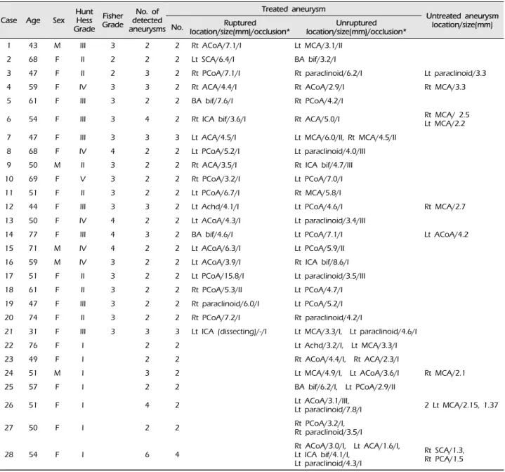
Three quarters of patients (75%) were diagnosed with acute ruptures. The Hunt and Hess grades at the time of treatment in the ruptured group were Grade II in 7 patients, Grade III in 8 patients, and Grade IV and V in 5 and 1 patient. The remaining 7 patients had multiple aneurysms without SAH (Table 1).
Sixty out of the total 71 aneurysms were success- fully embolized. Eleven aneurysms were unsuitable for endovascular treatment either because these were
Case Age Sex Hunt Hess Grade
Fisher Grade
No. of detected aneurysms
Treated aneurysm
Untreated aneurysm location/size(mm)
No. Ruptured
location/size(mm)/occlusion* Unruptured location/size(mm)/occlusion*
1 43 M III 3 2 2 Rt ACoA/7.1/I Lt MCA/3.1/II
2 68 F II 2 2 2 Lt SCA/6.4/I BA bif/3.2/I
3 47 F II 2 3 2 Rt PCoA/7.1/I Rt paraclinoid/6.2/I Lt paraclinoid/3.3
4 59 F IV 3 3 2 Rt ACA/4.4/I Rt ACoA/2.9/I Rt MCA/3.3
5 61 F III 3 2 2 BA bif/7.6/I Rt PCoA/4.2/I
6 54 F III 3 4 2 Rt ICA bif/3.6/I Rt ACA/5.0/I Rt MCA/ 2.5
Lt MCA/2.2
7 47 F III 3 3 3 Lt ACA/4.5/I Lt MCA/6.0/II, Rt MCA/4.5/II
8 68 F IV 4 2 2 Lt PCoA/5.2/I Lt paraclinoid/4.0/III
9 50 M II 3 2 2 Rt ACA/3.5/I Rt ICA bif/4.7/III
10 69 F V 3 2 2 Rt PCoA/3.2/I Lt PCoA/7.0/I
11 51 F II 3 2 2 Lt PCoA/6.7/I Rt MCA/5.8/I
12 44 F III 3 3 2 Lt Achd/4.1/I Lt PCoA/4.6/I Rt MCA/2.7
13 50 F IV 4 2 2 Lt ACoA/4.3/I Lt paraclinoid/3.4/III
14 77 F III 4 3 2 BA bif/4.6/I Lt PCoA/7.1/I Lt ACoA/4.2
15 71 M IV 4 2 2 Lt ACoA/6.3/I Lt PCoA/5.9/II
16 59 M IV 3 2 2 Lt ACoA/3.9/I Rt ICA bif/8.6/I
17 51 F II 3 2 2 Lt PCoA/15.8/I Lt paraclinoid/3.5/III
18 61 F II 3 2 2 Rt PCoA/5.3/II Lt PCoA/4.7/I
19 47 F III 3 2 2 Rt paraclinoid/6.0/I Lt PCoA/5.2/I
20 74 F II 3 2 2 Rt PCoA/7.2/I Rt paraclinoid/4.2/I
21 31 F III 3 3 3 Lt ICA (dissecting)/-/I Lt MCA/3.3/I, Lt paraclinoid/4.6/I
22 76 F I 2 2 Lt Achd/3.2/I, Lt MCA/3.3/I
23 49 F I 2 2 Rt ACoA/4.4/I, Rt ACA/2.3/I
24 51 M I 3 2 Lt MCA/4.9/I, Lt ACoA/3.6/I Rt MCA/2.1
25 57 F I 2 2 BA bif/6.2/I, Lt PCoA/2.9/II
26 51 F I 4 2 Lt ACoA/3.1/III,
Lt paraclinoid/7.8/I 2 Lt MCA/2.15, 1.37
27 50 F I 2 2 Rt PCoA/3.2/I,
Rt paraclinoid/3.5/I
28 54 F I 6 4 Rt ACoA/3.0/I, Lt ACA/1.6/I,
Lt ICA bif/4.1/I, Lt paraclinoid/4.3/I
Rt SCA/1.3, Rt PCA/1.5
*By Raymond classification; I: complete obliteration, II: residual neck, III: residual aneurysm. ACA= anterior cerebral artery; AChd= anterior choroidal artery; ACoA= anterior communicating artery; BA= basilar artery; bif= bifurcation; ICA= internal carotid artery; Lt= left; MCA=
middle cerebral artery; PCA= posterior cerebral artery; PCoA= posterior communicating artery; Rt= right; SCA= superior cerebellar artery.
Table 1. Summary of characteristics of 28 patients with multiple intracranial aneurysms treated by coil embolization in a sin- gle-session.
too small or the neck was broad. The number of aneurysms per locations were 16 in the posterior com- municating artery, 14 in the middle cerebral artery (MCA), 11 in the paraclinoid segment of the internal carotid artery (ICA), 10 in the anterior communicating artery, 7 in vertebrobasilar atery, 6 in the distal ante- rior cerebral artery, 4 in the ICA bifurcation, and 2 in
the anterior choroidal artery. The single remaining aneurysm was a blood-blister like aneurysm in the dorsal distal ICA. Multiple aneurysms were located in unilateral anterior circulation for 14 patients, bilateral anterior circulation for 11, and both the anterior and the posterior circulation for 3 patients. Immediate postembolization angiograms demonstrated total oc-
A B C D
E F G H
Fig. 1. (A) Computed tomogrphy scan demonstrates left frontal parenchymal hemorrhage, subarachnoid hemorrhage in the inter- hemispheric fissure, and intraventricular hemorrhage. (B and C) Left internal carotid angiogram shows a ruptured pericallosal aneur- ysm and an unruptured wide-neck aneurysm of the left middle cerebral artery bifurcation. (D) Right internal carotid angiogram shows a wide-neck aneurysm of the right middle cerebral artery bifurcation. (E,F and G) Angiogram obtained immediately after em- boization shows occlusion of the aneurysms preserving the parent arteries. (H) One-year follow-up magnetic resonance angiography shows continued occlusion of aneurysms and wide patency of the parent arteries.
clusion (Raymond classification I) in 49 (81.7%) aneur- ysms, neck remnant (Raymond classification II) in 6 (10%), and body filling (Raymond classification III) in 5 (8.3%).
Among the 60 aneurysms embolized, there were 2 procedure-related complications (3.3%). These con- sisted of an asymptomatic thromboembolic event and a partial coil protrusion without a subsequent throm- boembolic complication. The thromboembolic compli- cation happened for a case of ruptured anterior com- municating artery aneurysm. The immediate post-em- bolization angiography of this patient revealed a small thrombus at the coil in the aneurysm neck.
Mechanical thrombolysis was successfully done by us- ing a 10-sized microcatheter and microwire, and re- canalization was achieved for the parent artery. The other case involved partial coil protrusion in a rup-
tured posterior communicating artery aneurysm. A portion of the loop of previously deployed coil had protruded into the parent artery, but the coil loop was tiny and did not interfere with the blood flow that no additional procedure was done.
Out of the 28 total patients, 1 died, and this patient initially presented with a high grade (HHG IV) SAH.
Case illustration
A 47-year-old woman presented with a severe head- ache of sudden onset with a stuporous mentality (HHG III). The initial CT scan revealed diffuse SAH (Fisher grade III) in the basal cistern and dense focal intracranial hemorrhage (ICH) in the interhemispheric fissure with intraventricular hemorrhage (IVH) (Fig.
1A). The left internal carotid angiogram demonstrated a 4.5-mm pericallosal (A2) aneurysm with a relatively wide neck and a 6-mm sized middle cerebral artery
bifurcation aneurysm with a wide neck (Fig. 1B, 1C).
The right internal carotid angiogram showed a 4.5-mm sized MCA bifurcation aneurysm with a wide neck (Fig. 1D). We decided that the left pericallosal artery aneurysm was the cause of bleeding because of its proximity to the ICH. All 3 of these aneurysms were embolized in a single procedure. Under general anesthesia, a 6F guiding catheter was placed at the left ICA. The ruptured pericallosal aneurysm was se- lected with a microcatheter, and 4 detachable coils were delivered into the aneurysm lumen (Fig. 1E).
After confirming complete obliteration of the peri- callosal aneurysm, the left MCA bifurcation aneurysm was selected with the same microcatheter. Although the aneurysm was wide-necked, the initial coil de- ployed provided a stable frame for 5 additional de- tachable coils into the aneurysm sac. Repeated angio- gram showed a nearly complete embolization of the MCA bifurcation aneurysm and good patency of the MCA (Fig. 1F). Finally, we repositioned the same guiding catheter to the right ICA. Five detachable coils were enough for near-complete obliteration (Fig.
1G). The course of procedure and subsequent recov- ery was uneventful. The patient did not experience post-procedural contrast-induced complications. A fol- low-up magnetic resonance angiography confirmed continued occlusion of aneurysms and wide patency of the parent arteries (Fig. 1H).
DISCUSSION
The risk of rupture is higher for patients with multi- ple intracranial aneurysms than in patients with a sin- gle intracranial aneurysm.18) In addition, the outcomes are significantly poorer for those patients with multiple intracranial aneurysms.11)15) Previously published stud- ies have investigated the predictive factors for aneur- ysmal rupture, but the findings are not in complete agreement. Most studies demonstrated that risk factors for rupture are older age, female gender, larger size of aneurysms, cigarette smoking, and hypertension.5)9)10)13)
For the reason, it seems meaningful to consider pre- ventive intervention for the patients who have an epi- sode of SAH and its risk factors.
Neurosurgeons and neurovascular interventionists confront a special problem in the planning of the treatment of patients with multiple aneurysms. In the setting of SAH and multiple aneurysms, the ruptured aneurysm must be correctly identified using clinical, CT imaging, and angiographic findings.1) However, it is not uncommon to misidentify the bleeding aneurysm.7)
There are a number of treatment options of multiple intracranial aneurysms. The close observation without treatment of remaining unruptured aneurysms may be one option. Delayed elective endovascular coil em- bolization or microsurgical clipping is another way to manage unruptured aneurysms. Although multi- modality approaches (combined microsurgical and en- dovascular management) are best used for multiple intracranial aneurysms, simultaneous coil emboliza- tion of multiple intracranial aneurysms has some ben- efits over multimodality approaches. The most sig- nificant benefit is the elimination of the risk of a treat- ing the non-ruptured, non-bleeding aneurysm due to misidentification of the culprit lesion. Another major benefit is the decrease in future risk of intracranial hemorrhagic events associated with delayed treatment of multiple aneurysms. Although multiple unilateral aneurysms may be appropriate to a single stage mi- crosurgical clipping, many combinations of aneurysm locations (distal, axial, or bilateral distributions) may demand more careful surgical planning or multiple staged operations. On the contrary, coil embolizations are advantageous in that it can be performed in a sin- gle session for multiple aneurysms, regardless of ana- tomical distributions that may not all be exposed through a single craniotomy. Avoidance of a cumu- lative microinjury from brain retraction and arterial manipulation from microsurgical clipping may be an additional benefit of coil embolization.15) Therefore, single-session coil embolization for multiple intra- cranial lesions should be considered in cases involv-
ing large or irregular aneurysms with bimodal dis- tribution (bilateral or anterior-posterior), and/or when unruptured aneurysm to be treated is easy to access in the session of ruptured aneurysm treatment.
In this study, multiple intracranial aneurysms were coiled in single sessions without serious complications.
This low rate of procedure-related morbidity may be contributable to the carefully selected indication for coil embolization. Treatment of multiple aneurysms in a sin- gle session requires relatively prolonged treatment time and exposes the patients to increased risks of throm- boembolic events and contrast-induced nephrotoxicity.
Therefore, minimization of treatment time and addi- tional angiography should be considered for pre- vention of such complications. For this achievement, a thorough planning for procedure should precede each coiling session with a detailed review of the 3D X-ray rotational angiography (3DRA) before procedure. This planning should take into consideration the selection of aneurysms adequate for coil embolization and de- termination of proper working projection for each aneurysm.
Selection of aneurysms for coil embolization can be determined by the size of an aneurysm sac and the size of aneurysmal neck and its relationship with the parent artery. The order of aneurysms to be treated should be determined with the utmost emphasis for the aneurysm which must be treated first, with the subsequent coiling sequence decided upon accessi- bility of the secondary aneurysms. Lastly, pre-oper- ative prediction of proper technique (single catheter technique, multiple catheter technique and stent-as- sisted technique) for a given combination of aneur- ysm is important because this could facilitate treat- ment process by setting the device early.
However, there are limitations in this study. First, the number of patients with multiple intracranial aneurysms treated in a single-session coil emboliza- tion is small compared with the number of total pa- tients with multiple intracranial aneurysms treated in our institute. Nor this study permits comparisons to
multimodality approaches. We believe further studies comparing treatment outcome between single-session coil embolization and multimodality approach are necessary and the study could answer what the ap- propriate treatment method is for multiple intra- cranial aneurysms.
CONCLUSION
Multimodality approaches are usually used for pa- tients in whom multiple intracranial aneurysms can- not be addressed by a single treatment method. With careful evaluation of each individual aneurysm char- acteristics and configuration, however, multiple intra- cranial aneurysms can be safely treated in a sin- gle-session coil embolization.
REFERENCES
1. Almaani WS, Richardson AE. Multiple intracranial aneur- ysms: Identifying the ruptured lesion. Surg Neurol. 1978 May;9(5):303-5.
2. Chung J, Shin YS. Multiple intracranial aneurysms treat- ed by multiple treatment modalities. Neurosurgery. 2011 Oct;69(4):E1030-2.
3. Clatterbuck RE, Tamargo RJ. Contralateral approaches to multiple cerebral aneurysms. Neurosurgery. 2005 Jul;57(1 Suppl):160-3; discussion 160-3.
4. Dunn GP, Nahed BV, Walcott BP, Jung H, Tierney TS, Ogilvy CS. Dual ipsilateral craniotomies through a single incision for the surgical management of multiple intracranial aneurysms. World Neurosurg. 2012 Mar-Apr;77(3-4):502-6.
5. Ellamushi HE, Grieve JP, Jager HR, Kitchen ND. Risk factors for the formation of multiple intracranial aneurysms. J Neurosurg. 2001 May;94(5):728-32.
6. Fisher CM, Kistler JP, Davis JM. Relation of cerebral vasospasm to subarachnoid hemorrhage visualized by computerized tomographic scanning. Neurosurgery. 1980 Jan;6(1):1-9.
7. Hino A, Fujimoto M, Iwamoto Y, Yamaki T, Katsumori T. False localization of rupture site in patients with multiple cerebral aneurysms and subarachnoid hemorrhage. Neurosurgery. 2000 Apr;46(4):825-30.
8. Hunt WE, Hess RM. Surgical risk as related to time of intervention in the repair of intracranial aneurysms. J Neurosurg. 1968 Jan;28(1):14-20.
9. Inagawa T. Incidence and risk factors for multiple intra- cranial saccular aneurysms in patients with subarachnoid hemorrhage in Izumo City, Japan. Acta Neurochir (Wien). 2009 Dec;151(12):1623-30.
10. Juvela S. Risk factors for multiple intracranial aneurysms.
Stroke. 2000 Feb;31(2):392-7.
11. Kaminogo M, Yonekura M, Shibata S. Incidence and outcome of multiple intracranial aneurysms in a defined population. Stroke. 2003 Jan;34(1):16-21.
12. Lim YC, Shin YS, Chung J. Combined endovascular and microsurgical procedures as complementary approaches in the treatment of a single intracranial aneurysm. J Korean Neurosurg Soc. 2008 Jan;43(1):21-5.
13. Lu HT, Tan HQ, Gu BX, Wu W, Li MH. Risk factors for multiple intracranial aneurysms rupture: A retrospective study. Clin Neurol Neurosurg. 2013 Jun;115(6):690-4.
14. Ostergaard JR, Hog E. Incidence of multiple intracranial aneurysms. Influence of arterial hypertension and gender.
J Neurosurg. 1985 Jul;63(1):49-55.
15. Rinne J, Hernesniemi J, Niskanen M, Vapalahti M.
Management outcome for multiple intracranial aneurysms.
Neurosurgery. 1995 Jan;36(1):31-7; discussion 37-8.
16. Rinne J, Hernesniemi J, Puranen M, Saari T. Multiple in- tracranial aneurysms in a defined population: Prospective angiographic and clinical study. Neurosurgery. 1994 Nov;
35(5):803-8.
17. Roy D, Milot G, Raymond J. Endovascular treatment of unruptured aneurysms. Stroke. 2001 Sep;32(9):1998-2004.
18. Yasui N, Suzuki A, Nishimura H, Suzuki K, Abe T.
Long-term follow-up study of unruptured intracranial aneurysms. Neurosurgery. 1997 Jun;40(6):1155-9; discussion 1159-60.