서 론
Docetaxel은 paclitaxel과 구조 및 기능이 유사한 taxane계 물질로써 폐암뿐만 아니라 많은 고형암의 성장을 억제하 는 강력한 항암제로 잘 알려져 있다[1,2]. 최근 docetaxel이 세포의 미세관(microtubule)에 결합하여 중합반응을 유도 하고 탈중합반응을 방해하여 세포의 유사분열을 지연시 키며, 그 결과로 암세포의 세포사멸을 유도한다고 보고되 었으나, 아직은 그 기전이 명확하게 밝혀져 있지 않다[3,4].
암억제 유전자인 p53 유전자의 돌연변이는 암세포에서 일어나는 가장 흔한 유전학적 변화로, 암세포의 비정상적
인 증식을 유발하며, 폐암의 50% 이상이 p53 mutant임이 보고된 바 있다. 비소세포폐암에서 p53 유전자의 돌연변 이는 DNA cross-linking agent인 cisplatin이나 topoisomerase inhibitor 등에서 저항성을 가진다[5]. 그러나 taxene계 물질 인 paclitaxel이나 docetaxel이 p53 유전자 돌연변이인 암세 포에서 세포성장을 억제하는 것으로 나타났으나[6], 그 작 용 기전은 완전히 밝혀지지 않았다.
본 연구에서는 비소세포폐암 중 p53 유전자 돌연변이이 며 편평상피세포암인 NCI-H1703 세포주를 이용하여 doce- taxel을 처리한 뒤 세포사멸 유도기전을 규명하고자 하였다.
Docetaxel에 의한 비소세포폐암세포주 NCI-H1703의 세포사멸 유도기전
하현철*․김영대*․송승환*․박진수*․김종원*
Cell Death Induction Mechanism of Non-small Cell Lung Cancer Cell Line, NCI-H1703 by Docetaxel
Hyeon Cheol Ha, M.D.*, Yeong Dae Kim, M.D.*, Seung Hwan Song, M.D.*, Chin-Su Park, M.D.*, Jong Won Kim, M.D.*
Background: Docetaxel has been effectively used as an anti-cancer chemotherapuetic agent for various tumor treatments including lung cancer. However, the cell death induction mechanism(s) involved with docetaxel treatment in lung cancer cells has not been known yet. Material and Method: In the present study, the cellular and biochemical changes of NCI-H1703 cells (non-small cell lung cancer cell line, p53-mutant) after docetaxel treatment have been monitored by flow cytometry, fluorescence microscopy and western blot. Result: Docetaxel treatment significantly resulted in decrease of S phase as well as increase of G2 phase, and consequently evoked an increase of cell death in NCI-H1703 cells. After docetaxel exposure the activations of caspase-3 and caspase-9 were detected. Conclusion: Take together, it is suggested that the docetaxel induces NCI-H1703 cell death by caspase-9 and caspase-3 dependent mitochondrial apoptotic pathway.
(Korean J Thorac Cardiovasc Surg 2006;39:668-673) ꠏꠏꠏꠏꠏꠏꠏꠏꠏꠏꠏꠏꠏꠏꠏꠏꠏꠏꠏꠏꠏꠏꠏꠏꠏꠏꠏꠏꠏꠏꠏꠏꠏꠏꠏꠏꠏꠏꠏꠏꠏꠏꠏꠏꠏꠏꠏꠏꠏꠏꠏꠏꠏꠏꠏꠏꠏꠏꠏꠏꠏꠏꠏꠏꠏꠏꠏꠏꠏꠏꠏꠏꠏꠏꠏꠏꠏꠏꠏꠏꠏꠏꠏꠏꠏꠏꠏꠏꠏꠏꠏꠏ Key words: 1. Carcinoma, non-small cell, lung
2. Neoplasm biology 3. Cell death
*부산대학교 의과대학 흉부외과학교실
Department of Thoracic and Cardiovascular Surgery, College of Medicine, Pusan National University 논문접수일:2006년 5월 22일, 심사통과일:2006년 7월 12일
책임저자:김영대 (602-739), 부산시 서구 아미동 1가 10번지, 부산대학교병원 흉부외과 (Tel) 051-240-7822, (Fax) 051-243-9389, E-mail: [email protected]
본 논문의 저작권 및 전자매체의 지적소유권은 대한흉부외과학회에 있다.
대상 및 방법
1) NCI-H1703 cell의 배양
NCl-H1703 비소세포폐암 세포주는 American Type Cul- ture Collection (ATCC)에서 구입하였다. 10%의 fetal bovine serum (FBS; Gibco BRL, Grand Ishand, NY)와 1%의 항생 제(penicillin-streptomycin)가 포함된 RPMI 1640 배지에 2 mM L-glutamine, 1.5 g/L sodium bicarbonate, 4.5 g/L glucose, 10 mM HEPES와 1.0 mM sodium pyruvate (Sigma, St. Louis, MO)를 첨가하여 배양하였다. 5%의 CO2와 95%의 공기가 포함된 37oC의 배양기에서 배양하였다.
2) 시료의 처리
모든 시료의 처리는 6-well의 배양접시에 각각 1.3×10⁵ 개의 세포를 분주하여 48시간 동안 세포배양기에서 부착 시킨 후, 새로운 배양액으로 교체한 후 처리하였다. NCI- H1703 세포에 5 ng/mL docetaxel을 처리한 후, 37oC에서 48시간 배양시킨 뒤, 세포를 수획하여 실험을 수행하였다.
대조군에는 docetaxel의 용해에 사용한 ethanol (최종처리 농도; 0.05%, v/v)을 처리하였다. 모든 실험은 3번 이상 반 복 수행하였다.
3) 유세포측정기(flow cytometry)를 이용한 세포주기 분석 및 세포사멸률 측정
세포주기분석과 세포사멸률 측정을 위해서 수획된 세 포는 0.5%의 Tween 20 (Sigma, St. Louis, MO)이 들어 있 는 75% ethanol에서 냉장 상태로 24시간 동안 고정하였다.
고정된 세포의 핵은 propidium iodide (PI, 10μg/mL Sigma, St. Louis, MO) 용액으로 37oC에서 30분간 염색하였고, 염 색하는 동안 RNase A (100μg/m Sigma, St. Louis, MO)를 처리하여 세포 내의 RNA를 모두 제거하였다. 세포주기와 사멸률은 유세포 측정기로 분석하였다.
4) Hoechst staining을 이용한 핵 응축(nuclear con- densation) 관찰
수획한 세포는 PBS로 세척하여, 세포를 즉시 4% paraform- aldehyde로 고정하였다. Hoechst 33342 (Sigma, St. Louis, MO) 염색액을 4μg/mL의 농도로 PBS 용액에 희석하여 실온에서 2시간 동안 염색한 후, slide glass에 도말하여 핵 의 형태를 형광현미경으로 관찰하였다.
5) Western blot 분석
각각의 시료를 처리한 세포를 lysis buffer [50 mM Tris HCl (pH 7.6), 0.5% Triton X-100, 10 mM NaCl, 10 mM EDTA, protease inhibitor cocktail (104 mM AEBSF, 0.08 mM aprotinin, 2.1 mM leupeptin, 3.6 mM bestain 1.5 mM pepstatin)]로 4oC에서 40분간 용해시킨 다음, 4oC에서 15분 간 14,000 rpm으로 원심분리한 후 단백질이 포함된 상등 액만을 취하여 Bradford (Pierce, Rockford, IL) 방법으로 총 단백질을 정량하였다. 각각의 시료에서 단백질 70μg을 균 등하게 취하여 SDS-polyacrylamide gel (8∼15% gradient gel)에서 전기영동한 후, nylon membrane에 전기적으로 전 위(electrotransfer)시켰다. Anti-caspase-3 (Santa Cruz, CA), anti-cleaved caspase-3 (New England Biolabs, Beverly, MA), anti-cleaved caspase-9 (New England Biolabs, Beverly, MA), anti-caspase-9 (Santa Cruz, CA), anti-actin (Santa Cruz, CA) 등을 일차 항체로 하였고, enhanced chemiluminescence 방법 (ECL-plus Amersham Co.)으로 발색하였으며, LAS-1000PLUS (Fujifilm)로 섬광의 발생량을 측정함으로써 단백질의 발현 량을 분석하였다.
결 과
1) Docetaxel의 처리가 비소세포폐암 세포주 NCI- H1703의 세포사멸과 세포주기에 미치는 영향 Docetaxel의 처리가 세포주에 미치는 영향을 알아보기 위해 세포사멸률과 세포주기를 분석하였다. 5 ng/mL의 docetaxel을 NCI-H1703 세포에 48시간 동안 처리한 후, PI Fig. 1. Cell cycle distribution in NCI-H1703 cells incubated for 48 hours in the presence of 5 ng/mL docetaxel. Docetaxel treatment for 48 hours shows increase of sub-G1 (apoptotic) phase.
로 핵을 염색하여 유세포측정기로 세포사멸률을 측정하 였다. 48시간 처리군에서는 대조군과 비교해서 G1기가 10% 정도 감소하고 S기가 30% 정도 감소하는 양상을 보 였고, 대조군에 비해 세포사멸률이 25% 정도 증가한 것으 로 나타났다(Fig. 1).
2) 핵의 형태학적 변화
Docetaxel 5 ng/mL 처리군에 핵만을 특이적으로 염색하 는 Hoechst 33342 형광염색제와 propidium iodide (PI)로 세 포의 핵을 염색한 후 형광현미경으로 관찰하였으며 다량 의 핵조각과 응축된 형태의 핵이 관찰되었다. 그리고 다 핵의 형태가 관찰되었다(Fig. 2).
3) Caspase 의존적인 세포사멸
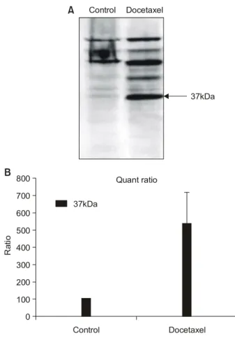
Caspase-9의 활성 정도를 Western blot 분석법으로 나타 냈을 때 대조군에서는 cleaved caspase-9가 검출되지 않았 으며, docetaxel 처리군에서 가장 강한 활성을 보이는 것으
로 나타났다(Fig. 3). Caspase-9의 활성화는 caspas-3를 활성 화시키게 되는데 대조군에서는 활성형 caspase-3이 검출되 지 않았으며, docetaxel 처리군에서 17 및 19 KDa 단백질 이 검출되어 강한 활성을 보였다(Fig. 4).
고 찰
폐암은 예후가 불량하여 치사율이 높은 암이며 5년 생 존율이 14%에 불과하다[7]. 특히 비소세포폐암은 전체 폐 암의 약 80%를 차지하고 있으며 항암화학요법에 대한 더 많은 연구가 필요한 실정이다[8]. 비소세포폐암은 치료경 과 중 항암제에 저항성을 보이므로 최근에는 세포사멸 경 로를 활성화시켜 비소세포폐암세포의 사멸을 유도하려는 시도가 이루어지고 있으며, 이에 대한 분자생물학적 기전 을 해명하려는 노력 또한 중요시되고 있다[9].
최근 강력한 항암제로 각종 암환자들에게 투여되고 있 는 taxane계 물질의 하나인 docetaxel이 폐암을 비롯한 유 Fig. 2. Nuclear morphology (chro- matin condensation) in NCI-H1703 cells incubated for 48 hours in the presence of 5 ng/mL doceta- xel. (A) Arrows indicate fragmated nuclei in Hoecsht 33342 stained cells, (B) Arrows indicate multinu- clei in propdium iodide stained cells.
A
B
Control Docetaxel
Control Docetaxel
방암, 전립선암 등 각종 고형암에서 암세포의 사멸을 유 도한다는 보고가 있으나 아직 암세포의 사멸유도 기전은 완전히 밝혀지지 않았다[10-12].
본 실험에서는 비소세포폐암 중 p53 돌연변이로써 편평 상피세포암 세포주인 NCI-H1703 세포주에 docetaxel이 유 도하는 암세포 사멸효과를 평가하였으며, 이들 처리에 의 해서 야기되는 세포사멸 기전을 연구하였다.
Docetaxel이 세포의 미세관에 결합하여 중합반응을 유 도하고 탈중합반응을 방해하여 세포주기 중 G2/M기 상태 를 지연시켜 세포의 유사분열을 억제시켜 그 결과로 암세 포의 세포사멸을 유도한다는 것은 유방암 세포 등에서 알 려져 있으나[13,14], 비소세포폐암 세포주인 NCI-H1703 세 포주에서는 아직 활발한 연구가 이루어지지 못하고 있다.
본 실험에서 docetaxel은 NCI-H1703 세포주에서 시간 의존 적으로 세포사멸을 유도했으며, 48시간에 44%의 세포사 멸을 나타냈다. 세포주기 분석결과 G2기에서 대조군에 비
해 45%의 강한 G2기 정지현상을 보였으며, 이 결과는 docetaxel이 NCI-H1703 세포주의 유사분열을 억제하여 세 포사멸을 유도했다는 것을 증명한다. Hoechst staining 결과 를 보면 docetaxel 처리군에서 세포사멸분체(apoptotic body) 라 불리는 핵조각이 관찰되며, 이것은 docetaxel이 NCI- H1703 세포주에서 전형적인 세포사멸을 유도하는 것이라 고 볼 수 있다. p53은 세포주기의 진행 과정에서 G1기 세 포주기 점검점(check point)의 조절자로써 잘 알려져 있으 나, p53 유전자의 돌연변이는 세포에 이상이 생겨도 G1기 를 조절하지 못하여 세포를 G2기로 진행시키게 된다[15].
최근 연구 보고에 따르면 p53 유전자 돌연변이를 가진 암 세포에 taxane계 물질을 처리하면 세포는 분열하지 못한 채로 세포주기를 G1기에서 S기로 계속 진행시키게 되며, 그 결과 4n, 8n, 16n 등의 거대다핵세포(polyploid giant cell)를 형성하게 되고, 이어서 세포를 사멸로 이끈다고 보 고되어 있다[16].
Fig. 3. Caspase-9 activation in NCI-H1703 cells treated with 5 ng/mL docetaxel for 48 hours. (A) Representative western blot of cleaved caspase-9 protein. (B) Quantitative analysis of active form of caspase-9 protein.
Ratio
0 100 200 300 400 500 600 700 800
37kDa
A Control Docetaxel
37kDa
Control Docetaxel
B Quant ratio
Fig. 4. Western blot analysis of caspase-3 activation in NCI-H1703 cells treated with 5 ng/mL docetaxel 48 hours. (A) Representative western blot of cleaved caspase-3, (B) Quantitative analysis of active forms of caspase-3 protein.
Ratio
0 500 1,000 1,500 2,000 2,500
32kDa
A Control Docetaxel
19kDa
Control Docetaxel
B
32kDa
17kDa
Quant ratio
19kDa 17kDa
Docetaxel은 또한 NCI-H1703 세포주에서 caspase-9을 활 성화시킴으로 최종적으로 caspase-3를 활성화시키는 전형 적인 세포사멸기전을 보여주고 있다.
또한 docetaxel은 p53 유전자 돌연변이인 NCI-H1703 비 소세포폐암의 G2/M기를 정지시킴으로써 세포의 유사분열 을 억제하여 다핵을 유도하는 것을 확인하였다. 그리고 caspase-9을 직접적으로 활성화시키며, 최종적으로는 caspase- 3의 활성화를 통해 세포사멸과정을 결정짓는 것으로 생각 된다.
Weigel 등[17]의 연구에서는 같은 taxen계 항암제인 pa- clitaxel을 여러 폐암세포주에 처리하였을 때 caspase-3 활 성도가 증가함을 보여 본 연구와 유사한 결과를 보여주었 다. 그러나 Huisman 등[18]은 NCI-H460 세포주에 pacli- taxel을 처리한 결과 caspase와는 독립적인 세포사멸기전이 있음을 발견하였다. 이는 폐암 세포주마다 같은 항암제라 도 다른 세포사멸 기전이 조직함을 시사하며 향후 이에 대한 추가 연구가 필요할 것으로 생각된다.
결 론
Docetaxel의 NCI-H1703 세포주에의 처리는 세포주기 중 세포분열과정을 억제함으로써 세포사멸을 apoptosis의 형 태로 유도하는 것으로 보이며 특히, docetaxel은 NCI-H1703 의 세포사멸 유도 시, caspase-3의 활성화와 더불어 세포사 멸 억제단백질인 XIAP의 양이 감소하는 것으로 보아, caspase-3 의존성 세포사멸기전을 나타냄을 알 수 있으며 caspase-8의 활성이 없고 caspase-9의 활성화가 존재하는 것으로 보아 사립체 관련성 세포사멸기전으로도 apop- tosis가 일어나는 것으로 생각된다.
참 고 문 헌
1. Langer CJ. Advanced non-small cell lung carcinoma: the emerging role of docetaxel. Invest New Drugs 2000;18:
17-28.
2. Calderoni A, Cerny T. Taxanes in lung cancer. A review with focus on the European experience. Crit Rev Oncol Hematol 2001;38:105-27.
3. Herbst RS, Khuri FR. Mode of action of docetaxel - a basis for combination with novel anticancer agents. Cancer Treat Rev 2003;29:407-15.
4. Ganansia-Leymarie V, Bischoff P, Bergerat JP, Holl V. Signal transduction pathways of taxanes-induced apoptosis. Curr Med Chem Anti-Canc Agents 2003;3:291-306.
5. King TC, Akerley W, Fan AC, et al. p53 mutations do not
predict response to paclitaxel in metastatic nonsmall cell lung carcinoma. Cancer 2000;89:769-73.
6. Kolfschoten GM, Hulscher TM, Duyndam MC, Pinedo HM, Boven E. Variation in the kinetics of caspase-3 activation, Bcl-2 phosphorylation and apoptotic morphology in un- selected human ovarian cancer cell lines as a response to docetaxel. Biochem Pharmacol 2002;63:733-43.
7. Yoon SJ, Lee H, Shin Y, Kim YI, Kim CY, Chang H.
Estimation of the burden of major cancers in Korea. J Korean Med Sci 2002;17:604-10.
8. Cullen M. Lung cancer. 4: chemotherapy for non-small cell lung cancer: the end of the beginning. Thorax 2003;58:
352-6.
9. Ferreira CG, Tolis C, Span SW, et al. Drug-induced apop- tosis in lung cnacer cells is not mediated by the Fas/FasL (CD95/APO1) signaling pathway. Clin Cancer Res 2000;6:
203-12.
10. Zelivianski S, Spellman M, Kellerman M, et al. ERK in- hibitor PD98059 enhances docetaxel-induced apoptosis of androgen-independent human prostate cancer cells. Int J Cancer 2003;107:478-85.
11. Masuda A, Maeno K, Nakagawa T, Saito H, Takahashi T.
Association between mitotic spindle checkpoint impairment and susceptibility to the induction of apoptosis by anti- microtubule agents in human lung cancers. Am J Pathol 2003;163:1109-16.
12. Chang JC, Wooten EC, Tsimelzon A, et al. Gene expression profiling for the prediction of therapeutic response to docetaxel in patients with breast cancer. Lancet 2003;362:
362-9.
13. Georgoulias V. Docetaxel (taxotere) in the treatment of non- small cell lung cancer. Curr Med Chem 2002;9:869-77.
14. Wang H. Combined effect of docetaxel and cisplatin for non- small cell lung cancer cell lines in vitro. Nagoya J Med Sci 2000;63:129-37.
15. Morris SM. A role for p53 in the frequency and mechanism of mutation. Mutat Res 2002;511:45-62.
16. Illidge TM, Cragg MS, Fringes B, Olive P, Erenpreisa JA.
Polyploid giant cells provide a survival mechanism for p53 mutant cells after DNA damage. Cell Biol Int 2000;24:
621-33.
17. Weigel TL, Lotze MT, Kim PK, Amoscato AA, Luketich JD, Odoux C. Paclitaxel-induced apoptosis in non-small cell lung cancer cell lines is associated with increased caspase-3 activity. J Thorac Cardiovasc Surg 2000;119:795-803.
18. Huisman C, Ferreira CG, Broker LE, et al. Paclitaxel triggers cell death primarily via caspase-independent routes in the non-small cell lung cancer cell line NCL-H460. Clin Cancer Res 2002;8:596.
=국문 초록=
Taxene계 화학요법제로서 항암치료에 사용되고 있는 docetaxel은 폐암을 포함한 다양한 종양에서 효 과적인 항암 치료제로 사용되고 있다. Docetaxel에 의한 폐암세포의 사멸 유도 기전은 정확히 알려져 있지 않으며, 기전을 연구하기 위해 docetaxel로 처리한 NCI-H1703 세포의 세포주기 및 형태적 변화 를 유세포측정기, 형광현미경, western blot 분석법을 통하여 확인하였다. 그 결과 docetaxel은 의미 있 게 S기를 감소시키고 G2기를 증가시킴으로써 NCI-H1703 세포의 사멸을 증가시켰다. Docetaxel에 노 출되었을 때 caspase-3와 caspase-9의 활성이 증가되었다. 이들 결과를 종합해볼때, docetaxel은 H1703 의 세포사멸 유도 시 caspase-3 의존성 미토콘드리아 관련 세포사멸기전으로 apoptosis를 일으키는 것 으로 생각한다.
중심 단어:1. 비소세포폐암 2. 종양생물학 3. 세포사멸