• 검색 결과가 없습니다.

A Case of Erdheim-Chester Disease Developed during Treatment of Leukemiain a Child

N/A
N/A
Protected

Academic year: 2021

Share "A Case of Erdheim-Chester Disease Developed during Treatment of Leukemiain a Child"

Copied!
5
0
0

로드 중.... (전체 텍스트 보기)

전체 글

(1)

소아 백혈병 환자에서 치료 중 발생한 에드하임-체스터 병

홍준표ㆍ안원기ㆍ임주연ㆍ정조은ㆍ한승민ㆍ한정우ㆍ유철주

연세대학교 의과대학 소아과학교실

A Case of Erdheim-Chester Disease Developed during Treatment of Leukemia in a Child

Joon Pyo Hong, M.D., Won Ki Ahn, M.D., Joo Yeon Lim, M.D., Jo Eun Jung, M.D., Seung Min Hahn, M.D., Ph.D., Jung Woo Han, M.D., Ph.D. and Chuhl Joo Lyu, M.D., Ph.D.

Division of Pediatric Hematology and Oncology, Department of Pediatrics, Yonsei University College of Medicine, Seoul, Korea

Erdheim-Chester disease (ECD) is a rare non-Langerhan’s cell histiocytosis disorder char- acterized by replacement of normal tissue by lipid-laden histiocytes affecting various organs. A few pediatric cases have been reported worldwide. Here we present a child with leukemia who was diagnosed as ECD. A 2-year and 11-month old boy diagnosed with high risk acute lymphoblastic leukemia (ALL) at the age of 17 months, received allogeneic hematopoietic stem cell transplantation (HSCT) at the age of 2 years old. Six months after the transplantation, the patient was admitted to the hospital with palpable left calf nodules. Bone marrow study suggested ECD without leukemia with complete chimerism status. Excisional biopsy of the left calf nodule showed ‘aggregation of non-Langerhan’s cell type epitheloid histiocytes’; clinically suggestive of ECD. The pa- tient was started on vinblastine and corticosteroid treatment.

pISSN 2233-5250 / eISSN 2233-4580 https://doi.org/10.15264/cpho.2018.25.1.66 Clin Pediatr Hematol Oncol 2018;25:66∼70

Received on March 26, 2018 Revised on April 4, 2018 Accepted on April 15, 2018

Corresponding Author: Chuhl Joo Lyu Division of Pediatric Hematology and Oncology, Department of Pediatrics, Yonsei University College of Medicine, 50-1 Yonsei-ro, Seodaemun-gu, Seoul 03722, Korea

Tel: +82-2-2228-2060 Fax: +82-2-393-9118 E-mail: CJ@yuhs.ac

ORCID ID: orcid.org/0000-0001-7124-7818

Key Words: Erdheim-Chester disease, Acute lymphoblastic leukemia, Child

Introduction

Erdheim-Chester disease (ECD) is an infiltrative form of histiocytosis characterized by replacement of normal tissue by lipid-laden histiocytes. ECD was first described in 1930 by Jacob Erdheim and Wiliam Chester [1]. ECD is catego- rized as a non-Langerhans cell histiocytosis affecting various organs with a wide range of manifestations and its in-

cidence is extremely rare; as of January 2013, approx-

imately 500 cases had been reported worldwide, but these

numbers have recently shown a rapid increase, with more

than 300 new cases published in the last 10 years [2]. ECD

involves xanthomatous or xanthogranulomatous infiltration

of tissues by foamy histiocytes, “lipid laden” macrophages,

or histiocytes, surrounded by fibrosis. ECD is, therefore,

mostly diagnosed by histological analysis; polymorphic

granuloma infiltrated with CD68-positive and CD1a-negative

(2)

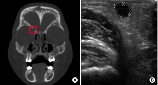
Fig. 1. PNS CT scan. (A) The red

square outlines the 1.8 cm mass- like soft tissue infiltration on the inferior orbital wall. Ultrasono- graphy. (B) Marked area shows the small 0.7 cm cystic mass lesion in the subcutaneous layer of left medial popliteal space.

foamy histiocytes [2,3]. Recent findings suggest that ECD is a clonal disorder, marked by recurrent BRAF V600E muta- tions (>50% of patients diagnosed with ECD) activating MAPK signaling, in which there is not only inflammation driven by aberrant immune activation, but also myeloid ne- oplasm as a potentially important pathogenesis of disease [4]. The usual onset of ECD is middle age, and it is rarely seen in pediatric patients [2]. Here we present a case of 2-year-old patient diagnosed with ECD who underwent al- logeneic hematopoietic stem cell transplantation (HSCT) due to acute lymphoblastic leukemia (ALL).

Case Report

A 2-year and 1-month-old Korean boy was born without any complications and grew up without any health problems. On November 2014, he was admitted to our de- partment with a pale face and leukocytosis of 171,500/ L on complete blood cell count (CBC). The 1

st

bone marrow study showed early pre-B cell ALL with no abnormal gene translocations on PCR, and 46, XY normal male karyotype with hyperdiploidy on chromosome test. Since he had se- vere leukocytosis on initial CBC, we started high-risk ALL induction chemotherapy and planned for allogeneic HSCT.

After induction and consolidation chemotherapy, the fol- low-up bone marrow study showed complete remission.

However, after interim-maintenance chemotherapy, a bone marrow study showed blast cells constituting 2.8% of abso-

lute neutrophil count and aggregation of epithelioid histio- cytes and foamy macrophages, which highly suggested ECD or disseminated juvenile xanthogranuloma. However, the pateint did not show any systemic lesions or symptoms correlated with ECD and we decided to continue with regu- lar observation. The patient then underwent allogeneic HSCT and a bone marrow study 1 month after the trans- plantation and still showed solid aggregates of large atyp- ical histiocytoid cells with mixed chimerism state. On January 2016, 7 months after the transplantation, the patient was admitted to the emergency department with fever, low- er eyelid swelling and palpable left calf nodules.

Laboratory test results including CBC, erythrocyte sed-

imentation rate (ESR), C-reactive protein (CRP), electrolyte

and culture studies were relatively normal with mild ele-

vations of ESR and CRP. Paranasal sinus (PNS) Computed

tomography (CT) scan showed orbital cellulitis with possi-

ble involvement of Lt. nasolacrimal duct and a 1.8 cm exo-

phytic mass lesion (Fig. 1). A whole body bone scan was

done to evaluate leg pains and it showed diffusely in-

creased uptake in the entire skeleton. The bone marrow

study was performed to exclude leukemic or malignant in-

volvement, and aggregation of epithelioid histiocytes was

seen, which is highly suggestive of ECD with complete a

chimerism state of the bone marrow (Fig. 2A). The patient

subsequently underwent excisional biopsy of the left calf

mass lesion. The pathology results were ‘aggregation of

non-Langerhans cell type epithelioid histiocytes with acute

(3)

Fig. 2. Bone marrow biopsy. (A) Aggregation of epithelioid histiocytes is seen in the bone marrow study. The encircled cytosol-rich

cells are foamy macrophages shown in the bone marrow (×200). Left calf nodule biopsy. (B) Aggregation of non-Langerhans cell type epithelioid histiocytes with acute inflammation are seen. The long and relatively thin cells seen in the red circle are histiocytes (×200). (C) Histiocytes circled, long and thin band-like cells are observed at in most of the site (×400).

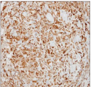
Fig. 3. Immunostaining of left calf nodule biopsy shows CD68

and CD163 positive cells.

inflammation with CD163+, CD68+ and BRAF mutations (+)’ clinically suggestive of ECD (Fig. 2B, C and Fig. 3).

The patient started weekly vinblastine chemotherapy and corticosteroids after pathologic confirmation of ECD. Within 2 weeks of treatment with vinblastine, the patient’s symp- toms of fever and bone pain started to disappear. After 2 months of treatment with vinblastine, a follow-up bone marrow study and whole body bone scan were performed.

The bone marrow study revealed no pathologic evidence of ECD with complete chimerism state and the bone scan showed decreased intensity of diffuse uptake throughout the whole skeleton. The patient is still undergoing vinblas-

tine treatment.

Discussion

ECD is a rare, non-Langerhans cell histiocytic disease

with various clinical manifestations. ECD typically affects

the long bones and often shows symmetrical sclerosis in

the diametaphyseal areas. Extra-skeletal involvement such

as lung, cardiovascular, liver, spleen, retroperitoneum and

central nervous system, can be seen in ECD and the extent

of disease affects its prognosis [2]. Treatment of ECD has

not been standardized yet. The types of treatment include

corticosteroids, cyclosporine, chemotherapy (e.g., vinblas-

tine, cyclophosphamide, etoposide, 6-mercaptopurine, and

doxorubicin), interferon-alpha, surgical resection and radio-

therapy, all of which have variable responses [5]. Among

these treatment choices, interferon-alpha seems to be the

best choice for the initial prolonged treatment of ECD con-

sidering the survival rate [5]. However, recent studies have

shown that 57 to 82% of patients with ECD harbor a BRAF

V600E mutation [4]. Of these studies, two researches, one

from the French Histiocytosis Study Group and one from

Memorial Sloan Kettering Cancer Center (MSKCC), have

shown that targeted therapies (vemurfenib; BRAF inhibitor)

in adult patients with BRAF-mutated histiocytosis have a

dramatic and highly consistent efficacy [6,7]. Along with

these two studies, there are some other reports worldwide

that suggest targeted therapies with BRAF inhibitors (e.g.,

(4)

vemurfenib, dabrafenib) are effective for treatment of ECD patients with BRAF mutations [8]. Moreover, MSKCC has re- cently shown that MEK inhibitors (trametinib, cobimetinib) are rapidly effective in patients with histiocytic neoplasm, which led to the development of clinical trials targeting BRAF wild-type adult histiocytosis [9]. In our case, we are treating the patient with weekly vinblastine and corticos- teroids. Interferon alpha had been our first treatment con- sideration, but steroids and vinblastine was tried first due to the patient’s refusal of a long hospital stay. Though in continuation, treatment seems to be quite effective so far.

According to numerous recent reports and studies of target- ing BRAF, MEK, and mTOR in histiocytosis treatment, muta- tion targeted therapy might be an efficient and standard treatment for ECD in the future. However, a large, mul- ti-center approach of using BRAF inhibitors or MEK in- hibitors in pediatric patients has not been studied yet. In terms of safety along with efficacy, more clinical trials and studies of treating ECD child patients with BRAF/MEK in- hibitors are needed to establish a standard treatment guide- line for children with ECD.

Our case also shows the concurrence of ECD with ALL, which is extremely rare. The first time histiocytes were not- ed in bone marrow biopsy was right after treated with inter- im-maintenance chemotherapy before allogeneic HSCT. We just decided to have careful observation without any further treatment since no other symptoms were present. However after allogeneic HSCT, symptoms mentioned in the case re- port have appeared and found that ECD was the cause.

Since ECD is a very rare disease, we carefully think that ECD seen in the bone marrow after allogeneic HSCT is of recipient origin rather than donor origin. Only two cases of ECD in a child with acute lymphoblastic leukemia (ALL) have been reported so far; a 14-year-old girl who was in remission with pre-B acute lymphocytic leukemia, sub- sequently diagnosed with ECD [10] and a 6-year-old girl di- agnosed with ECD during maintenance therapy for B-cell ALL [11]. Although extremely rare, considering Langerhans cell histiocytosis (LCH), which has been reported to have a close relationship with several malignancies [12], ECD might also have coincidences with some malignancies.

Though it is rare, it is reported that acute leukemia is the

most common malignancy associated with LCH [12]. Several reports suggest that coincident LCH and ALL is due to the fact that they share the same mutation or have an identical T-cell receptor or immunoglobulin rearrangement [13].

Moreover, there is a recent report of high prevalence of myeloid neoplasm in adults with non-Langerhans cell his- tiocytosis [14]. They say that driver mutations in myeloid neoplasm (such as JAK2V617F and CALR mutations) have been frequently detected with coexisting mutations (such as BRAFV600E and MAP2K1 mutations) which are known to be characteristics of histiocytosis. Unfortunately, we did not examine the cytogenetic studies of this patient with ECD aside from the BRAF mutation. Therefore, we could not identify the pathogenesis of this concurrence of ECD and ALL. It is difficult to say whether there is a consid- erable chance of occurrence of ECD during or after ALL treatment at this time. However, because more than half of the ECD patients harbor BRAF mutations and reports of recurrent RAS and PIK3CA mutations found in ECD patients [15], ECD might occur with some malignancies including acute leukemia, owing to the fact that they share same mu- tations or genetic rearrangement finding in cytogenetic studies.

References

1. Chester W. Über Lipoidgranulomatose. Virchows Arch Pathol Anat 1930;279:561-602.

2. Haroche J, Arnaud L, Cohen-Aubart F, et al. Erdheim-Chester disease. Curr Rheumatol Rep 2014;16:412.

3. Veyssier-Belot C, Cacoub P, Caparros-Lefebvre D, et al.

Erdheim-Chester disease. Clinical and radiologic character- istics of 59 cases. Medicine (Baltimore) 1996;75:157-69.

4. Hervier B, Haroche J, Arnaud L, et al. Association of both Langerhans cell histiocytosis and Erdheim-Chester disease linked to the BRAFV600E mutation. Blood 2014;124:1119-26.

5. Jeon IS, Lee SS, Lee MK. Chemotherapy and interferon-alpha treatment of Erdheim-Chester disease. Pediatr Blood Cancer 2010;55:745-7.

6. Haroche J, Cohen-Aubart F, Emile JF, et al. Dramatic efficacy of vemurafenib in both multisystemic and refractory Erdheim-Chester disease and Langerhans cell histiocytosis har- boring the BRAF V600E mutation. Blood 2013;121:1495-500.

7. Hyman DM, Puzanov I, Subbiah V, et al. Vemurafenib in mul-

tiple nonmelanoma cancers with BRAF V600 mutations. N

(5)

Engl J Med 2015;373:726-36.

8. Váradi Z, Bánusz R, Csomor J, et al. Effective BRAF inhibitor vemurafenib therapy in a 2-year-old patient with sequentially diagnosed Langerhans cell histiocytosis and Erdheim-Chester disease. Onco Targets Ther 2017;10:521-6.

9. Diamond EL, Durham BH, Haroche J, et al. Diverse and tar- getable kinase alterations drive histiocytic neoplasms. Cancer Discov 2016;6:154-65.

10. Krishna VV, James TE, Chang KT, Yen SS. Erdheim-Chester disease with rare radiological features in a 14-year old girl with pre-B acute lymphocytic leukemia and diabetes mellitus.

J Radiol Case Rep 2014;8:7-15.

11. Vallonthaiel AG, Mridha AR, Gamanagatti S, et al. Unusual presentation of Erdheim-Chester disease in a child with acute

lymphoblastic leukemia. World J Radiol 2016;8:757-63.

12. Egeler RM, Neglia JP, Puccetti DM, Brennan CA, Nesbit ME.

Association of Langerhans cell histiocytosis with malignant neoplasms. Cancer 1993;71:865-73.

13. Feldman AL, Berthold F, Arceci RJ, et al. Clonal relationship between precursor T-lymphoblastic leukaemia/lymphoma and Langerhans-cell histiocytosis. Lancet Oncol 2005;6:435-7.

14. Papo M, Diamond EL, Cohen-Aubart F, et al. High prevalence of myeloid neoplasms in adults with non-Langerhans cell histiocytosis. Blood 2017;130:1007-13.

15. Emile JF, Diamond EL, Hélias-Rodzewicz Z, et al. Recurrent

RAS and PIK3CA mutations in Erdheim-Chester disease. Blood

2014;124:3016-9.

수치

Fig. 1. PNS CT scan. (A) The red  square outlines the 1.8 cm mass-  like soft tissue infiltration on the  inferior orbital wall
Fig. 3. Immunostaining of left calf nodule biopsy shows CD68  and CD163 positive cells.

참조

관련 문서

It considers the energy use of the different components that are involved in the distribution and viewing of video content: data centres and content delivery networks

After first field tests, we expect electric passenger drones or eVTOL aircraft (short for electric vertical take-off and landing) to start providing commercial mobility

In this group of high-risk patients with severe COVID-19 pneumonia, treatment with lenzilumab was associated with a significantly shorter time to clinical improvement compared

1 John Owen, Justification by Faith Alone, in The Works of John Owen, ed. John Bolt, trans. Scott Clark, "Do This and Live: Christ's Active Obedience as the

The purpose of the present study was to elucidate the effect of photography and reporting activities conducted at a day care center on 5-year-old

- distributes indexes across multiple computers and/or multiple sites - essential for fast query processing with large numbers of documents - many variations:

Perspectives on allogeneic hematopoietic stem cell transplantation in Myeloma..

3) A comparison of the stoichiometric equation with the experimental kinetic expression can suggest whether or not we are dealing with an elementary reaction. 4) If one