Vol. 3, No. 2, pp 80~88, 2009
Copyright � 2009 by Korean Society of Spine Surgery
This is an open-access article distributed under the terms of the Creative Commons Attribution License (http://creativecommons.org/licenses/by/3.0), Which permits unrestricted use, distribution, and reproduction in any medium, provided the original work is properly cited.
Asian Spine Journal�pISSN 1976-1902 eISSN 1976-7846
Received Dec 29, 2008; 1st revised Sep 15, 2009; 2nd revised Oct 11, 2009; accepted Oct 12, 2009 Corresponding author:Kee-Yong Ha, MD
Department of Orthopaedic Surgery, Seoul St. Mary’s Hospital, The Catholic University of Korea College of Medicine 505 Banpo-dong, Seocho-gu, Seoul 137-040, Korea
Tel: +82-2-590-1464, Fax: +82-2-535-9834, E-mail: [email protected]
Kyphotic Angle Progression of Thoracic and Thoracolumbar Tuberculous Spondylitis after Surgical Treatment: Comparison with Predicted Kyphosis Outcome after Conservative Treatment
Soon-Eok Kwon*, Jae-Hyuk Shin�, Ki-Ho Na�, Yoon-Chung Kim�, Kee-Yong Ha�
*Department of Orthopaedic Surgery, The Chungju St. Mary’s Hospital, Chungju, Korea,
�Department of Orthopaedic Surgery, Hangang Sacred Heart Hospital, Hallym University College of Medicine, Seoul, Korea,
�Department of Orthopaedic Surgery, St. Paul’s Hospital, The Catholic University of Korea College of Medicine, Seoul, Korea,
�Department of Orthopaedic Surgery, Seoul St. Mary’s Hospital, The Catholic University of Korea College of Medicine, Seoul, Korea
S
Sttuuddyy DDeessiiggnn:: Retrospective comparative study P
Puurrppoossee:: To compare the progression of the kyphotic angle (KA) in a surgically treated group with the predicted outcome of a conservatively treated group.
O
Ovveerrvviieeww ooff LLiitteerraattuurree:: Late onset kyphosis is a complication of tuberculous spondylitis making its prevention a major goal of surgery.
M
Meetthhooddss:: Twenty six consecutive patients underwent an anterior reconstruction and posterior instrumented fusion in con- junction with antituberculous chemotherapy. The mean follow up was 56 months (range, 28 to 112 months). The patients were divided into subgroups based on the involved region of the thoracic and the thoracolumbar spine, initial KA, and the initial vertebral body loss (VBL(x)). The predicted KA (KAPd) was calculated using the formula, KAPd=5.5+30.5 VBL(x), to predict the final gibbus deformity. Kyphotic angle progression (ΔKA) based on the radiographic measurements after surgery (ΔKAR), and the predicted outcome of conservative treatment (ΔKAP) with chemotherapy were compared.
R
Reessuullttss:: Among the subgroups of the regions involved and initial KA, the ΔKA was radiographically superior with a reduced amount of kyphogenesis in the surgery group than the predicted outcome of the conservatively treated patients (p
�0.05). The radiographic ΔKA was similar (p�0.05) with VBL(x)≤0.5 in the VBL(x) subgroup.
C
Coonncclluussiioonnss:: These results showed that in the VBL(x) subgroup, an initial VBL(x)≤0.5 is an indication of conservative anti- tuberculous chemotherapy without surgery.
Key WWords: Tuberculous spondylitis, Kyphosis, Initial vertebral body loss
Introduction
Kyphosis and paralysis are the major outcomes of spinal tuberculosis (TB). Spinal TB can stabilize spontaneously
without progression of the deformity. However, the devel- opment of a kyphotic deformity is a serious complication. It is not only cosmetically unpleasant but also impairs the car- diopulmonary function, causes pain from nerve impinge- ment between the ribs and pelvis, and/or causes late-onset
paraplegia. A severely deformed kyphosis can be treated only by corrective surgery. However, this is a challenging option with a high complication rate1. A kyphotic deformity can be prevented if spinal TB is diagnosed early and antitu- berculosis chemotherapy is administered promptly. Howev- er, some kyphotic deformity may still develop even with adequate medical treatment. The sequela of a kyphotic deformity has been a subject of research into the treatment of patients with spinal TB.
The fifth report of the Medical Research Council Work- ing Party on Tuberculosis of the Spine (MRC)2stated the following: 1) a kyphotic deformity develops most severely in the thoracic spine region, which is followed in order by the thoracolumbar, lumbar and lumbosacral spine after treatment with standard TB medications; 2) the greater the pretreatment kyphotic deformity, the less likely the kypho- sis will progress further after initiating treatment; and 3) further collapse is appreciable when the initial vertebral body loss upon admission was small (< 2 vertebrae), while the initial vertebral body loss of two or three segments showed no further collapse on average2.
Rajasekaran and Shanmugasundaram3 reported that kyphosis progresses gradually in the thoracic and thora- columbar spine above L2 during conservative treatment with anti-tuberculous chemotherapy. Regarding the degree of kyphogenesis, the initial vertebral body loss (VBL(x)) was considered to be the determining factor, while the initial kyphotic angle (KA) was not. A predicted KA (KAPd) was formulated as ‘KAPd=5.5+30.5VBL(x)’ with a correlation coefficient of 0.83 and an accuracy of 90%. This formula applies to the thoracic and thoracolumbar region above L2, which forms a normal kyphotic curve. In the lumbar spine, the lordotic curvature contributes to the progression of the deformity in the thoracic spine4.
This study examined the change in the sagittal profile after surgical treatment in tuberculous (Tbc) spondylitis patients, and compared it with the predicted change of the sagittal profile after chemotherapy alone. It is believed that the indications for surgery or conservative treatment might be better determined using this information.
Materials and Methods
1. Patients
This study was approved by the institutional review
board. Twenty six patients (M:F=12:14) were examined after undergoing surgery for thoracic and thoracolumbar TB between 1992 and 2001. The mean follow up period and age was 56 months (range, 28 to 112 months) and 41.7 years (range, 18 to 73 years), respectively. The regional subgroups included 12 and 14 cases in the thoracic (T: from T2 to T10) and thoracolumbar (TL: from T11 to L2), respectively.
2. Operation method
Anterior radical surgery with an autogenous iliac bone graft was performed in all cases, and posterior instrumenta- tion (TSRHTM; Texas Scotish Rite Hospital, Medtronic- Sofamor Danek, Memphis, TN, USA) and fusion were per- formed using an autogenous cancellous bone graft.
3. Measurement
The KA progressions (ΔKA)’s were obtained. ΔKA fol- lowing surgery (ΔKAR) was measured by comparison of the difference in the kyphotic angle of the last follow up standing lateral radiograph (KAFinal) to the preoperative standing lateral radiograph (KAPre) (Table 1). The radi- ographic measurements were performed by two indepen- dent spine surgeons that did not participate in the surgery.
Each surgeon performed the measurements twice; the mean of the measurements was used as the final value. The KAPd, or the final gibbus deformity, was calculated using the for- mula ‘KAPd=5.5+30.5 VBL(x)’ by measuring the VBL(x).
The estimated KAPd was calculated presuming that only antituberculous chemotherapy was administered as conserv- ative treatment. Predicted kyphotic angle progression (Δ KAP) was calculated by difference of KAPd and KAPre
(Table 1).
(1) Measurement of the kyphotic angle
The measured KA angle was the Cobb’s angle between the upper border of the upper normal vertebra and the lower border of the lower normal vertebra on a simple standing lateral radiograph of the lesion, as previously reported by the MRC2,5,6.
(2) Measurement of the preoperative VBL(x)
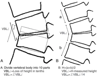
Rajasekaran and Shanmugasundaram3assessed the loss of the vertebral body, which we regarded as VBL1, using the following method: divide the lateral height of each vertebra
into 10 equal parts on a lateral radiograph; measure the loss of height in tenths from each vertebra; and add the loss of height proportions from each vertebra. The sum of the destroyed portions (ΣVBLι.
) from the affected vertebrae was the VBL1(Fig. 1).
Jain et al.7proposed a different method for assessing the loss of the vertebral body, which we regarded as VBL2. Pre- sumed normal anterior height (H) of the affected vertebral body was arithmetic mean of the anterior heights of the upper and lower normal vertebral bodies. The loss of the anterior height was calculated by subtracting the measured height from the H. The anterior height loss per affected ver- tebral body, was then summed to obtain the total height loss (ΣVBLι.
). The VBL2was the ratio of ΣVBLι.
to H. In this study, the arithmetic mean of the above mentioned two cal- culations was used as the initial vertebral body loss:
VBL(x) = (VBL1+VBL2)/2 (Fig. 1).
4. Patient assessment by subgroup
The patients were divided into three independent sub- groups. The first subgroup was based on the region affect- ed: thoracic (T) (n=12), thoracolumbar (TL) (n=14) and combined (T+TL) (n=26). The second subgroup was based on the KAPre: KAPre≤30。(n=22), KAPre≤25。(n=19), KAPre≤20。(n=15), and KAPre≤15。(n=10). The third sub- group was based on the VBL(x): VBL(x)≤ 1.0 (n=23), VBL(x)≤0.75 (n=20), and VBL(x)≤ 0.5 (n=12). The
‘T+TL’ group refers to all patients. The range was set by the authors to include a gradual increase in the range in an attempt to identify the threshold values to use as guidelines, assuming that that the increase in KA and VBL(x) were Table 1. Patient demographics
Patients Age Gender FU
Level Involved KAPre KAPost KAFinal KAPd
VBL(x) ΔKAR ΔKAP
(month) vertebra (�) (�) (�) (�) (KAFinal-KAPre) (KAPd-KAPre)
1 65 M 39 T 7,8 20 15 22 23.5 0.59 2 3.5
2 43 M 29 T 5,6 15 11 15 19.2 0.45 0 4.2
3 54 M 45 T 3,4,5 38 20 25 37.2 1.04 -13 -0.8
4 29 F 47 T 6.7 25 16 35 26.5 0.69 10 1.5
5 17 M 27 T 2,3 35 30 35 37.5 1.05 0 2.5
6 26 F 49 T 5,6,7 30 20 25 26.9 0.7 -5 -3.2
7 36 F 56 T 7,8 7 5 10 11.6 0.2 3 4.6
8 28 M 52 T 8,9 25 15 20 27.5 0.72 -5 2.5
9 29 M 26 T 4,5 22 20 23 25.3 0.65 1 3.3
10 73 F 26 T 6,7 15 10 13 23.2 0.58 -2 8.2
11 73 F 43 T 7,8,9 20 20 30 38.7 1.09 10 18.7
12 16 F 36 T 8,9 10 6 10 13.4 0.26 0 3.4
13 52 F 25 TL 12,1 18 12 14 14.0 0.28 -4 -4.0
14 69 F 27 TL 11,12 34 24 26 34.8 0.96 -8 0.8
15 37 M 65 TL 1,2 10 6 10 15.9 0.34 0 5.9
16 25 F 112 TL 12,1 30 15 22 21.7 0.53 -8 -8.3
17 29 F 48 TL 1,2 20 5 10 26.5 0.69 -10 6.5
18 32 M 25 TL 11,12,1 15 10 13 16.5 0.36 -2 1.5
19 61 M 36 TL 1,2 5 2 8 19.8 0.47 3 14.8
20 38 M 66 TL 1,2 8 1 9 10.1 0.15 1 2.1
21 30 M 33 TL 1,2 22 18 26 19.8 0.47 4 -2.2
22 41 M 62 TL 11,12 ,1,2 30 24 28 31.7 0.86 -2 1.7
23 38 F 81 TL 1,2 17 7 15 13.1 0.25 -2 -3.9
24 24 F 87 TL 12,1 15 7 11 11.6 0.2 -4 -3.4
25 64 F 49 TL 11,12 34 26 25 35.1 0.97 -9 1.1
26 44 F 68 TL 10,11,12,1 15 10 7 16.8 0.37 -8 1.8
FU: follow up, KA: kyphotic angle, VBL(x): initial vertebral body loss, KAPre: preoperative KA, KAPost: immediate postoperative KA, KAFinal: final postoperative KA, KAPd: predicted KA by formula ‘KAPd=5.5+30.5 VBL(x)’ after conservative treatment with anti-tuberculous chemotherapy, ΔKAR: radiographically measured kyphotic angle progression, ΔKAP: predicted kyphotic angle progression by formula-calculation.
associated with the progression of the disease.
For each subgroup, the ΔKARand ΔKAPwere com- pared using a paired t-test. A p-value<0.05 was considered significant (SPSS ver. 10.0; SPSS Inc., Chicago, IL, USA).
Results
A mean of 2.4(2-4) vertebral bodies were affected by bone destruction. Preoperative Pott’s paraplegia and para-
paresis were observed in 8 of the 26 patients. Table 1 lists the patient demographic information.
1. Regional subgroups
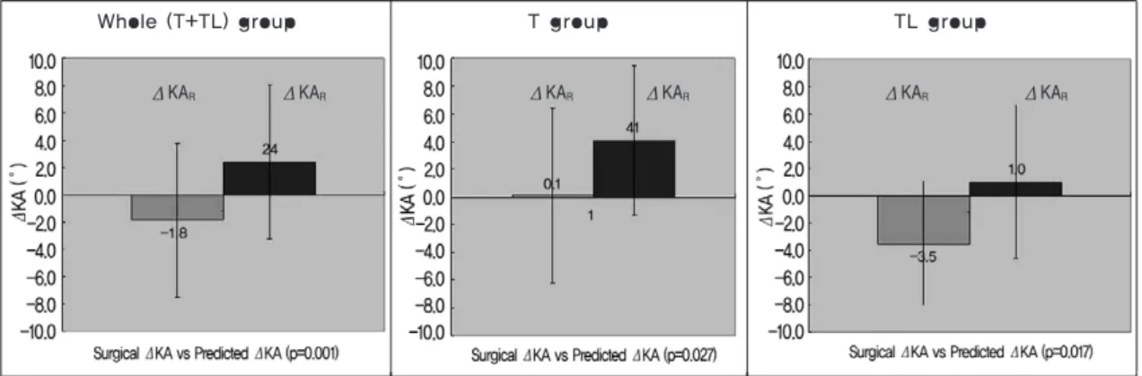
In the T group, the KAPre, KAFinaland KAPdwas 21.8。± 9.4。, 21.9。±8.8。and 25.9。±8.8。, respectively. The radi- ographically measured kyphotic angle progression (ΔKAR) and predicted kyphotic angle progression (ΔKAP) were sig- nificantly different (p=0.027) (Fig. 2, Table 2).
In the TL group, the KAPre, KAFinaland KAPdwas 19.5。± 9.4。, 16.0。±7.7。and 20.5。±8.4。, respectively. A compari- son of the kyphotic angle progression between ΔKARand ΔKAPshowed a significant difference (p=0.017) (Fig. 2, Table 2).
For the T-TL group, which represents the entire cohort, the KAPre, KAFinaland KAPdwas 20.6。±9.3。, 18.7。±8.6。
and 23.0。±8.8。, respectively. The ΔKARwas significantly smaller than the ΔKAP(p=0.001) (Fig.2, Table 2).
2. KA subgroup
For the KAPre≤30。group, the KAPreKAFinaland KAPdwas 17.9。±7.4。, 17.1。±8.1。, and 20.6。±7.3。, respectively (Table 2). The ΔKARwas significantly smaller than the Δ KAp(p=0.001). Each subgroup of KA was analyzed to deter- mine if the ΔKARwas significantly smaller than the pre- dicted outcome, a kyphogenesis of ΔKAPd(Fig.3, Table 2).
3. VBL(x) subgroup
In the VBL(x)≤1.0 group, the KAPreKAFinaland KAPdwas 19.2。±8.6。, 17.3。±7.8。, and 21.1。±7.4。, respectively. The Table 2. The mean (±SD) of the results
Level subgroup N KAPre (�) KAPost(�) KAFinal (�) KAPd (�) VBL(x) p-value (ΔKAR Vs ΔKAPd )
T Regional 12 21.8±9.4 15.7±7.0 21.9±8.8 25.9±8.8 0.67±0.29 0.027
TL Regional 14 19.5±9.4 11.9±8.3 16.0±7.7 20.5±8.4 0.49±0.28 0.017
T+TL (whole) Regional 26 20.6±9.3 13.7±7.8 18.7±8.6 23.0±8.8 0.57±0.29 0.001
T+TL KA1≤30� 22 17.9±7.4 11.6±6.4 17.1±8.1 20.6±7.3 0.50±0.24 0.009
T+TL KA1≤25� 19 16.0±5.9 10.3±5.8 15.8±8.0 19.6±7.3 0.46±0.24 0.015
T+TL KA1≤20� 15 14.0±4.9 8.5±4.9 13.1±6.0 18.3±7.5 0.42±0.24 0.002
T+TL KA1≤15� 10 11.5±.4.0 6.8±3.5 10.6±2.5 15.8±4.2 0.34±0.14 0.003
T+TL VBL≤1.0 23 19.2±8.6 12.4±7.2 17.3±7.8 21.1±7.4 0.50±0.24 0.005
T+TL VBL≤0.75 20 17.2±7.2 10.6±5.7 15.9±7.5 19.1±5.8 0.45±0.19 0.024
T+TL VBL≤0.5 12 13.1±5.1 7.9±4.6 12.3±5.1 15.2±3.4 0.32±0.11 0.071
KAPre: preoperative KA, KAPost: immediate postoperative KA, KAFinal: final postoperative KA, KAPd: predicted KA by formula, VBL(x): initial vertebral body loss, ΔKAR: radiographically measured kyphotic angle progression, T: thoracic, TL: Thoracolumbar.
Fig. 1. The diagram shows the method used to assess the preop- erative initial vertebral body loss (VBL(x)). (A) VBL1was defined as the fractional loss of the vertebral body area. When more than one vertebra was involved, the sum of each loss (∑VBLi) was calculated as the VBL1. (B) VBL2was defined as the fractional loss of the anterior vertebral body height, assuming that the original anterior vertebral height (H) was the arithmetic mean of the anterior height of the upper (a) and lower (b) normal vertebral bodies. In this study the arithmetic mean of VBL1and VBL2was used as the ‘VBL(x)’.
A B
a
b
A: Divide vertebral body into 10 parts VBLι∙=Loss of height in tenths VBL1=ΣVBLι∙
B: H=(a+b)/2
VBLι∙=H-measured height VBL2=ΣVBLι∙/ H VBLι∙
VBLι∙
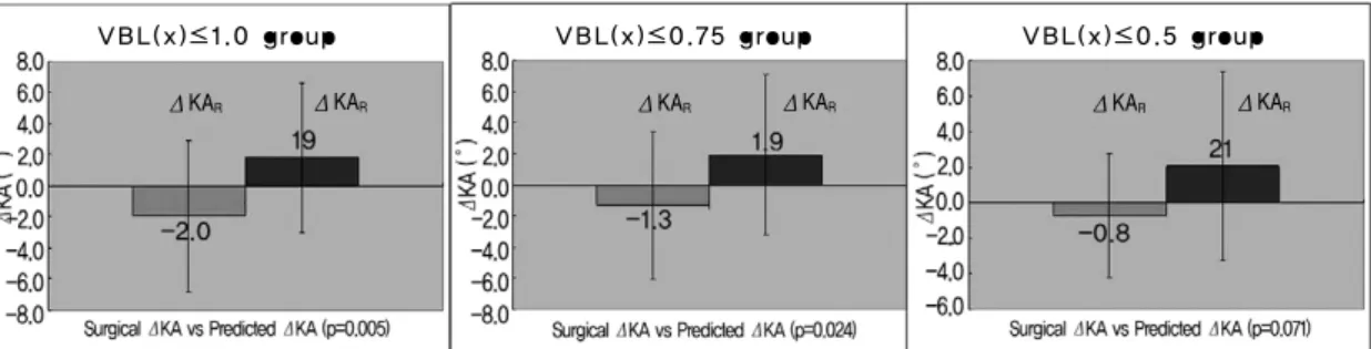
ΔKARwas significantly smaller than the ΔKAP(p=0.005).
In the VBL(x)≤ 0.75 group, the ΔKARwas significantly smaller than the ΔKAP(p=0.024) (Fig. 4, Table 2).
However, in the VBL(x)≤0.5 group, the KAPreKAFinal
and KAPdwas 13.1。±5.1。, 12.3。±5.1。and 15.2。±3.4�, respectively (Table 2). The ΔKARand ΔKApwere similar (p=0.071) (Fig. 4, Table 2).
Discussion
The fifth MRC report of a 5-year follow-up2showed that ambulatory antituberculous chemotherapy was successful when compared with combined bed rest for six months, or a plaster-of-Paris jacket for nine months. Antituberculous medication was used for 18 months, and the regimen included isoniazid and para-aminosalicylic acid with or without additional streptomycin during the first three months2. Tull and Kumar8, Tuli9reported that only 19% of patients showed progression of their kyphotic deformity>10。after rest and conservative treatment with antituberculous drugs.
The sixth MRC report of a 5-year follow-up10showed similarly favorable results with ambulatory chemotherapy versus debridement, as well as with debridement versus rad- ical treatment with autologous bone graft reconstruction.
The favorable results included the following: return to phys- ical activity, absence of neurological abnormalities, absence of abscess formation, absence of the development of sinus tracts, and the presence of radiographically quiescent spinal lesions10.
The eighth MRC report after a 10-year follow-up6com- pared a radical anterior autologous bone reconstruction with
debridement of the abnormal tissue leaving the apparently unaffected bone. The sagittal profile was superior after the radical series compared to after the debridement cases. Ver- tebral body loss increased by 0.05 for the radical cases, while it increased by 0.23 for the debridement cases. The KA decreased by 1.4。for the radical cases, whereas for the debridement cases, it increased by 9.8。in the thoracic and thoracolumbar lesions. The KA decreased by 0.5。in the radical cases, whereas it increased by 7.6。for the debride- ment cases with lumbar lesions.
The bone loss and increase in KA usually occurred within the first 18 months from the initial diagnosis and treatment.
Upadhyay et al.11 reported that anterior column support resulted in less kyphotic deformity than simple anterior debridement. Rajasekaran and Shanmugasundarm12reported less kyphogenesis with little bone loss in the single-segment lesions than in multi-segment lesions.
Firm fixation is essential for the healing of bone infec- tions and bone fusion, and cast immobilization has been used to treat vertebral infections. The development of instruments has led to successful treatment by instrumented fusion for vertebral infections. Moon et al.13 reported that the kyphosis could be corrected and maintained effectively in adult spinal TB by adding posterior instrumented fusion.
Sundararaj et al.14 reported the following advantages of adjuvant posterior instrumented fusion: prevention of graft dislodgement; correcting and maintaining the kyphosis;
resulting in a superior outcome for intervertebral bone fusion. No additional risk was found for persistent infec- tions due to the posterior instrumentation14,15. For pyogenic infections, internally fixed metal appliances facilitate the attachment and proliferation of microbes, forms a biofilm
Fig. 2. Comparison of the kyphotic angle (KA) progression (ΔKA) in the subgroups: thoracic (T), thoracolumbar (TL), and combined (T+TL) groups. The surgical ΔKA (ΔKAR) was significantly smaller than the predicted ΔKA (ΔKAP) which is the predicted outcome of conservative treatment: p=0.027 (T); p=0.017 (TL); and p=0.001 (T+TL).
Whole (T+TL) group T group TL group
ΔKAR ΔKAR ΔKAR ΔKAR ΔKAR ΔKAR
on the surface of the metal and prevents antibiotics from reaching the infection and/or inflammation and thereby healing. This occasionally necessitates removal of the metal implants16. Ha et al.17performed an in-vitro experiment and reported much greater adherence and multiplication of biofilm formation on the metal segments with Staphylococ-
cus epidermidis strains than in Mycobacterium tuberculosis strains. They demonstrated a more favorable environment for instrumentation in the treatment of Tbc spondylitis.
Instrumentation to the posterior side, which is opposite to the initial infection, is not considered to be harmful17.
For measuring the VBL(x), Rajasekaran and Shanmuga-
Fig. 4. Comparison of ΔKA in the VBL(x) subgroups (‘VBL(x)≤1.0’, ‘VBL(x)≤0.75’, and ‘VBL(x)≤0.5’). In the ‘VBL(x)≤0.5’ group, the ΔKAR was not significantly different from the ΔKAPwhich is the predicted KA after conservative treatment (p=0.071). In the other groups, ΔKARwas significantly smaller than ΔKAP, indicat- ing smaller amount of kyphogenesis in the surgical group: p=0.005 (VBL(x)≤1.0); and p=0.024 (VBL(x)≤0.75).
Fig. 3. Comparison of the ΔKA by the preoperative KA (KAPre) subgroups. For the subgroups of ‘KAPre≤30�’,
‘KAPre≤25�’, ‘KAPre≤20�’, and ‘KAPre≤15�’, the surgical ΔKA (ΔKAR) was significantly smaller than the pre- dicted ΔKA (ΔKAP): p=0.009 (KAPre≤30�); p=0.015 (KAPre≤25�); p=0.002 (KAPre≤20�); and p=0.003 (KAPr
≤15�), respectively.
KAPer≤30。group KAPer≤25。group
KAPer≤1 5。group KAPer≤20。group
ΔKAR ΔKAR ΔKAR
ΔKAR ΔKAR
ΔKAR ΔKAR ΔKAR ΔKAR ΔKAR ΔKAR
ΔKAR ΔKAR
ΔKAR
VBL(x)≤1.0 group VBL(x)≤0.75 group VBL(x)≤0.5 group
sundaram3divided each affected vertebral body into 10 parts (Fig. 1). This calculation led to frequent ambiguity and arbitrary measurements because severely damaged ver- tebrae often do not have definite borders7. On the other hand, Jain et al.7used the arithmetic mean from the anterior heights of the upper and lower vertebrae, which were nor- mal, as the ‘presumed normal anterior height’ at the affect- ed level. The ‘anterior height loss’ is the difference between the presumed normal height and actual height measured. In lesions at multiple levels, the anterior height losses were added to obtain the ‘total height loss’. The ‘initial vertebral body loss’ is the ratio between the ‘total height loss’ to the
‘presumed normal anterior height’. Jain et al.7suggested an approximate 76% accuracy using their method for conserv- ative treatment. However, this method may not reflect the precise vertebral body loss if the anterior cortical bone is spared selectively in the lesion distribution. In this study, the arithmetic mean of the above two calculations for the VBL (x was used) to ameliorate the shortcomings of each (Fig. 1).
The results showed significantly less thoracic KA pro- gression after surgery than the predicted outcome after con- servative treatment: (ΔKAR=0.1。±6.3。vs. ΔKAP=4.1。± 5.4v; p=0.006). In addition, the thoracolumbar KA progres- sion was significantly less after surgery than after the pre- dicted-conservative treatment: (ΔKAR=-3.5。±4.6。vs. Δ KAP=1.0。±5.6。; p=0.000). The KA progression of the entire cohort including the thoracic and thoracolumbar spine subgroups was significantly less after surgery than after the predicted outcome with conservative treatment:
(ΔKAR=-1.8。±5.6。vs. ΔKAP= 2.4。±5.6。; p=0.001). The results suggest that surgical treatment was justified based on the sagittal profile outcome because the ΔKAR, which rep- resents the kyphotic angle progression after surgery, was significantly smaller than the ΔKAP, which represents the kyphotic angle progression in the formula for predicting the outcome.
For the KA subgroup, all subgroups with a ‘KA≤30�’,
‘KA≤20。’ and ‘KA≤15。’ showed a significantly smaller ΔKARthan ΔKAP, suggesting less kyphotic angle progres- sion after surgical treatment than after the predicted out- come of conservative treatment-alone. The same conclusion in all KA subgroups confirms the efficacy of surgical treat- ment. However, the KA subgroup results did not suggest a range in which conservative treatment-alone might be justi- fied.
For the VBL(x) subgroup, the ΔKARand the ΔKAP
were not significantly different in the ‘VBL(x)≤0.5’ sub- group (p=0.071). This suggests that the surgical treatment group did not have a significantly superior sagittal profile compared to the predicted outcome of conservative treat- ment. As the progression of kyphosis was the main concern for the surgical treatment of Tbc spondylitis, conservative treatment might be indicated initially in patients identified with a VBL(x)≤0.5. The other VBL(x) subgroups showed that the ΔKARwas significantly smaller than the ΔKAP, justifying surgical treatment for improved outcome on the sagittal profile. The surgical treatment used in all cases was an anterior-posterior combined surgery (anterior decom- pression, interbody fusion, and posterior instrumented
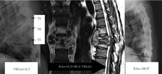
Fig. 5. A 26-year-old female patient with T 5-7 Tbc spondylitis, with a VBL(x) of 0.7: the KAPrewas 30�before surgery and final postoperative KA (KAFinal) was 25�. The predicted KA (KAPd) was 26.9�.
fusion with bone graft) (Fig. 5). The KA was corrected sub- stantially by the combined anterior column support and pos- terior instrumentation and the correction was maintained.
The limitations of this study include the following. The control group consisted of theoretical, calculated, results.
The accuracy of the ‘equation’ requires further statistical validation to be considered a reliable control group. Howev- er, in a limited cohort with a small number of patients, dif- ferent patients with the same KA and VBL(x) might include a range of factors, such as co-morbidities that reduce the homogeneity of the group, which bias the comparison. This study compared the surgical and predicted results on the same patient. The VBL(x) was calculated by taking the mean of the two methods but this value requires further val- idation. In addition to the sagittal profile discussed in this study, the presence of neurological complications would be an additional independent factor indicating a need for surgi- cal treatment. Subsidence at the anterior strut graft would be likely to occur due to endplate injury either by the disease process or by the surgical procedure, even though the disc space is destroyed less aggressively compared to the patho- genesis of pyogenic spondylitis18. This endplate -associated subsidence might explain some of the variance of the subsi- dence observed in our data. However, the statistical analysis still leads to certain conclusions based on a comparison of the two groups. In subgroup analysis, conservative treat- ment appears to be warranted with an initial VBL(x) of ≤ 0.5. Nevertheless, the KA subgrouping did not suggest guidelines for conservative treatment. Further study with a larger cohort would enable statistical analysis to improve the categorization of patients, as well as a comparison of surgically and medically treated groups.
Conclusions
For patients with thoracic and thoracolumbar Tbc spondylitis, surgical treatment was more effective for kyphosis correction than the predicted outcome of conserv- ative treatment. An initial VBL(x) of ≤0.5 on the sagittal profile after surgical treatment was not significantly superi- or to the outcome predicted by conservative treatment. The KA is not a parameter that can be used to determine whether to perform surgical treatment. These results suggest that a VBL(x) of ≤ 0.5 is an indication for conservative antituberculous chemotherapy.
REFERENCE
01. Tuli SM: Severe kyphotic deformity in tuberculosis of the spine. Int Orthop 1995; 19: 327-331.
02. A five-year assessment of controlled trials of in-patient and out-patient treatment and of plaster-of-Paris jackets for tuberculosis of the spine in children on standard chemotherapy. Studies in Masan and Pusan, Korea. Fifth report of the Medical Research Council Working Party on tuberculosis of the spine. J Bone Joint Surg Br 1976; 58:
399-411.
03. Rajasekaran S, Shanmugasundaram TK: Prediction of the angle of gibbus deformity in tuberculosis of the spine. J Bone Joint Surg Am 1987; 69: 503-509.
04. Moon MS, Moon YW, Moon JL, Kim SS, Sun DH: Con- servative treatment of tuberculosis of the lumbar and lum- bosacral spine. Clin Orthop Relat Res 2002; (398): 40-49.
05. Konstam PG, Blesovsky A: The ambulant treatment of spinal tuberculosis. Br J Surg 1962; 50: 26-38.
06. A 10-year assessment of a controlled trial comparing debridement and anterior spinal fusion in the management of tuberculosis of the spine in patients on standard chemotherapy in Hong Kong. Eighth Report of the Medical Research Council Working Party on Tuberculosis of the Spine. J Bone Joint Surg Br 1982; 64: 393-398.
07. Jain AK, Aggarwal PK, Arora A, Singh S: Behaviour of the kyphotic angle in spinal tuberculosis. Int Orthop 2004;
28: 110-114.
08. Tull SM, Kumar S: Early results of treatment of spinal tuberculosis by triple drug therapy. Clin Orthop Relat Res 1971; (81): 56-70.
09. Tuli SM: Results of treatment of spinal tuberculosis by
“middle-path” regime. J Bone Joint Surg Br 1975; 57: 13- 23.
10. Five-year assessments of controlled trials of ambulatory treatment, debridement and anterior spinal fusion in the management of tuberculosis of the spine. Studies in Bul- awayo (Rhodesia) and in Hong Kong. Sixth report of the Medical Research Council Working Party on Tuberculosis of the Spine. J Bone Joint Surg Br 1978; 60: 163-177.
11. Upadhyay SS, Sell P, Saji MJ, Sell B, Hsu LC: Surgical management of spinal tuberculosis in adults. Hong Kong operation compared with debridement surgery for short and long term outcome of deformity. Clin Orthop Relat Res 1994; (302): 173-182.
12. Rajasekaran S, Soundarapandian S: Progression of
kyphosis in tuberculosis of the spine treated by anterior arthrodesis. J Bone Joint Surg Am 1989; 71: 1314-1323.
13. Moon MS, Woo YK, Lee KS, et al: Posterior instrumen- tation and anterior interbody fusion for tuberculous kypho- sis of dorsal and lumbar spines. Spine (Phila Pa 1976) 1995; 20: 1910-1916.
14. Sundararaj GD, Behera S, Ravi V, et al: Role of posteri- or stabilisation in the management of tuberculosis of the dorsal and lumbar spine. J Bone Joint Surg Br 2003; 85:
100-106.
15. Oga M, Arizono T, Takasita M, Sugioka Y: Evaluation of the risk of instrumentation as a foreign body in spinal tuberculosis. Clinical and biologic study. Spine (Phila Pa 1976) 1993; 18: 1890-1894.
16. Gracia E, Fernandez A, Conchello P, et al: Adherence of Staphylococcus aureus slime-producing strain variants to biomaterials used in orthopaedic surgery. Int Orthop 1997;
21: 46-51.
17. Ha KY, Chung YG, Ryoo SJ: Adherence and biofilm for- mation of Staphylococcus epidermidis and Mycobacterium tuberculosis on various spinal implants. Spine (Phila Pa 1976) 2005; 30: 38-43.
18. Ha KY, Shin JH, Kim KW, Na KH: The fate of anterior autogenous bone graft after anterior radical surgery with or without posterior instrumentation in the treatment of pyo- genic lumbar spondylodiscitis. Spine (Phila Pa 1976) 2007;
32: 1856-1864.