142
목 적 :회전근 개의 파열과 동반된 견관절 질환은 상지의 통증 및 장애를 일으키는 원인 중 가장 흔한 질환이며 파열은 전층 파열 보다는 부분 파열인 경우가 많다. 실제 임상 치료시 대부분의 환자에서 보존적 요법으로 증상의 호전을 얻을 수 있으며, 여러 저자들에 의해 회전근 개의 자연 치유력에 대한 연구가 진행 되었으나 명확히 밝혀지지는 않았다. Matrix metalloproteinases (MMPs)는 다양한 결체 조직 및 염증 세포들로부터 생성되고 분비되며 정상 조직 또는 병적 상태의 조직에 작용하여 조직의 변성 및 재형성에 관여하는 것으로 알려져 있다. 저자들은 동물 실험을 통해 회전근 개의 치유 과정에서 MMP-2의 발현을 알아보았다.
대상 및 방법 : 실험 동물로는 성숙한 흰쥐(Sprague-Dawley rat)를 이용하였다. 20마리는 실험군으로, 나머지 20마리는 대조군으로 하여 실험군에서는 회전근 개의 부분 파열을 만들어 4주 후에 모두 희생시켜 조직학적 검사 및 역전사 중합효소 연쇄반응을 시행하였다.
결 과 : 육안 검사에서 인위적인 결손부위는 육아조직으로 덮였다. 조직학적 소견에서는 섬유 아세포 및 혈관의 활발한 증식을 보여 어느 정도의 치유력
이 있을 것이라고 생각되었다. 면역 조직화학적 검사 결과에서 MMP-2는 회전근 개 결손이 있던 부위에 진하게 염색되었고, 역전사 중합효소 연쇄반응에 서도 MMP-2의 발현이 관찰되었다.
결 론 :이상의 실험의 결과로 미루어 볼 때 회전근 개의 재형성 및 치유 과정은 MMP-2와 연관성이 있을 것으로 추정된다.
색인 단어 : 회전근 개, 치유, MMP-2
142
흰쥐의 급성 회전근 개 파열 치유과정에서 MMP-2의 발현
강수용∙이한준∙박제성*
중앙대학교 의과대학 정형외과학교실, 생화학교실*
회전근 개 파열과 동반된 견관절 통증은 상지의 관절통증 중 비교적 흔하다2,7). 회전근 개 파열은 전층 파열보다는 부분 파열 이 많으며5) 회전근 개 부분 파열은 재활 치료 및 운동 요법과 같은 비수술적 요법으로 증상의 호전을 얻을 수 있다. 따뜻한
물찜질이나 초음파와 같은 온열 치료는 심부 연부 조직의 혈액 양을 증가시켜 건조직의 치유를 촉진하며12,23), 회전근개 강화운 동은 견관절 근육 불균형으로 발생한 증상들을 호전시킬 수 있 다고 알려 졌으나4,12), 회전근 개 부분 파열의 치유력에 대해서
142 142 통신저자 : 강 수 용
서울시 중구 필동2가 82-1 중앙대학교 부속 필동병원 정형외과 TEL: 02-2260-2403∙FAX: 02-2271-1710 E-mail: [email protected]
*본 논문은 2001년도 중앙대학교 교내 연구비 지원에 의해 이루어졌음.
Expression of MMP-2 in the Healing Process of Acute Tear of the Rotator Cuff Tendon in Rats
Soo-Yong Kang, M.D., Han-Jun Lee, M.D., and Je-Sung Park, M.D.*
Department of Orthopaedic Surgery, College of Medicine, Chung-Ang University, Seoul; Department of Medical Biochemistry and Molecular Biology*, College of Medicine, Chung-Ang University, Seoul, Korea
Purpose :Rotator cuff diseases combined with cuff tear are the most common causes of pain and disability of the upper extremity.
Most rotator cuff tears are of partial thickness and respond well to conservative treatment. Many authors have addressed the capacity of cuff tendon healing, but little is known of the precise healing process. The MMPs (matrix metalloproteinases) are synthesized and secreted from connective tissue cells and are known to participate in the degradation and remodeling of organs in both normal and pathologic states. Using a rat model, we studied the healing process of rotator cuff tendon tear with reference to the expression of MMP-2.
Materials and Methods :Forty Mature Sprague-Dawley rats were used in this anima study. Twenty rats were assigned to the experimen- tal group, the other twenty to the control group. In the experimental group, a partial thickness tear of the rotator cufft tendon was made and rats were examined 4 weeks after surgery.
Results :On gross inspection, the defects were covered with granulation tissue, and on microscopic evaluation, proliferation of fibrob- lasts and vessels at the defect demonstrated the healing capacity of rotator cuff tendon. The expression of MMP-2 was determined by immunohistochemistry and RT-PCR. Immunohistochemistry demonstrated MMP-2 positively stained cells at the defect site. Also, RT- PCR demonstrated MMP-2 expression.
Conclusion :These results suggest that the remodeling and healing processes of injured rotator cuff tendon are associated with the expression of MMP-2.
Key Words :Rotator cuff tendon, Healing, MMP-2
Address reprint requests to Soo-Yong Kang, M.D.
Department of Orthopaedic Surgery, Phil-Dong Hospital, Chung-Ang University, 82-1 Phildong 2ga, Junggu, Seoul 110-272, Korea
Tel : +82.2-2260-2403, Fax : +82.2-2271-1710 E-mail: [email protected]
는 많은 논란이 있다8,9,17,26).
근육 및 건의 치유과정에는 다양한 효소들의 작용에 의한 기 질의 변성 및 재생이 필요하게 되며 이 과정에 관여하는 중요한 효소가 matrix metalloproteinases (MMPs)로 알려지고 있다27). MMPs는 세포외 기질(extracellular matrix)을 분해하는 아연 의존성의 단백 분해 효소(Zinc dependent endopeptidase)로써 작용하는 기질에 따라 collagenase, gelatinase, stromelysin의 세 종류로 구분되며 병적 조직뿐 아니라 정상 조직에도 작용하 여 그 조직의 분화 및 형태 변화에 중요한 역할을 담당하는 것 으로 보고되고 있다24).
이중 MMP-2는 다양한 결체조직 세포들로부터 생성되며 변 성된 I, II, III형 교원질 섬유와 정상상태의 IV, V 교원질 섬유에 작용한다. 정상 상태의 회전근 개의 교원 섬유는 I형이 주성분이 나, 건의 퇴행성 변화 또는 파열이 있을 경우에는 III형 교원 섬 유가 증가한다18,21). MMP-2가 작용하는 기질에 회전근 개의 교 원질 섬유형이 포함되고, 이 효소는 변성된 교원질 섬유와 교원 섬유 분절들에 대한 작용이 빠른 것으로 보고되고 있어22), 이 효소들이 회전근 개의 치유에 관여할 수 있을 것이라는 가정하 에 본 연구를 시행하게 되었다.
MMPs에 대하여 슬관절 에서는 비교적 광범위한 연구가 이 루어졌으나1,6,16), 견관절 회전근 개에 대해서는 연구가 미흡한 실정이다.
본 연구의 목적은 면역 조직 화학적 염색법과 역전사 중합효 소 연쇄 반응(reverse transcription-polymerase chain reaction, RT-PCR)을 이용하여 흰쥐의 급성 회전근 개 파열 치유과정에 서 MMP-2의 발현을 알아보는데 있다.
연구 대상 및 방법
1.연구 대상성별에 관계없이 체중 300 g 내외의 성숙 흰쥐(Sprague- Dawley)를 이용하였다. 실험군 및 대조군을 각각 30마리씩으로 하였으나 수술 조작 후 살아 남은 20마리씩을 연구 대상으로 하 였다.
2.연구 방법 1) 실험 원안
수술 후 4주에 실험 동물을 모두 희생시켜 5마리에서 얻은 조 직에서는 조직학적 검사 및 면역조직화학적 검사를, 나머지 15 마리에서 얻은 조직으로는 역전사 중합효소 연쇄반응을 시행하 였다. 정상 대조군도 같은 수의 흰쥐로 동일한 실험을 하였다.
흰쥐의 희생 시기는 예비 실험에서 육안 검사 및 조직학적 검사 상 결손 부위의 섬유 조직 증식이 활발한 시점인 4주로 정했다.
2) 수술방법
흰쥐를 ketamine 50 mg/kg의 근육내 주사로 마취후 무균 조작하에 견봉의 외측에서 1.0 cm 상방에서 상완골 방향으로
약 1.5 cm의 피부절개를 가한 뒤 삼각근을 견봉으로부터 분리 하고, 상완골을 따라 절개하여 견봉하 공간을 노출시켰다. 노출 된 견봉하공간에서 극하근의 상완골 부착부위에 약 3×3 mm 정도의 부분 결손을 전기소식자(electrocautery)를 이용해서 1 mm의 깊이로 만든뒤(Fig. 1), 삼각근 및 피부를 봉합 한 후 gentamycin 4 mg/kg을 1회 근육내 주사했다. 회전근 개에 부 분 파열을 만들 때 수술칼을 이용하려 했으나 조직의 두께가 너 무 얇아 기술적인 어려움이 있어 전기 소식자를 이용했으며, 극 상근에 부분 결손을 만들려고 하였으나 공간이 너무 좁고 견봉 골의 골절이 발생하는 등의 문제점이 있어 극하근에 결손을 만 들었다. 정상 대조군은 위장 수술(sham operation)로서 절개와 노출은 동일하게 하고 회전근 개 결손은 만들지 않았으며, 같은 용량의 항생제를 근육내 주사 했다.
3) 회전근 개 조직 채취
수술 후 4주에 흰쥐를 모두 희생하여 결손부위의 조직을 11번 수술칼을 이용하여 채취한 뒤 RT-PCR을 위한 조직은 질소 용 기에 보관하여 조직의 손상을 최소화하였다.
4) 조직학적 검사 (1) H-E 검사
파라핀을 4 m 두께로 박절하여 유리 슬라이드에 부착한 후 56℃ incubator에서 파라핀을 녹여 조직절편을 유리 슬라이드 에 완전히 부착시킨 후에 Hematoxylin-Eosin (H-E) 염색하여 광학 현미경으로 섬유 아세포의 증식 및 혈관의 증식 정도 등을 관찰하였다.
(2) 면역 조직화학 염색
파라핀에 포매된 조직을 5 m 두께로 박절하여 통상의 탈파 라핀 및 함수과정을 거친 뒤, 3% 과산화 수소로 5분간 처리하 여 내인성 과산화 수소의 작용을 차단하였다. 증류수와 Tris 완 Fig. 1.The rotator cuff tendon defect in the rat. The Needle tip indi- cates the defect.
충액으로 세척한 후 일차 항체인 MMP-2 (Oncogene, Boston, USA)를 1:50으로 희석하여 표본에 가한 후 상온에서 2시간 반 응 시켰다. 이차 항체로 LSAB kit (Histostatin plus kit, Zymed, South San Francisco, USA)를 사용하고, DAB (3,3 - diaminobenzidine tetrahydrochloride, Zymed, South San Francisco, USA)로 발색한 후 Methyl green으로 대조 염색하 고 알코올 탈수과정을 거친 후 Canada balsam으로 봉입하고 광학 현미경으로 관찰하였다.
5) 역전사 중합효소 연쇄 반응 (1) 전체RNA 분리
적출된 조직으로부터 전체 RNA를 RNeasy mini kit (Qiagen, Hilden, Germany)를 사용하여 분리하였다. 실험에 앞서 RNA 분리에 사용되는 모든 초자류는 0.1% DEPC (Diethyl pyro- carbonate, Duchefa, Haarlem, Netherlands)에 담그고 37℃ 에서 12시간 동안 처리한 후 고압 소독(autoclave)하여 사용하 였다. plastic tube나 pipette tip은 RNase free 제품을 사용하 였고, 실험시에는 항상 비닐 장갑을 착용하여 RNase의 오염을 막았다. 먼저, 액체 질소에 동결보관 했던 각각의 실험군 조직을 막자사발 내에서 곱게 갈았다. 분쇄한 조직을 QIA shredder (Qiagen, Hilden, Germany)로 homogenization 시킨 후, 조직 20 mg 당 -mercaptoethanol (Sigma, St. Louis, USA)이 0.001% 포함된 350 L의 buffer RLT에 신속히 넣어서 완전 히 용해시킨 다음, 3분간 원심분리 후에 상층액만을 취하여 동 량의 70% ethanol을 넣고 잘 섞어준 뒤, 700 L씩의 상층액을 RNeasy mini column에 분주하여 8,000×g에서 15초간 원심분 리하고 하층액을 버렸다. 그리고 column에 700 L의 buffer RW1을 넣고 다시 8,000×g에서 15초간 원심분리하여 column 을 세척한 후, 500 L의 buffer RPE를 넣고 8,000×g에서 15 초간 원심분리하는 과정을 2회 반복하였다. column에 남아있는 ethanol을 제거하기 위해, 15,000×g에서 2분간 원심분리를 하 고 RNase-free water로 column의 총(total) RNA를 용해시켰 다. 이렇게 분리된 총 RNA는 UV-visible spectrophotometer (Varian, Mulgrave, Australia)을 사용하여 260 nm (A260)와 280 nm (A280)에서의 흡광도를 측정하여 MMP-2의 농도와 순 도(purity)를 측정하여 이후의 실험에 사용하였다.
(2) MMP-2의 RT-PCR 분석
분리한 RNA를 정량하여 각각 0.5 g의 총 RNA를 RNA PCR Kit Ver.2.1 (Takara, Otsu, Japan)을 사용하여 RT- PCR을 수행하였다. 먼저 cDNA를 합성하기 위하여 25 mM의 MgCl2 4 L, 10×RNA PCR buffer 2 L, 10 mM의 dNTP Mixture 2 L, RNase inhibitor 0.5 L (20 units), reverse transcriptase 1 L (2.5 units), Oligo dT primer 1 L (2.5 pmol)를 넣고 0.5 g의 total RNA를 넣고 반응의 총 양은 RNase-free water로 20 L되게 맞추었다. 이 반응액을
thermal cycler (GeneAmp PCR system 9600, Perkin Elmer, Foster City, USA)에서 42℃로 30분동안 reverse transcriptase에 의해 mRNA를 cDNA로 전환시킨 후, 99℃에 서 5분간 효소를 불활성화시켜 이후의 PCR 반응에 사용하였다.
이렇게 합성된 20 L의 cDNA에 25 mM의 MgCl2 6 L, 10×RNA PCR buffer 8 L, 2.5 units의 Taq polymerase를 넣고, MMP-2 및 MMP-9의 시발체(primer)를 각각 20 pmol 되게 넣었고 100 L의 총 양은 멸균된 증류수로 맞추었다. PCR 반응은 94℃에서 1분간 1 cycle과 94℃에서 25초, 57℃에서 30 초, 72℃에서 40초의 35 cycle을 통하여 합성된 cDNA로부터 MMP-2와 MMP-9을 증폭시켰다. 이 반응물은 1.5% agarose gel (TAE)에서 전기영동(electrophoresis)하고 ethidium bromide (Sigma, St. louis, USA)로 염색하여 UV-illuminator (Vilber lourmat, Marne-la-vallee, France) 상에서 그 증폭 정도를 확 인하였다.
(3) MMP-2의 반정량적(semi-quantitative) RT-PCR분석 대조군과 실험군 조직의 MMPs의 발현 정도를 정량적으로 분석하기 위하여 반정량적 RT-PCR 방법을 사용하였다. MMP- 2의 발현 정도를 각 조직마다 비교하기 위하여 internal control 로 -actin을 661 염기쌍(base pairs, bp)으로 증폭할 수 있는 시발체(Table 1)를 RT-PCR 반응에 첨가하여, PCR 과정을 12 cycle 부터 35 cycle까지 cycle 수를 다르게 조정하여 수행 한 후, 증폭된 DNA 밴드의 density를 수치화 하여 MMP-2와 -actin이 PCR 과정을 거치면서 일정한 비율로 증폭되는 단계 를 지나 점점 증폭의 효율이 떨어지는 plateau 단계를 배제하여 반정량적 RT-PCR에 적용하기 위한 최적의 PCR 구간으로 15 cycle부터 31 cycle까지 4 cycle 간격으로 PCR 반응을 실험군 마다 각각 수행하였고, 이 결과를 분석하여 -actin과 MMP-2 의 증폭이 일정하게 유지되는 23 cycle에서 MMP-2의 발현 정 도를 각 실험군에서 비교하였다. 실험의 정확성을 위해서 반정 량적 RT-PCR은 3회 반복하여 실험하였으며, 23 cycle의 PCR 과정을 통해서 증폭된 DNA 밴드는 image gauge V.3.12 (Fuji film, Tokyo, Japan) 프로그램을 사용하여 수치화 시킨 다음, 증폭된 MMPs와 -actin 사이의 비율을 계산하였다. 통 계학적 검정에는 Wilcoxon의 순위합 검정법(rank sum test)을 이용하였다.
Product size (bp) -actin sense 5 -TGACGGGGTCACCCACACTGTGCC-3
anti-
sense 5 -TAGAAGCATTTGCGGTGGACGATG-3 661 MMP2 sense 5 -CCCATACTTTACTCGGACCA-3
anti-
sense 5 -TGACCTTGACCAGAACACCA-3 420 Table 1.PCR primer and product size
Gene Primer sequences
결 과
1.육안적 소견수술 4주 후 모든 흰쥐를 희생 시켜 조직을 채취하기 전에 관 찰한 결과 부분 파열을 만들었던 부위는 전례에서 육아 조직으 로 덮여 있으며 정상 부위와 어느 정도의 연속성을 보여 주었다 (Fig. 2).
2.조직학적 소견 1) H&E소견
정상군의 인대를 구성하고 있는 교원질 섬유는 일정한 방향을 유지하고 있는(Fig. 3A) 반면에 실험군에서는 교원질 섬유가 불규칙하게 배열되어 있으며, 두께가 일정하지 않고 침착된 교 원질의 양도 정상군에 비하여 적은 미성숙 교원질 섬유의 형성 이 관찰되었으며 염증세포의 침윤과 함께 미세혈관의 증식과 섬 유아세포의 활발한 증식이 보였다(Fig. 3B).
2) 면역 조직화학적 염색에 의한MMP-2의 발현 양상
정상군에서 MMP-2는 일부의 혈관내피세포에서 미약하게
발현되는 비특이적인 소견을 보였지만(Fig. 3C), 실험군에서는 섬유아세포의 세포질과 섬유아세포가 생성한 미성숙 교원질 섬 유 및 증식된 미세혈관 내피세포의 세포질에서 진한 갈색으로
Fig. 2. Defect of the tendon is covered with granulation tissue at 4 weeks after the operation (arrow).
C D
A B
Fig. 3.Histologic and immunohistochemical findings of both groups. (A) H-E staining of normal tendon tissue (control group) (×200). (B) Proliferation of fibroblast and of small vessels are shown in the artificial defect site (×100). (C) The expression of MMP-2 in the control group. Non specific staining is shown (×100). (D) Immunohistochemical staining shows dense staining in and around fibroblasts and endothelium of small vessels (×100).
염색이 되는 양성 반응을 보였다(Fig. 3D).
3.역전사 중합효소 연쇄반응에 의한 MMP-2의 발현 1) MMP-2
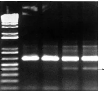
RT-PCR 실험에서 대조군 및 실험군 모두 MMP-2가 증폭 이 된 것을 볼 수 있었다(Fig. 4). 여기서 대조군은 실험군보다 그 띠(band)의 두께가 얇았으나 실험의 정확성을 위해 반정량 적 RT-PCR을 23 cycle에서 세 차례 더 시행 하였다(Fig. 5).
Image gauge V.3.12 (Fuji film, Tokyo, Japan)프로그램을 사용해서 -actin과 MMP-2의 발현정도를 수치화 하였으며 MMP-2의 발현을 정량하기 위해 internal control로 쓰인 - actin의 값을 기준으로 해서 MMP-2/ -actin의 값을 구하여 Wilcoxon의 순위합 검정법으로 검정 결과 유의한 차이(p<0.05)가 있었다(Table 2, 3).
고 찰
회전근 개의 치유력에 대해 여러저자들이 다양한 보고를 하고 있으나 명확히 밝혀지지는 않았다. Viidic 등26)은 손상 받은 건 은 뼈와는 달리 정상 구조나 강도로 회복될 수 없다고 했으며,
Kumagai 등17)은 손상된 회전근 개에서의 치유는 처음에는 type III Collagen이 생성되며 나중에는 type I Collagen으로 점점 대 치되면서 치유된다고 하였다. Fukuda 등8)은 설사 부분 파열이 라 할지라도 완전한 자연치유는 관찰할 수 없었다고 하였다.
본 연구에서의 육안적 소견 및 조직학적 소견은 여러 저자들 과 비슷한 결과를 나타낸 것으로 생각되며 회전근 개의 완전한 자연 치유력이 있다고 말할 수는 없을 것으로 판단되나 어느 정 도의 치유력은 있을 것으로 추정된다.
근육이나 건의 치유 및 재생에 관여하는 효소의 한 종류인 MMPs는 1962년 처음 발견된 후로 약 20종 이상이 발견되었고11), 창상의 치유과정, 치주염, 종양의 침습이나 전이과정 등 생체의 다양한 조직반응에 관여하는 것으로 알려졌으며3,15,19), 정형외과 영역에서는 척추 추간판의 퇴행성 변화, 인공 관절의 무균성 해 리, 근육의 치유과정, 관절염의 진행과정 등에서 그 작용이 알려 지기 시작했다1,6,10,14,20,24,25).
이중 MMP-2는 퇴행성 관절염에서 연골의 변성(denatura- tion)에 관여하지만, 퇴행성 질환의 진행에 있어 중요한 분해과 정(catabolic process)보다는 연골의 재형성(remodeling)에 관 Fig. 4.RT-PCR for expression of MMP-2. Note: line 1-control;
line 2-experimental group (arrow indicated 420 bp MMP-2)
1 2
Fig. 5.Semi-quantitative RT-PCR for MMP-2 expression. Thick bands indicate the internal control ( -actin). Expression of MMP-2 is represented by lines 3 and 4. Note: lines 1 and 2-con- trol; lines 3 and 4-experimental group (arrow indicated 420 bp MMP-2)
1 2 3 4
Control group Experimental group
RT-PCR1 MMP-2 4630 11710
-actin 97900 99800
RT-PCR2 MMP-2 3570 13080
-actin 95200 99100
RT-PCR3 MMP-2 4640 11240
-actin 100700 100500
Table 2.Raw expression of values of -actin and MMP-2 by image gauge V.3.12.
*: Significant difference of the value (p<0.05) against the control group.
Control group Experimental group RT-PCR1 MMP-2/ -actin 0.0437 0.1382*
RT-PCR2 MMP-2/ -actin 0.0437 0.1382*
Table 3.The ratio of MMP-2 expression to -actin expression
RT-PCR3 MMP-2/ -actin 0.0427 0.1433*
Mean value 0.0425 0.1355*
여하는 것으로 알려져 있다13). 또한 MMP-2 유전자 발현의 조 절은 다른 MMPs와는 다른점이 있다. MMP-2는 다른 종류의 MMPs처럼 cytokine이나 성장 인자(growth factor) 등에 의 해 발현되지 않으며 세포막형 MMPs (membrane type- MMPs)에 의해 발현되거나13), 유기 수은 물질(organomercuri- als) 등에 의해 발현된다24). 본 연구에서 MMP-2의 발현은 4주 에도 관찰되어 급성 염증기 이후에도 생성이 되는 것을 보여주 었다.
그러나 실험 방법이 RT-PCR이므로 messenger RNA의 발 현이 곧 MMP-2의 활성도를 의미할 수는 없지만, MMP-2의 발현 양상을 고려할 때 효소의 활성을 나타낼 수 있을 것으로 생각된다. 그러나 보다 정확한 MMPs의 활성도를 보기 위해서 는 zymography나 western blot에 의한 연구가 필요할 것으로 판단되며13)앞으로의 연구 과제로 생각 된다.
본 실험에서 MMP-2의 발현을 연구한 이유는 MMP-2가 작 용하는 기질에 회전근 개의 주요 성분인 I형 및 III형 교원질 섬 유가 포함되며, 이 효소들이 특히 변성된 교원질 섬유와 교원질 섬유 분절들에 대한 작용이 빠르다고 알려졌고22), 따라서 건의 치유과정에 관여할 수 있다고 생각했기 때문이다. 발현된 MMP- 2가 연골 세포에서처럼 작용할 지는 알 수 없으나 MMP-2의 작용을 고려한다면 건의 재형성에서도 중요한 역할을 담당할 수 있을 것으로 추정된다.
MMP-2의 발현을 흰쥐의 회전근 개 조직에서 정량적으로 알 아 본 세번의 semi-quantitative RT-PCR 실험에서 MMP-2 의 의미 있는 발현 증가를 나타내었다. 이 결과는 한 개의 세포 에서 전체 RNA의 양이 같다고 가정한 실험이며, 대조군과 실 험군 모두 같은 양의 전체 RNA를 사용했기 때문에, 실제로 한 개의 세포에서 발현되는 MMP-2의 messenger RNA의 양은 대조군에서 실험군 보다 극히 미미한 상태임을 알 수 있었다.
다만 인체에 있어 회전근 개 질환은 만성적인 경과를 취하는 데 반하여 본 연구는 4주의 단기간의 과정에서 얻은 결과이므로 만성 회전근 개 파열에서도 같은 발현을 보일지는 검증을 요하 지만, 치유과정 중 발현된 MMP-2에 대해 더 많은 연구가 진 행된다면 회전근 개 파열의 치유과정 규명에 도움이 될 수 있을 것으로 사료된다.
결 론
흰쥐의 회전근 개 파열의 치유 과정에서 면역화학 조직학적 및 역전사 효소 중합반응으로 의미 있게 발현된 MMP-2는 임 상적으로 직접적인 연관성이 있다고 하기는 어려우나 초기 회전 근 개 조직의 치유 과정 및 재형성에 관여할 것으로 추정된다.
참고문헌
1. Ahrens D, Koch AE, Pope RM, Stein-Picarella M and Niedbala MJ: Expression of matrix metalloproteinase 9 (96-kd gelatinase B) in human rheumatoid arthritis. Arthritis Rheum, 39: 1576-1587, 1996.
2. Binder A, Parr G, Hazleman B and Fitton-Jackson S: Pulsed electro- magnetic field therapy of persistent rotator cuff tendinitis. Lancet, 1: 695- 698, 1984.
3. Birkedal RT and Quigley JP: Role of matrix metalloproteinases in human periodontal disease. J Periodontol, 64: 474-484, 1993.
4. Burkhead WZ Jr, Burkhart SS, Gerber C, et al: Symposium : The rotator cuff, debridement versus repair. part I. Contemp. Orthop, 31: 262- 271, 1995.
5. Carpenter JE, Thomopoulos S, Flanagan CL, DeBano CM and Soslowsky LJ: Rotator cuff defect healing: A biomechanical and histolog- ic analysis in an animal model. J Shoulder Elbow Surg, 7: 599-605, 1998.
6. Clark IM, Powell LK, Ramsey S, Hazleman BL and Cawston TE:
The measurement of collagenase, tissue inhibitor of metalloproteinase (TIMP), and collagenase-TIMP complex in synovial fluids from patients with osteoarthritis and rheumatoid arthritis. Arthritis Rheum, 36: 372- 379, 1993.
7. Cofield RH: Current concepts review: Rotator cuff disease of the shoulder.
J Bone Joint Surg, 67-A: 974-979, 1985.
8. Fukuda H, Hamada K, Nakajima T and Tomonaga A: Pathology and pathogenesis of the intratendinous tearing of the rotator cuff viewed from en bloc histologic sections. Clin Orthop, 304: 60-67, 1994.
9. Fukuda H, Hamada K, Nakajima T, Yamada N, Tomogana A and Goto M: Partial-thickness tears of the rotator cuff: A clinicopathlogical review based on 66 surgically verified cases. International Orthopedics, 20: 257-265, 1996.
10. Goupille P, Jayson MIV, Valat JP and Freemont AJ: Matrix metal- loproteinases: The clue to intervertebral disc degeneration. Spine, 23: 1612- 1626, 1998.
11. Gross J and Lapiere CM: Collagenolytic activity in amphibian tissues:
a tissue culture assay. Proc Natl Acad Sci USA, 54: 1197-1204, 1962.
12. Iannoti JP: Rotator cuff disorder: Evaluation and treatment. 1st ed, Am Acad of Orthop Surg, 18-33, 1991.
13. Ijima Y, Kobayashi M and Kubota E: Role of interleukin-1 in induc- tion of matrix metalloproteinases synthesized by rat temporomandibular joint chondrocytes and disc cells. Eur J Oral Sci, 109: 50-59, 2001.
14. Ishiguro N, Ito T, Ito H, et al: Relationship of matrix metalloproteinas- es and their inhibitors to cartilage proteoglycan and collagen turnover:
Analyses of synovial fluid from patients with osteoarthritis. Arthritis Rheum, 42: 129-136, 1999.
15. Kawahara E, Okada Y, Nakanishi I, et al: The expression of invasive behavior of differentiated squamous carcinoma cell line evaluated by an in vitro invasion model. Jpn J Cancer Res, 84: 409-418, 1993.
16. Kenji T, Randal SG, Fred H, Toshikazu K, Yasusuke H and David A:The effects of hyaluronan on matrix metalloproteinase-3 (MMP-3), inter- leukin-1 (IL-1 ), and tissue inhibitor of metalloproteinase-1 (TIMP-1) gene expression during the development of osteoarthritis. Osteoarthritis
and Cartilage, 7: 182-190, 1999.
17. Kumagai J, Uhthoff HK, Sarkar K and Murnaghan JP: Collagen type III in rotator cuff tears: An immunohistochemical study. J Shoulder Elbow Surg, 1: 187-192, 1992.
18. Kumagai J, Sarkar K and Uhthoff HK: The collagen type in the attachment zone of rotator cuff tendons in the eldery: An immunohisto- chemical study. J Rheumatol 21: 2096-2100, 1994.
19. Matrisian LM: Metalloproteinases and their inhibitors in matrix remod- eling. Trends Genet, 6: 121-125, 1990.
20. Matsui Y, Maeda M, Nakagami W and Iwata H: The involvement of matrix metalloproteinases and inflammation in lumbar disc herniation.
Spine, 15: 863-868, 1998.
21. Riley GP, Harral RL and Constant CR: Tendon degeneration and chronic shoulder pain: Changes in collagen composition of the human rota- tor cuff tendons in rotator cuff tendinitis. Ann Rheum Dis 53: 359-366, 1994.
22. Ronald T, Aimes and James PQ: Matrix metalloproteinase-2 is an interstitial collagenase. J Biol Chem, 270: 5872-5876, 1995.
23. Rocks JA: Intrinsic shoulder pain syndrome. Physical Therapy 59: 153- 159, 1979.
24. Sonia K, Chantal L, Michele D, et al: Expression of matrix metallopro- teinases 2 and 9 in regenerating skeletal muscle: A study in experimental- ly injured and mdx muscles. Developmental Biology, 205: 158-170, 1999.
25. Takagi M, Santavirta S, Ida H, Ishii M, Mandelin J and Kontti- nen YT: Matrix metalloproteinases and tissue inhibitors of metallopro- teinases in loose artificial hip joints. Clin Orthop, 352: 35-45, 1998.
26. Viidik A, Mow VC, Ratcliffe A and Woo SL-Y: Structure and func- tion of normal and healing tendons and ligaments: Biomechanics of Diarthrodal Joints. New York, Springer-Verlag vol 1, 1990.
27. Woessner JF Jr: Matrix metalloproteinases and their inhibitors in connec- tive tissue remodeling. FASEB J, 5: 2145-2154, 1991.