Ann Hepatobiliary Pancreat Surg 2018;22:136-143
https://doi.org/10.14701/ahbps.2018.22.2.136
Case Report
Management of very late peritoneal metastasis of hepatocellular carcinoma 10 years after liver transplantation: Lessons from two cases
Abdulwahab A Alshahrani1,2, Shin Hwang1, Gi-Won Song1, Deok-Bog Moon1, Dong-Hwan Jung1, Chul-Soo Ahn1, Ki-Hun Kim1, Tae-Yong Ha1, Gil-Chun Park1, Su-Min Ha1, Yo-Han Park3, and Sung-Gyu Lee1
1Division of Hepatobiliary Surgery and Liver Transplantation, Department of Surgery, Asan Medical Center, University of Ulsan College of Medicine, Seoul, Korea, 2Multi Organ Transplant Center, King Fahad Specialist University Hospital, Dammam, Saudi Arabia, 3Department of Surgery, College of Medicine Inje University, Busan
Paik Hospital, Busan, Korea
Recurrence of hepatocellular carcinoma (HCC) 10 years after liver transplantation (LT) is very rare. Here, we present two cases of peritoneal metastasis of HCC that occurred 10 and 12 years after LT. A 77-year-old male who had under- gone deceased-donor LT 10 years earlier showed slow progressive elevation of tumor marker levels over 6 months.
Close observation with frequent imaging studies and monthly tumor marker analyses revealed a solitary peritoneal seeding mass. Imaging studies revealed that the mass was highly likely to be metastatic HCC. After excision of the mass, all tumor markers returned to the normal range. Over past 10 months, the patient has received everolimus monotherapy and half-dose sorafenib, and has shown no evidence of HCC recurrence. In the second case, marginally elevated tumor marker levels were detected in a 65-year-old male who had undergone living-donor LT 12 years earlier.
After observation for 3 months, follow-up studies revealed a peritoneal seeding mass. Thorough imaging studies re- vealed that the mass was highly likely to be metastatic HCC. Two mass lesions were excised, and the patient was administered low-dose calcineruin inhibitor, sirolimus, and full-dose sorafenib. Subsequently, the tumor marker levels increased again and growth of new peritoneal seeding nodules was observed; therefore, sorafenib was stopped after 2 years of administration. During 6 years since HCC recurrence diagnosis, the patient has experienced slowly growing tumors, but has been doing well. For very late peritoneal metastasis of HCC, the therapeutic modalities include surgical resection if possible, everolimus monotherapy, and long-term use of sorafenib. (Ann Hepatobiliary Pancreat Surg 2018;22:136-143)
Key Words: Hepatocellular carcinoma; Recurrence; Metastasis; Sorafenib; Resection
Received: March 7, 2018; Revised: March 20, 2018; Accepted: April 10, 2018 Corresponding author: Shin Hwang
Department of Surgery, Asan Medical Center, University of Ulsan College of Medicine, 88 Olympic-ro 43-gil, Songpa-gu, Seoul 05505, Korea Tel: +82-2-3010-3930, Fax: +82-2-3010-6701, E-mail: [email protected]
Copyright Ⓒ 2018 by The Korean Association of Hepato-Biliary-Pancreatic Surgery
This is an Open Access article distributed under the terms of the Creative Commons Attribution Non-Commercial License (http://creativecommons.org/
licenses/by-nc/4.0) which permits unrestricted non-commercial use, distribution, and reproduction in any medium, provided the original work is properly cited.
Annals of Hepato-Biliary-Pancreatic Surgery ∙ pISSN: 2508-5778ㆍeISSN: 2508-5859
INTRODUCTION
Liver transplantation (LT) is an established treatment for patients with liver cirrhosis and/or hepatocellular car- cinoma (HCC). Patient selection according to institutional eligibility criteria contributes to reduction of HCC re- currence after LT, but a considerable number of LT recip- ient deaths are still associated with HCC recurrence.1-5 HCC recurrence usually happens during the first a few years after LT, although very late recurrence has been re- ported sporadically. Because of the rarity of very late HCC recurrence that occurs at least 10 years after trans- plantation,6,7 early detection is difficult and a therapeutic
strategy for such tumors has not yet been established.
Currently, there are no clear recommendations or con- sensuses for the management of recurrent HCC, partic- ularly peritoneal metastasis. Here, we describe two cases of peritoneal metastasis of HCC that occurred 10 and 12 years after LT and discuss a therapeutic strategy for such patients.
CASE
Case 1
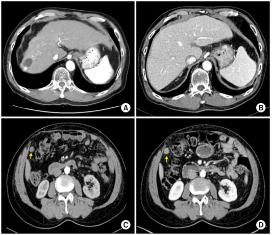
A 67-year-old male underwent deceased-donor LT for hepatitis B virus-associated liver cirrhosis and HCC (Fig.
Fig. 1. Computed tomographic images of Case 1. (A) Pre-trans- plant finding. (B) Early post- transplant finding. (C) Image taken 2 months before the diag- nosis of HCC recurrence. The arrow indicates the metastatic lesion. (D) Image taken at the time of diagnosis of HCC recurrence. The arrow indicates the metastatic lesion.
1A and 1B). Before LT, the patient underwent two ses- sions of transarterial chemoembolization and one session of radiofrequency ablation. The extent of HCC within the explanted liver met the Milan criteria. The pre-transplant serum -fetoprotein (AFP) level was 7 ng/ml.
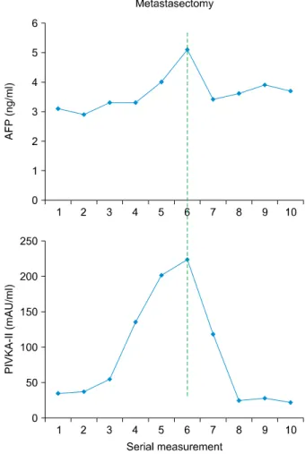
At the age of 77 years (10 years after LT), the patient showed slow elevation of tumor marker levels during 6 months of outpatient clinic follow-ups. Because of slow but progressive elevations of the levels of AFP and pro- tein induced by vitamin K antagonist or absence-II (PIVKA-II) (Fig. 2), the patient was closely observed by bimonthly computed tomography (CT) analyses of the chest and abdomen-pelvis, as well as monthly tumor marker tests. After observation for 6 months, a single 2 cm-sized mass was found around the transverse colon (Fig. 1C and 1D). The lesion was visible on the CT scan taken 2 months previously, but we had missed it at that time (Fig. 1C). There was only a slight growth of the mass during the 2 month period. A positron emission to- mography (PET) scan using [18F]-fluorodeoxyglucose re- vealed that the mass showed hypermetabolic uptake (Fig.
3). During this work-up, serum levels of AFP and PIVKA-II were gradually elevated. These findings sug-
gested that the mass was likely to be metastatic HCC.
Open laparotomy was performed, and the mass was ex- cised with tumor-negative resection margins. A patho- logical analysis confirmed that the mass was metastatic HCC. After excision, the patient’s tumor marker levels rapidly returned to normal ranges (Fig. 2). Considering his age of 77 years, the patient was prescribed everolimus monotherapy and half-dose sorafenib therapy (200 mg twice per day). Over the past 10 months, he has been do- ing well and has not shown any serious adverse side-ef- fects or signs of HCC recurrence. In this patient, tumor marker testing is highly diagnostic and the current surveil- lance protocol comprises tumor marker tests every 2 months and CT scans every 6 months.
Case 2
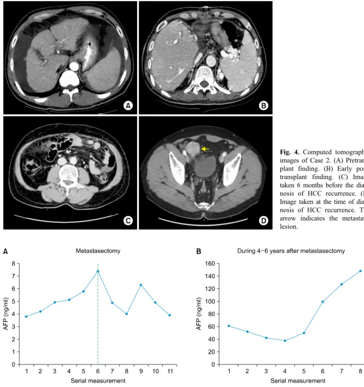
A 53-year-old male underwent living-donor LT for hep- atitis B virus-associated liver cirrhosis and HCC (Fig. 4A and 4B). Before LT, he did not receive any HCC treatment. The resected liver had a single 1.5 cm-sized HCC without microvascular invasion, and therefore met the Milan criteria. The pretransplant serum AFP level was 16 ng/ml.
138 Ann Hepatobiliary Pancreat Surg Vol. 22, No. 2, May 2018
Fig. 2. Serial measurement of tumor marker levels in Case 1 before and after metastasectomy.
Fig. 3. Positron emission tomography analysis of Case 1 showing a mass with hypermetabolic uptake (arrow).
At the age of 65 years (12 years after LT), the patient showed very slow elevation of AFP levels, albeit within the normal range, over 3 months of outpatient clinic fol- low-ups (Fig. 5A). A CT scan of the abdomen was taken 6 months prior to the observed increase in AFP levels, but no abnormalities were detected (Fig. 4C). The pelvis was not examined at that time. A subsequent CT scan of the abdomen-pelvis was performed after detection of the slow rise in the AFP level and identified a 4 cm-sized mass at the pelvis (Fig. 4D). A PET scan using [18F]-fluorodeoxyglucose revealed that the mass showed hypermetabolic uptake (Fig. 6). At this time, the AFP lev- el was gradually elevated but was still within the normal range (Fig. 5A). These findings suggested that the mass was likely to be metastatic HCC.
Open laparotomy was performed, and two masses were excised with equivocal tumor-negative resection margins (Fig. 7). A pathological analysis confirmed that the mass- es were metastatic HCCs. After excision, the patient’s
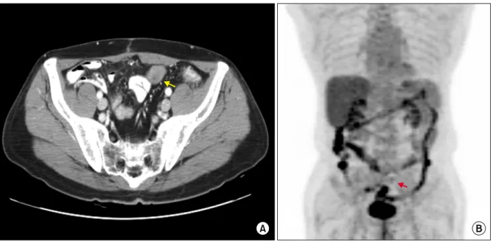
AFP level dropped rapidly (Fig. 5A); however, 6 months later, it increased again, although it was still within the normal range. Follow-up CT and PET scans revealed mul- tiple seeding nodules at the pelvis (Fig. 8). The patient underwent treatment with low-dose calcineurin inhibitor, sirolimus, and full-dose sorafenib, and displayed no seri- ous adverse side-effects. Growth of the peritoneal seeding nodules was visualized on follow-up CT scans, and sor- afenib therapy was stopped after 2 years of administration.
The patient changed to receive everolimus monotherapy because its Korean National Health Insurance coverage for LT recipients. During the 6 years since HCC re- currence was diagnosed, the patient has shown very slow growing tumors, alongside elevated AFP levels (Fig. 5B and Fig. 9), but has been doing well without significant deterioration of his quality of life. Recently, he has been hospitalized twice due to deterioration of his general condition. We expect that supportive care will prolong his life further.
DISCUSSION
Although the majority of HCC recurrence happens dur- ing the first a few years after LT, a small number of pa- tients display very late recurrence, sometimes as late as 10 years after transplantation. Advanced HCC beyond the Milan criteria is often associated with early HCC re- currence; however, like the two cases described here, the
Fig. 4. Computed tomographic images of Case 2. (A) Pretrans- plant finding. (B) Early post- transplant finding. (C) Image taken 6 months before the diag- nosis of HCC recurrence. (D) Image taken at the time of diag- nosis of HCC recurrence. The arrow indicates the metastatic lesion.
Fig. 5. Serial measurement of tumor marker levels in Case 2 before and after metastasectomy (A) and at 4-6 years after meta- stasectomy (B).
majority of patients showing late recurrence have HCCs within the Milan criteria.6 Because of its rarity, early diag- nosis of delayed or very late HCC recurrence is difficult to detect. In the real-world practice, such very late re- currence is often diagnosed after overt manifestation of symptoms, or is incidentally detected during routine fol- low-up studies. From 10 years post-transplantation on- wards, our institution performs surveillance imaging of
LT recipients once every 2 years. This interval period, which was determined based on cost-effectiveness and pa- tient compliance, makes it difficult to detect delayed HCC recurrence at an early stage.
Measurement of serum AFP levels is a simple test that can be done during outpatient clinic visits. In patients who had elevated AFP levels prior to LT, there is a high prob- ability of AFP elevation at the time of HCC recurrence.
140 Ann Hepatobiliary Pancreat Surg Vol. 22, No. 2, May 2018
Fig. 8. Follow-up computed tomography (A) and positron emission tomography (B) images of Case 2 showing metastatic lesions at the pelvis (arrows).
Fig. 7. Gross image of the resected metastatic mass with equivocal tumor-negative resection margins in Case 2.
Fig. 6. Positron emission tomography analysis of Case 2 showing a mass with hypermetabolic uptake (arrow).
In fact, we reported previously that the ability of AFP testing to detect post-transplant HCC recurrence is de- pendent on the pretransplant AFP levels; specifically, we reported HCC detection sensitivities of approximately 40%, 50%, and 90% in patients with pretransplant AFP levels of ≤20 ng/ml, 21-200 ng/ml, and >200 ng/ml, respectively.6 Detection of PIVKA-II levels plays a com- plementary role in the diagnosis of HCC, although the sensitivity and specificity of PIVKA-II testing are lower
than those of AFP testing.8-10 Concurrent measurement of AFP and PIVKA-II levels improves the sensitivity of HCC recurrence detection; therefore, we have measured the levels of these proteins during follow-up analyses of HCC patients, including LT recipients, since 2006. In a Japanese study of patients with HCC in the explanted liv- er, AFP and PIVKA-II levels were measured every 1-2 months after LT, but confirmation of HCC recurrence via imaging took 17-208 days after the observed increases in
Fig. 9. Magnetic resonance images showing intrahepatic metastasis (arrow) and computed tomography analyses showing multiple metastatic lesions at the pelvis (arrows).
the levels of tumor markers.11
Compared with those that recur early, post-transplant HCCs showing very late recurrence may have less ag- gressive tumor biology and may be more responsive to locoregional treatments. However, we emphasize that post-transplant HCC recurrence itself is a strong evidence of aggressive tumor biology. Thus, the therapeutic strat- egy for very late HCC recurrence is similar to that for early recurrence. Surgical resection of metastatic lesions is the most effective therapy for recurrent HCC in LT recipients. We previously reported a beneficial effect of surgical metastasectomy of metachronous pulmonary and adrenal metastases from HCCs on patient survival.12,13 There are only a small number of studies supporting re- section of tumors arising from peritoneal seeding of HCC, and resection of peritoneal metastases should only be con- sidered in patients whose primary liver neoplasm is under control and who have no metastases in other organs.14-16 Since there was no evidence of tumor recurrence other than localized peritoneal metastasis in the two cases de- scribed here, we decided to perform peritoneal meta- stasectomy in both patients.
In LT recipients with HCC recurrence, metastasectomy is not considered to be curative because it can be compat- ible with local control methods. There is a high proba- bility of further tumor recurrence from residual tumor cells, as well as inevitable immunosuppression. Thus, con- version to treatment with a mammalian target of rapamy-
cin (mTOR) inhibitor and systemic therapy with sorafenib should be considered in these patients. Alongside their im- munosuppressive effects, which are mediated via in- hibition of interleukin-2-mediated T-cell proliferation,17 mTOR inhibitors display antitumor effects by inhibiting cell growth and angiogenesis. Treatment with the mTOR inhibitor everolimus is associated with a low rate of HCC recurrence in LT recipients.18-20 However, to date, the ma- jority of clinical LT studies have focused on the pre- vention rather than the treatment of established HCC recurrence. In the cases described here, we switched the primary immunosuppressant therapy to mTOR inhibitor after HCC recurrence and de novo malignancy; however, there is still a lack of strong evidence supporting im- munosuppressive treatment regimens using mTOR inhibitors.18-21
The multikinase inhibitor sorafenib is an effective ther- apy for HCC.22,23 Although sorafenib noticeably improves the survival of patients with advanced HCC,24,25 its poten- tial mechanisms of action are not well known. It is not recommended to use sorafenib as an adjuvant therapy for HCC following hepatic resection or ablation,26 and its therapeutic effect on post-transplant HCC recurrence is still largely unknown.27,28 However, we reasoned that met- astasectomy of post-transplant recurrent HCC is asso- ciated with a high risk of further recurrence in LT recipi- ents; thus we administered sorafenib as an adjuvant che- motherapy in the two cases described here.
142 Ann Hepatobiliary Pancreat Surg Vol. 22, No. 2, May 2018
Combination therapy with sorafenib and mTOR in- hibitor, in addition to locoregional treatment, has been performed previously. A Korean single institution study found that 12 patients who received combination therapy with sorafenib and sirolimus had better post-recurrence survival outcomes than 27 patients receiving best suppor- tive care for post-transplant HCC recurrence.29 Furthermore, in a Spanish multi-center study involving 31 LT recipients with HCC recurrence, combination therapy resulted in a partial response in a single patient and stable disease in 13 patients, giving an overall clinical benefit rate of 53.8%.30
Lifelong surveillance is necessary for LT recipients be- cause HCC recurrence can occur after prolonged periods, as highlighted in the two cases described here. Since most recurrences are diagnosed subclinically, surveillance via frequent tumor marker testing is the cornerstone of fol- low-up studies to detect HCC recurrence in a timely manner.
In conclusion, very late peritoneal metastasis of HCC happens sporadically. We suggest that the therapeutic mo- dality for this condition includes local control through sur- gical resection if possible, everolimus monotherapy, and long-term use of sorafenib.
REFERENCES
1. Lee SG, Hwang S, Moon DB, Ahn CS, Kim KH, Sung KB, et al. Expanded indication criteria of living donor liver trans- plantation for hepatocellular carcinoma at one large-volume center. Liver Transpl 2008;14:935-945.
2. Mazzaferro V, Regalia E, Doci R, Andreola S, Pulvirenti A, Bozzetti F, et al. Liver transplantation for the treatment of small hepatocellular carcinomas in patients with cirrhosis. N Engl J Med 1996;334:693-699.
3. Hwang S, Lee SG, Joh JW, Suh KS, Kim DG. Liver trans- plantation for adult patients with hepatocellular carcinoma in Korea: comparison between cadaveric donor and living donor liver transplantations. Liver Transpl 2005;11:1265-1272.
4. Todo S, Furukawa H; Japanese Study Group on Organ Transplantation. Living donor liver transplantation for adult pa- tients with hepatocellular carcinoma: experience in Japan. Ann Surg 2004;240:451-459; discussion 459-461.
5. Yao FY, Ferrell L, Bass NM, Watson JJ, Bacchetti P, Venook A, et al. Liver transplantation for hepatocellular carcinoma: ex- pansion of the tumor size limits does not adversely impact survival. Hepatology 2001;33:1394-1403.
6. Hwang S, Moon DB, Ahn CS, Kim KH, Ha TY, Song GW, et al. Risk-based long-term screening for hepatocellular carcinoma recurrence after living donor liver transplantation. Transplant Proc 2013;45:3076-3084.
7. Park MS, Lee KW, Yi NJ, Choi YR, Kim H, Hong G, et al.
Optimal tailored screening protocol after living donor liver trans- plantation for hepatocellular carcinoma. J Korean Med Sci 2014;29:1360-1366.
8. Yamamoto K, Imamura H, Matsuyama Y, Kume Y, Ikeda H, Norman GL, et al. AFP, AFP-L3, DCP, and GP73 as markers for monitoring treatment response and recurrence and as surro- gate markers of clinicopathological variables of HCC. J Gastroenterol 2010;45:1272-1282.
9. Taketomi A, Sanefuji K, Soejima Y, Yoshizumi T, Uhciyama H, Ikegami T, et al. Impact of des-gamma-carboxy prothrombin and tumor size on the recurrence of hepatocellular carcinoma af- ter living donor liver transplantation. Transplantation 2009;87:
531-537.
10. Hwang S, Song GW, Lee YJ, Kim KH, Ahn CS, Moon DB, et al. Multiplication of tumor volume by two tumor markers is a post-resection prognostic predictor for solitary hepatocellular carcinoma. J Gastrointest Surg 2016;20:1807-1820.
11. Yamashiki N, Sugawara Y, Tamura S, Tateishi R, Yoshida H, Kaneko J, et al. Postoperative surveillance with monthly serum tumor markers after living-donor liver transplantation for hep- atocellular carcinoma. Hepatol Res 2010;40:278-286.
12. Hwang S, Kim YH, Kim DK, Ahn CS, Moon DB, Kim KH, et al. Resection of pulmonary metastases from hepatocellular carcinoma following liver transplantation. World J Surg 2012;36:
1592-1602.
13. Ha TY, Hwang S, Ahn CS, Kim KH, Lee YJ, Moon DB, et al. Resection of metachronous adrenal metastasis after liver re- section and transplantation for hepatocellular carcinoma. Dig Surg 2014;31:428-435.
14. Hashimoto M, Sasaki K, Moriyama J, Matsuda M, Watanabe G.
Resection of peritoneal metastases in patients with hepatocellular carcinoma. Surgery 2013;153:727-731.
15. Kneuertz PJ, Cosgrove DP, Cameron AM, Kamel IR, Geschwind JF, Herman JM, et al. Multidisciplinary management of recurrent hepatocellular carcinoma following liver transplantation. J Gastrointest Surg 2012;16:874-881.
16. Chua TC, Morris DL. Exploring the role of resection of extra- hepatic metastases from hepatocellular carcinoma. Surg Oncol 2012;21:95-101.
17. Augustine JJ, Bodziak KA, Hricik DE. Use of sirolimus in solid organ transplantation. Drugs 2007;67:369-391.
18. Cholongitas E, Goulis I, Theocharidou E, Antoniadis N, Fouzas I, Giakoustidis D, et al. Everolimus-based immunosuppression in liver transplant recipients: a single-centre experience. Hepatol Int 2014;8:137-145.
19. Jeng LB, Thorat A, Hsieh YW, Yang HR, Yeh CC, Chen TH, et al. Experience of using everolimus in the early stage of living donor liver transplantation. Transplant Proc 2014;46:744-748.
20. Cholongitas E, Mamou C, Rodríguez-Castro KI, Burra P.
Mammalian target of rapamycin inhibitors are associated with lower rates of hepatocellular carcinoma recurrence after liver transplantation: a systematic review. Transpl Int 2014;27:1039-1049.
21. Kang SH, Hwang S, Ha TY, Song GW, Jung DH, Kim KH, et al. Tailored long-term immunosuppressive regimen for adult liv- er transplant recipients with hepatocellular carcinoma. Korean J Hepatobiliary Pancreat Surg 2014;18:48-51.
22. Wilhelm SM, Carter C, Tang L, Wilkie D, McNabola A, Rong H, et al. BAY 43-9006 exhibits broad spectrum oral antitumor activity and targets the RAF/MEK/ERK pathway and receptor tyrosine kinases involved in tumor progression and angiogenesis.
Cancer Res 2004;64:7099-7109.
23. Carlomagno F, Anaganti S, Guida T, Salvatore G, Troncone G, Wilhelm SM, et al. BAY 43-9006 inhibition of oncogenic RET mutants. J Natl Cancer Inst 2006;98:326-334.
24. Llovet JM, Ricci S, Mazzaferro V, Hilgard P, Gane E, Blanc JF, et al. Sorafenib in advanced hepatocellular carcinoma. N Engl J Med 2008;359:378-390.
25. Cheng AL, Kang YK, Chen Z, Tsao CJ, Qin S, Kim JS, et al.
Efficacy and safety of sorafenib in patients in the Asia-Pacific region with advanced hepatocellular carcinoma: a phase III rand- omised, double-blind, placebo-controlled trial. Lancet Oncol 2009;10:25-34.
26. Bruix J, Takayama T, Mazzaferro V, Chau GY, Yang J, Kudo M, et al. Adjuvant sorafenib for hepatocellular carcinoma after resection or ablation (STORM): a phase 3, randomised, double-blind, placebo-controlled trial. Lancet Oncol 2015;16:1344-1354.
27. de'Angelis N, Landi F, Nencioni M, Palen A, Lahat E, Salloum C, et al. Role of sorafenib in patients with recurrent hep-
atocellular carcinoma after liver transplantation. Prog Transplant 2016;26:348-355.
28. Yoon DH, Ryoo BY, Ryu MH, Lee SG, Hwang S, Suh DJ, et al. Sorafenib for recurrent hepatocellular carcinoma after liver transplantation. Jpn J Clin Oncol 2010;40:768-773.
29. Na GH, Hong TH, You YK, Kim DG. Clinical analysis of pa- tients with hepatocellular carcinoma recurrence after living-donor liver transplantation. World J Gastroenterol 2016;22:5790-5799.
30. Gomez-Martin C, Bustamante J, Castroagudin JF, Salcedo M, Garralda E, Testillano M, et al. Efficacy and safety of sorafenib in combination with mammalian target of rapamycin inhibitors for recurrent hepatocellular carcinoma after liver transplantation.
Liver Transpl 2012;18:45-52.