J Korean Surg Soc 2012;82:45-49
http://dx.doi.org/10.4174/jkss.2012.82.1.45
CASE REPORT
Journal of the Korean Surgical Society
JKSS
pISSN 2233-7903ㆍeISSN 2093-0488
Received June 13, 2011, Revised June 21, 2011, Accepted June 24, 2011 Correspondence to: In Kyu Lee
Department of Surgery, Yeouido St. Mary's Hospital, The Catholic University of Korea School of Medicine, 62 Yeouido-dong, Yeongdeungpo-gu, Seoul 150-713, Korea
Tel: +82-2-3779-2235, Fax: +82-2-786-0802, E-mail: cmcgslee@catholic.ac.kr
cc Journal of the Korean Surgical Society is an Open Access Journal. All articles are distributed under the terms of the Creative Commons Attribution Non-Commercial License (http://creativecommons.org/licenses/by-nc/3.0/) which permits unrestricted non-commercial use, distribution, and reproduction in any medium, provided the original work is properly cited.
Inflammatory myofibroblastic tumor in colon
Eun Young Kim, In Kyu Lee, Yoon Suk Lee, Naery Yang, Dong Jin Chung
1, Kwang-il Yim
2, Jin Il Kim
3, Seung Taek Oh
Departments of Surgery, 1Radiology, 2Hospital Pathology and 3Internal Medicine, The Catholic University of Korea School of Medicine, Seoul, Korea
Inflammatory myofibroblastic tumor (IMT) is an uncommon mesenchymal solid tumor commonly documented in children and young adults. Here, we report a case of IMT in colon confirmed pathologically after laparoscopic anterior resection. A 35-year-old man presented with anal bleeding after defecation for 2 weeks. Colonoscopy demonstrated a mass with shallow ulceration in the central area and irregular margin accompanied by intact mucosa in the descending colon. Computer tomog- raphy showed a well-demarcated and homogenous solitary mass in the descending colon. We performed laparoscopic ante- rior resection. This case was diagnosed as IMT after microscopic examination. The tumor was composed of a proliferation of spindle-shaped cells arranged in the hyaline material with chronic inflammatory cells, composed mainly of plasma cells and lymphocytes. Immunohistochemically, tumor cells were positive for smooth muscle actin, and vimentin, and negative for desmin, CD117 (c-kit), anaplastic lymphoma kinase-1.
Key Words: Inflammatory myofibroblastic tumor, Colon
INTRODUCTION
Inflammatory myofibroblastic tumor (IMT) is an un- common mesenchymal solid tumor commonly docu- mented in children and young adults [1,2]. Although it oc- curs primarily in the lung, IMTs in many organs including stomach, small intestine, large intestine, liver, mediasti- num, retroperitoneum and bladder have also been docu- mented [1,2]. The clinical presentation is determined by the site of origin and the effects of the mass. Thus, IMT presents with non-specific clinical symptoms and its diag- nosis can be difficult. IMT derived from the gastrointesti- nal (GI) tract presents with clinical symptoms of anemia,
GI obstruction, fecal occult blood positive or intussus- ception. Those are not specific symptoms in IMT because other GI tract tumor may also present with similar symp- toms [1,3,4]. IMT has been synonymously referred to as in- flammatory pseudotumor, pseudosarcomatous myofibro- blastic lesion, pseudosarcomatous fibromyxoid lesion or plasma cell granuloma. These words are synonymous and have been in common use, all sharing a key pathologic dif- ferentiation; a dominant spindle cell proliferation with a variable inflammatory component. These spindle cells are in fact myofibroblasts, thus, the preferred term for refer- ence should be IMT reflective of the pathologic characters despite the persistence of others [5].
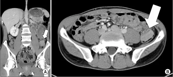
Fig. 2. Trans-axial view (A) and coronal view (B) show a 4.0 cm sized homogeneous enhancing intra-luminal mass (arrow) in descending colon.
Fig. 1. Endoscopic examination reveals a 4 cm sized irregularly margined mass with intact mucosa (margin) and shallow ulcer (central) in descending colon.
We report an uncommon case of 35-year-old man pre- senting with complaints of anal bleeding and histopatho- logically diagnosed with descending colonic IMT after operation.
CASE REPORT
A 35-year-old man presented with anal bleeding after defecation for 2 weeks. It is not associated with the symp-
toms of abdominal pain, fever or weight loss. He had taken oil mixed with a trace of benzopyrene for 3 years because of his job of checking oil type in the edible oil plant. He had been treated for variant angina and underwent hernior- rhaphy for inguinal hernia 12 years ago. Vital signs, phys- ical examination, and peripheral blood analysis (hemoglobin 14.5 g/dL, hematocrit 43.2%) were in the nor- mal range. Colonoscopy demonstrated a 4.0 cm sized-mass with shallow ulceration in the central area and irregular margin accompanied by intact mucosa (Fig. 1).
Microscopic examination of biopsy specimens showed chronic inflammation. Computer tomography showed a well-demarcated and homogenous solitary mass in the de- scending colon. On contrast enhanced view, the mass had enhanced homogeneously in delayed phase (Fig. 2A, B).
Colonoscopy was performed for clipping and tattooing and the patient underwent a laparoscopic anterior resection. On laparoscopy, a considerable amount of as- cites (about 30 mL) and enlarged mesocolic lymph nodes were shown. The lymph nodes were resected and frozen biopsy concluded all lymph nodes were negative for malignancy. Because the main tumor was found to arise from descending colon, left colic branch of inferior mesen- teric artery (IMA) and inferior mesenteric vein were li- gated while IMA was preserved, then laparoscopic ante- rior resection was performed. After the main mass was re- sected, hand-sewing colocolic anastomosis was
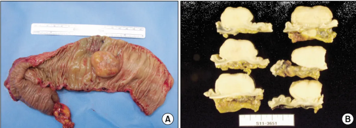
Fig. 3. Gross examination reveals a 3.9 × 3.8 cm sized, fungating, white to yellow colored and hard mass in descending colon. Mass involves muscularis propria.
Fig. 5. Tumor cells stain strongly for vimentin and variably with myoid markers including smooth muscle actin, muscle-specific actin and desmin (A, vimentin, ×200; B, actin, ×200).
Fig. 4. Tumor is composed of cytologically bland spinle cells arranged in hyaline stroma with scattered inflammatory cells. Inflammatory cells are composed of lymphocytes, histiocytes and plasma cells (A, H&E, ×40; B, H&E, ×400).
performed. The 3.9 × 3.8 cm sized mass grossly presented a fungating surface, white to yellow color and invaded muscularis propria (Fig. 3). Consistency of the mass was
harder than adenocarcinoma. The histopathologic exami- nation of the specimens showed the tumor was composed of a proliferation of spindle-shaped cells arranged in hya-
line material with chronic inflammatory cells, composed mainly of plasma cells and lymphocytes, not neutrophils.
The specimens did not have celluar atypia or hyper- chromatism in the cells (Fig. 4A, B). Immunohistochem- ical staining suggested that fibroblastic tissue was of my- ofibroblastic nature. Immunohistochemically, tumor cells were positive for smooth muscle actin, and vimentin, and negative for desmin, CD117 (c-kit), anaplastic lymphoma kinase (ALK)-1 (Fig. 5A, B). These findings were compat- ible with an IMT.
The patient’s postoperative course was uneventful, and he was discharged on the 7th postoperative day. He has been free of complication after 1 month of follow-up.
DISCUSSION
IMT was originally described in the lungs in 1937, and since then has been reported at various sites such as mes- entery, stomach, small intestine, large intestine, liver, me- diastinum, retroperitoneum and bladder [1,2,6]. The most common site of IMTs is the lung and the most common sites of extrapulmonary inflammatory myofibroblastic tu- mor are the mesentery and omentum [1,2,6,7]. Among ex- trapulmonary IMT, 43% arose in the mesentery and omen- tum [7]. Although it occurs primarily in children and young adults (mean age, approximately 10 years), in more recent years a broad age range has been documented [1].
There is no difference in incidence between females and males, though each report has a difference in mean age and gender ratio [2,5].
The etiology of IMT is still unknown. But some people think that development of IMT occurs after trauma, sur- gery or infection such as Epstein-Barr virus and human herpes virus related with reactive cytokine production [6,7]. A recent study reports that these lesions may possess chromosomal aberrations with resultant monoclonality and may frequently demonstrate locally aggressive behavior. As such, this entity should better be considered a neoplastic process. A chromosomal rearrangement in- volving 2p23, the site of the ALK gene, is present in a subset of these tumors [5]. These findings have recently shown that chromosomal abnormalities may be suggestive of clo-
nal origin, not merely a reactive process. IMT should be considered as a true neoplasm [5].
Several cases with IMT in the colon have been con- firmed in other countries. A total of 5 cases have been re- ported during the last ten-year period. The patients with IMT presented with anemia, intussusception, fecal occult blood positive, abdominal pain, chest pain, fever or weight loss. Except 1 case, all cases reported were in males.
From 32-month-old to 79-year-old, a broad age range of IMT has been documented. The most common site of documented IMT in the colon was the right colon (ascending colon, 2 cases; cecum, 2 cases). IMT in the trans- verse colon (1 case) was also reported [1,3,4]. A retro- spective review of the medical records was conducted at two major academic institutions over a 15-year period by Kovach et al. [5]. Review of the records from two in- stitutions yielded 44 cases of pathologically confirmed IMT. Among these cases, only 3 cases occurred in colon [5].
Differential diagnosis of IMT includes malignancy and submucosal tumor from endoscopic findings. In this case, on endoscopic examination, we need to distinguish IMT from lymphoma and submucosal tumor composed of in- tact mucosal margin. Radiologically differential diagnosis of abdominal IMT includes solitary fibrous tumor and fi- bromatoses (desmoid tumor).
Inflammatory fibroid polyp, fibromatoses (desmoid tu- mor), gastrointestinal stromal tumors (GIST), leiomyoma, leiomyosarcoma, and schwannoma have similar patho- logical findings with IMT. Inflammatory fibroid polyp is typically submucosal and consists of a mixture of small granulation tissue-like vessels, spindle cells, and in- flammatory cells (particulary eosinophils). Fibromatoses are composed of bland spindled or stellate cells, arranged in parallel with evenly spaced blood vessels and a collage- nous background. Fibromatoses often have mitotic activ- ity, but cytologic pleomorphism is generally not seen.
GIST stains for CD34 and CD117 (c-kit). Leiomyoma and leiomyosarcoma stain positively for desmin and actin and negatively for CD117 and CD34. Schwannoma stains strongly for S-100 (nuclear and cytoplasmic) and is CD117 negative [1,6,8]. Immunohistochemically, strong diffuse cytoplasmic reactivity for vimentin is typical for virtually all IMT. Reactivity for smooth muscle actin and muscle
specific actin varies from a focal to a diffuse pattern in the spindle cell cytoplasm, and desmin is identified in many cases [7]. Immunohistochemical cytoplasmic positivity for ALK using a variety of monoclonal antibodies is shown in approximately 50% of IMTs. However, ALK positivity is not specific for IMT [7]. Because IMT is rare and sometimes similar with malignancy, even after various examinations, it is important to distinguish IMT from other colonic tumors. IMT cannot be diagnosed by preoperation biopsy.
Also, the tumor occasionally undergoes malignant trans- formation if not surgically excised, therefore surgical re- section is the treatment of choice. We cannot exclude ma- lignancy completely preoperatively, so it is hard to de- termine surgical resection margin. We have to understand IMT accurately and be careful to prevent over diagnosis and unnecessary operation and treatment.
Chemotherapy, radiation treatment, nonsteroidal an- ti-inflammatory drug (NSAID), steroid and cyclosporin- A have been used as treatment modalities, but surgical re- section is considered as treatment of choice [5,6]. It rarely presents recurrence, metastasis or malignant transforma- tion, and is classified as intermediate neoplasm in the World Health Organization histological typing, but it may recur locally or manifest systemic symptoms. It is for this reason that regular follow-up is necessary even though surgical resection was done [2,7].
Several studies have reported that ALK positive tumors have a less aggressive clinical course, although no clear re- lationship exists between ALK expression and prognosis in IMT [2,7,9,10]. In addition, there have been some studies that reported a correlation between p53 expression and prognosis [2,10]. The role of p53 reactivity in predicting the behavior of IMTs is uncertain [2,10]. The etiology of IMT is still unknown, neither reactive process nor neo- plasm. And it is unclear about natural course of IMT, un- less surgical resection is done.
Most IMTs behave in a benign manner after surgical re- section, but it presents recurrence, metastasis or malignant transformation in some cases. It is unclear what kind of factor is associated with prognosis. Also, it is unclear what the best way of treatment of recurrence or metastasis is during long-term follow-up. We have to follow-up pa- tients diagnosed IMT even if they have undergone surgi-
cal resection.
CONFLICTS OF INTEREST
No potential conflict of interest relevant to this article was reported.
REFERENCES
1. Tanaka A, Hirabayashi K, Sadahiro S, Maeda Y, Suzuki T, Ogoshi K. Inflammatory myofibroblastic tumor of the as- cending colon in adults manifested by positive fecal occult blood test. Gastrointest Endosc 2010;71:214-6.
2. Coffin CM, Hornick JL, Fletcher CD. Inflammatory myofi- broblastic tumor: comparison of clinicopathologic, histo- logic, and immunohistochemical features including ALK expression in atypical and aggressive cases. Am J Surg Pathol 2007;31:509-20.
3. Saleem MI, Ben-Hamida MA, Barrett AM, Bunn SK, Huntley L, Wood KM, et al. Lower abdominal inflam- matory myofibroblastic tumor -an unusual presentation- a case report and brief literature review. Eur J Pediatr 2007;166:679-83.
4. Salameh M, Sultan I, Barbar M, Al Hussaini M, Jameel A, Ghandour K, et al. Inflammatory myofibroblastic tumor causing unexplained anemia in a toddler: a case report. J Med Case Reports 2011;5:69.
5. Kovach SJ, Fischer AC, Katzman PJ, Salloum RM, Etting- hausen SE, Madeb R, et al. Inflammatory myofibroblastic tumors. J Surg Oncol 2006;94:385-91.
6. Karnak I, Senocak ME, Ciftci AO, Cağlar M, Bingöl- Koloğlu M, Tanyel FC, et al. Inflammatory myofibroblastic tumor in children: diagnosis and treatment. J Pediatr Surg 2001;36:908-12.
7. Coffin CM, Fletcher JA. Inflammatory myofibroblastic tumour. In: Fletcher CD, Unni KK, Mertens F, editors.
World Health Organization classification of tumours Pa- thology and genetics of tumours of soft tissue and bone.
Lyon: IARC Press; 2002. p.91-3.
8. Greenson JK. Gastrointestinal stromal tumors and other mesenchymal lesions of the gut. Mod Pathol 2003;16:366- 75.
9. Chun YS, Wang L, Nascimento AG, Moir CR, Rodeberg DA. Pediatric inflammatory myofibroblastic tumor: ana- plastic lymphoma kinase (ALK) expression and prognosis.
Pediatr Blood Cancer 2005;45:796-801.
10. Jiang YH, Cheng B, Ge MH, Cheng Y, Zhang G. Compar- ison of the clinical and immunohistochemical features, in- cluding anaplastic lymphoma kinase (ALK) and p53, in in- flammatory myofibroblastic tumours. J Int Med Res 2009;
37:867-77.