Journal of Korean Spine Surg.
Vol. 12, No. 4, pp 269~274, 2005
Address reprint requests to Jeong Gook Seo, M.D.
Department of Orthopaedic Surgery, Seoul Paik Hospital, Inje University, 85, 2-GA, Jur-Dong, Jung-Gu, Seoul, Korea
Tel: 82-2-2270-0028, Fax: 82-2-2270-0023, E-mail: [email protected]
✽ 본 논문의 요지는 2004년도 대한정형외과학회 추계학술대회에서 발표되었음.
경추 전방 유합술 후 발생하는 인접분절의 퇴행성 변화 -수술 전 인접분절 추간판 MRI 소견과의 상관관계-
김정훈・서정국・장석환・조승현・이승강
인제대학교 의과대학 정형외과학교실
Degenerative Changes of Adjacent Segment Following Cervical Anterior fusion -Correlation with Preoperative MRI Findings of Adjacent Disc-
Jung Hoon Kim, M.D., Jeong Gook Seo, M.D., Suk Hwan Jang, M.D., Seung Hyun Cho, M.D., Kang Lee, M.D.
Department of Orthopedic Surgery, Seoul Paik Hospital, Inje University, Seoul, Korea
– Abstract –
Study Design: We retrospectively reviewed the preoperative and postoperative radiographs of patients who underwent anterior cervical discectomy and fusion.
Objectives: We wanted to determine whether the preoperative Magnetic Resonance Imaging (MRI) findings of the levels adja- cent to the level of fusion correlated with the postoperative degenerative changes seen on X-ray after anterior cervical discec- tomy and fusion.
Summary of Literature Review: Anterior cervical fusion causes acceleration of the degenerative changes at the levels below or above the fused segment. These changes may be accelerated if preoperative MRI shows degenerative changes at the levels adjacent to the segment to be fused.
Materials and Methods: Twenty-two patients (forty-four adjacent levels) who underwent anterior cervical discectomy and fusion from January 1998 to August 2002 (average follow up: 2 years and 6 months, range: 2 to 4 years) were enrolled in this study. Preoperatively, all the patients had no degenerative changes at adjacent levels on the plain radiographs, but they had at least one adjacent level with degenerative findings on MRI. The patients were grouped according to the findings of the adjacent levels seen on MRI: low signal changes on the T2 weighted image (group A), disc bulging on the sagittal and axial images (group B), annular tear seen on the axial image (group C), osteophyte formation (group D), and no abnormalities (group E).
Results: Out of 44 cases of 22 patients, 14 cases (31.8%) showed degenerative changes. 2 out of 7 in group A, 6 out of 11 in group B, 3 out of 4 in group C, 2 out of 3 in group D and 1 out of 19 in group E showed degenerative changes on X-rays at the final follow up.
Conclusion: Our findings suggest that abnormalities on the levels adjacent to the level to be fused, as seen on preoperative MRI, predispose these levels to degenerative changes postoperatively.
Key Words: Anterior cervical fusion, Adjacent level, Degenerative change
서 론
경추부의 전방 감압과 유합술은 1 9 5 5년 Smith 와 Robinson1)에 의해 처음 보고된 이래, 비교적 술기가 안 전하고 척수에 대한 직접적인 감압과 골유합을 동시에 시행할 수 있는 장점이 있어, 현재까지 많이 사용되며 양호한 결과들을 보고하고 있다.
그러나 장기 추시에서, 인접 분절의 퇴행성 변화의 발 생은 비교적 높은 빈도로 나타나며2,3),이것에 대한 유발 인자나 예방법 혹은 진단 기준 등은 아직 확실치 않으 며, 저자마다 의견이 분분하다. 특히 인접 분절이 수술 전에 이미 퇴행성 변화가 있는 상태에서는, 유합술 시행 이 인접 분절의 퇴행성 변화를 더욱 빠르게 진행시킨다 는 것은 널리 알려져 있다4,5,6,7).
최근에는 MRI가 수술 전 진단 방법의 하나로 보편화 되어 사용된 이후, 일반 방사선 사진에서 발견할 수 없 는 추간판의 초기 퇴행성 변화까지 확인이 가능해졌으 며, 이에 저자들은 퇴행성 경추 질환으로 본원에서 전방 유합술을 시행한 환자 중, 수술 전 인접 상, 하 추간판이 방사선사진 소견은 정상이나 MRI 소견상 추간판 퇴행 성 변화가 있는 경우에도, 수술 후 발생하는 인접 분절
의 퇴행성 변화의 빈도가 높을 것으로 예상하고 이 연구 를 시작하였다.
연구대상 및 방법
1. 연구대상
본원 정형외과에서 1 9 9 8년 1월부터 2 0 0 2년 8월까지, 단일 전문의에 의해 경추 전방 유합술을 시행 받은 환자 중 수술 전 일반 방사선 사진 소견상 인접 추간판에 퇴 행성 변화의 소견이 없으며, MRI 소견에서는 상, 하의 인접 추간판 중 한 부위라도 퇴행성 변화의 소견을 보인 환자 22명을 대상으로 하여, 상, 하 인접 추간판 44개에 대하여 조사하였다.
수술 후 2년 이상 4년 이하까지 추시된 환자를 대상으 로 하였으며, 그 외의 군은 연구 대상 환자간의 기간 차 이가 많아 제외했다. 남자가 14명, 여자가 8명이었으며, 수술 당시 환자들의 평균 나이는 42세였다. 수술 전 진 단은 모두 추간판 질환으로 신경근병증이 18예, 척수증 이 4예 이었다.
Fig. 1. Degenerative findings of adjacent levels seen on MRI (A) Group A. Disc herniation at C3-4 with low signal change seen at lower adjacent level, C4-5, on T2 weighted image. (B) Group B. Disc herniation at C4-5 with disc bulging at lower adjacent level, C5-6. ( C) Group C. Annular tear shown on axial image. (D) Group D. Disc herniation at C5-6 with osteophyte forma- tion at upper adjacent level, C4-5.
유합 부위는 한 분절이 19예, 두 분절이 3예 이었으며, 수술 시 자가골 이식 후 모든 환자에서 금속판 고정술을 병행하였다. 수술 후 2 ~ 3일간은 Philladelphia 보조기, 3 개월 까지는 soft collar를 착용시켰다.
2. 연구방법
수술 전 MRI 및 단순 방사선사진 소견과, 최종 추시 경추부 굴곡 신전 측면 방사선사진을 비교 조사하였다.
수술전 MRI 에서 확인되는 상, 하 인접 추간판의 퇴행 성 소견을 다음과 같이 5가지 군으로 분류하였다( F i g . 1A-D).
A군: MRI 소견상 T2 강조영상에서 저신호 강도만 보 이는 경우,
B군: MRI 소견상 시상면, 축상면 상에서 추간판의 팽 윤 또는 돌출 소견이 보이는 경우,
C군: MRI 축상면 상에서 섬유륜의 파열이 있는 경우, D군: 단순 방사선 사진에서는 잘 보이지 않는 후방 골 극이 관찰된 경우,
E군: 이상 소견이 없는 경우
최종 추시 경추부 굴곡-신전 측면 방사선사진 소견과 수술 전 사진을 비교하여,인접 분절에서의
1) 1 mm 이상의 추간판 높이의 감소, 2) 추체의 전방 또는 후방 골극 형성,
3) 굴곡 및 신전 사진상 시상면에서 3 mm 이상 전위 또는 인접 분절의 각 운동이 Dvorak8)등의 정상 각운동 20°이상의 운동을 보인 경우를 확인하여 3가지 소견 중 한가지라도 나타나면 퇴행성 변화가 발생한 것으로 정 하였다.
결 과
총 2 2명 환자의 상, 하 추간판 4 4례 중 1 4례에서
(31.8%) 퇴행성 변화가 발생하였으며, 이중 유합 부위
상분절이 6례, 하분절이 7례, 양쪽에서 일어난 경우도 1 례로 상, 하간의 발생 빈도 차이는 없었다.
A군에서는 7례 중 2례, B군에서는 11례 중 6례, C군에 서는 4례 중 3례, D군에서는 3례 중 2례에서 퇴행성 병 변이 나타났으며, 정상 MRI 소견의 E군에서는 19례 중 1례에서만 퇴행성 변화가 발생하였다(Table 1). 즉, 수술 전 MRI소견상 퇴행성 소견이 있었던 A,B,C,D 군 25예의 추간판 중 1 9예(76%) 에서 퇴행성 변화가 발생했으나, 정상 M R I소견의 E군 1 9예의 추간판에서는 1례에서만 퇴행성 변화가 발생하였다(Fig. 2A-D, Fig. 3A-D).
Table 1. Degenerative changes in each group
A B C D E
1) Disc space narrowing 1 2 1
2) Spur formation 1 3 2 1 1
3) Changes in motion segment 1 1
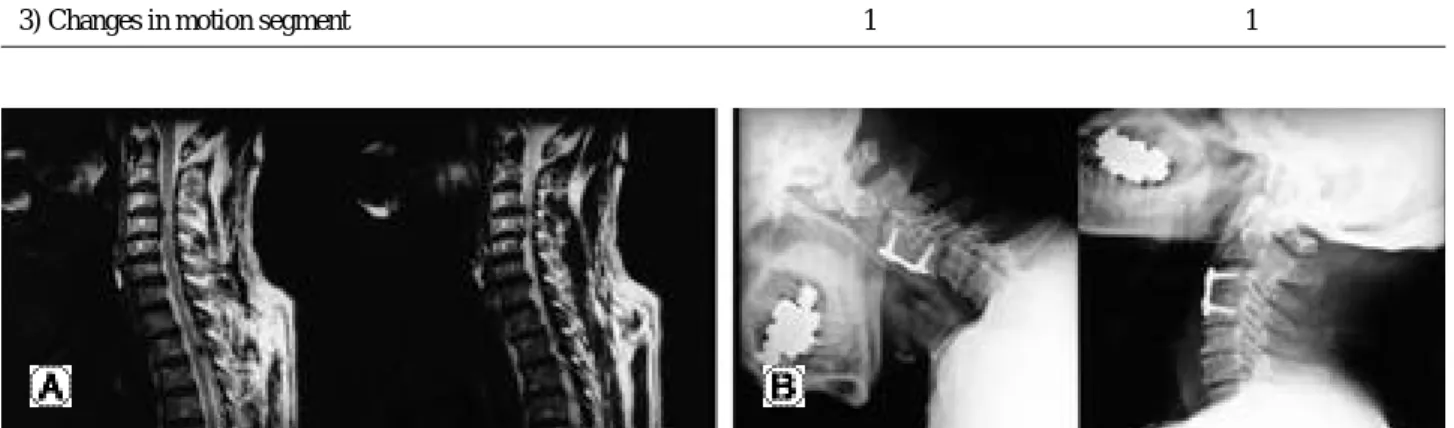
Fig. 2. This 67-year old male patient underwent anterior cervical discectomy and fusion for herniated intervertebral disc at C3-4. Pre- operative X-ray showed no degenerative changes at adjacent levels of C3-4 (A) But preoperative T2 weighted sagittal MR image showed protrusion of the disc at C3-4. Adjacent to this level, C4-5 disc showed bulging, which was categorized as group B. Disc at C2-3 showed no degenerative changes and subsequently were categorized as group E. (B) Twenty-seven months after anterior cervical discectomy and fusion was done using autogenous iliac bone graft and ORION plate, X-ray shows that degenerative changes have occurred at lower adjacent level, C4-5, with narrowing of the disc space and spur for- mation. The upper adjacent level, C2-3, showed no degenerative changes.
고 찰
경추 전방 유합술에 시행되는 전방 도달법은 1 9 5 5년 Smith와 Robinson1)에 의해 소개된 이래 지난 40여 년 동 안 퇴행성 병변에 의한 척수병증이나 신경근증의 치료 에 널리 이용되고 있으며 좋은 결과를 보고하고 있다3,8,9).
추간판을 제거하고 추체 유합술을 위한 자가골 이식 을 한 후, 부가적으로 사용하는 금속판 및 나사못의 고 정술은, 수술부의 일차적인 안정성을 부여하여, 간단한 보조기의 착용만으로 조기 활동을 가능하게 함으로 간 호의 용이함과 입원 기간의 단축등의 장점이 있다10).또 한 이식골의 전이를 막고 생리적인 전만곡을 유지할 수 있으며, 높은 골유합율을 얻을 수 있다고 보고된다11,12).
척추 유합술 시행 후 추시 중, 경부 동통이 발생하는 경우는 여러 가지 원인이 있을 수 있으나, 인접분절의 이차적인 퇴행성 변화도 흔한 원인 중 하나로 알려져 있 다13).퇴행성 변화의 출현과 증상의 발현에 대한 고찰은 추후 연구 후 게재할 예정이다.
이러한 퇴행성 변화에서 보이는 방사선 사진 소견으 로, 추간판의 높이가 감소하게 되며, 퇴행성 변화가 진 행함에 따라 주위의 골조직도 이에 병행하는 변화를 동 반하게 되어 추체 종판(end plate) 의 경화상, 골극 형성 그리고 후관절의 비후 등이 나타난다14,15,16).또한 추간판 의 퇴행은 이차적으로 척추 후관절(facet joint)의 손상을 가져와 분절 불안정 소견을 보일 수 있다17).
이러한 단순 방사선 소견상의 변화는 전산화 단층 촬 영(CT)이나 자기 공명 영상(MRI)에 비해 한정된 정보를 제공하지만 촬영의 용이함 및 경제적 유용성으로 인하 여 수술 후 정기적 추시에 많이 시행되고 있으므로, 단 순 방사선 소견에서 나타나는 인접 관절의 퇴행성 변화 판정이 중요한 정보를 제공할 수 있다고 판단되어진다.
인접 분절의 퇴행성 병변의 원인에 대해서는 확실히 밝혀진 바 없다. 척추 유합 후 부정정렬이 유합 인접분 절의 퇴행성 병변을 촉진시킨다는 보고들이 있으며18,19), Katsuura20)등은 한 분절의 유합보다는 다분절의 유합을 시행한 경우에 인접분절의 퇴행성 변화에 영향을 미친 다고 하였다. Cinerontgenography를 이용한 촬영에서 보 면 경추 유합분절 상하 모든 분절에서 운동이 증가하고 인접 분절에서 최대로 증가하는 것을 알수 있으며 이러 한 증가된 분절 운동이 퇴행성 변화를 촉진 시킨다고 생
각한다21,22).또한 개인적 성향 및 생활 습관 등도 퇴행성
변화를 유발시키는 요인으로 생각된다20).
많은 학자들은 전방유합술 후에, 경추에 나타나는 변 화에 세심한 주의가 필요하다고 주장하나23,24)수술전 이 를 예측할 수 있는 지표에 대한 보고는 많지 않다. Wu6) 등은 수술 전 및 수술 후의 자기 공명 영상 촬영을 하여 유합 인접분절의 퇴행성 변화가 발생함을 보여 주었으 나, 수술 전의 인접분절의 이상 소견과 수술 후의 퇴행 성 변화와 연관성을 부여하지는 못하였다.
결 론
경추부 질환의 치료에 이용되고 있는 전방 유합술의 경우, 수술 전 유합 인접분절의 퇴행성 변화가 일반 방 사선사진 소견상 나타나지 않더라도, MRI 소견에서 인 접 추간판의 신호 강도의 변화, 팽윤 및 돌출, 섬유륜의 파열, 미세한 골극 형성 등의 퇴행성 변화 소견이 확인 될 시에는 수술 후 퇴행성 변화 진행의 가능성이 높다.
따라서, 이러한 소견이 확인되는 경우, 인접 관절의 예 상되는 퇴행성 변화에 대해 수술 전에 미리 환자에게 인 지 시켜야 될 것이며, 수술 후 경부 근육 강화 운동 강조
Fig. 3. This 44-year old male patient underwent anterior cervical discectomy and fusion for herniated intervertebral disc at C5-6. Pre- operative X-ray showed no degenerative changes at C4-5 or at C6-7 (A) But preoperative T2 weighted sagittal MR image showed protrusion of the disc at C5-6. Adjacent to this level, C4-5 disc showed annular tear on T2 weighted axial image, which was categorized as group C. Disc at C6-7 showed no degenerative changes and subsequently were categorized as group E. (B) Twenty-six months after anterior cervical discectomy and fusion was done using autogenous iliac bone graft and ORION plate, X-ray shows that degenerative changes have occurred at upper adjacent level, C4-5, with narrowing of the disc space and spur formation. The lower adjacent level, C6-7, showed no degenerative changes.
및 자세 교육 등의 재활교육과 장기적인 정기 추시가 필 요하다고 사료된다.
참고문헌
01) Robinson RA and Smith GW: Anterolateral cervical disc removing and interbody fusion for cervical disc syn - drome. Bull John Hopkins Hospital,1955; 96:223-224.
02) Braunstein EM, Hunter LY, Bailey RW: Long term radiographic changes following anterior cervical fusion.
Clin Radiol 1980; 31:201-203.
03) Gore DR, Sepic SB : Anterior cervical fusion for degener - ated or protruded discs: A review of one hundred forty-six patients. Spine 1984; 9:667-671.
04) Gore DR, Sepic SB: Anterior discectomy and fusion for painful cervical disc disease. A report of 50 patients with an average follow-up of 21 years. Spine 1998; 23:2047- 2051.
05) Shinomiya K, Okamoto A, Kamikozuru M, Furuya K, Yamaur I: An analysis of failures in primary cervical anterior spinal cord decompression and fusion. J Spinal Disord 1993; 6:277-288.
06) Wu W, Thuomas KA, Hedlund R, Leszniewski W, Vavruch L: Degenerative changes following anterior cer - vical discectomy and fusion evaluated by fast spin-echo MR imaging. Acta Radiol 1996; 37:614-617.
07) Yonenobu K, Okada K, Fuji T, Fujiwara K, Yamashita K, Ono K: Causes of neurologic deterioration following surgical treatment of cervical myelopathy. Spine 1986;
11:818-823
08) Goto S, Mochizuki M, Kita T et al: Anterior surgery in four consecutive technical phases for cervical spondylotic myelopathy. Spine 1993; 18:1968-1973.
09) Bohlman HH, Emery SE, Goodfellow DB, Jones PK:
Robinson anterior cervical discectomy and arthrodesis for cervical radiculopathy: Long-term follow-up of one hun - dred and twenty-two patients. J Bone Joint Surg Am 1993;
75:1298-1307.
10) Robinson RA, Walker AE, Ferlic DC, Wiecking DK:
The results of anterior interbody fusion of the cervical spine. J Bone Joint Surg 1962; 44:1569-1587.
11) Ahn JS, Lee JK, Yang JY, Lee HH: Change of the lor - dosis on cervical spine after anterior interbody fusion with antogenous iliac strut boen graft. J Kor Spine Surg 2001;
8:468-474.
12) Park HJ, Rha JH, Yoon YS: Anterior cervical interbody fusion using cervical locking plate (CSLP). J Kor Orthop Assoc 1996; 31:52-57
13) Harris RI, Wiley JJ: Acquired spondylosis as a sequel to spine fusion. J Bone Joint Surg Am 1963; 45:1159-1170.
14) Baba H, Furusawa N, Imura S, Kawahara N, Tsuchiya H, Tomita K: Late radiographic findings after anterior cervical fusion for spondylotic myeloradiculopathy. Spine 1993; 18:2167-2173.
15) Gore DR, Gardner GM, Sepic SB, Murray MP : Roentgenographic findings following anterior cervical fusion. Skeletal Radiol 1986; 15:556-9.
16) Hunter LY, Braunstein EM, Bailey RW: Radiographic changes following anterior cervical fusion. Spine 1980; 5:
399-401.
17) Gregorius FK, Estrin T, Crandall PH: Cervical spondy - lotic radiculopathy and myelopathy. A long-term follow- up study. Arch Neurol 1976; 33:618-625.
18) Jackson RP, McManus AC: Radiographic analysis of sagittal plane alignment and balance in standing volun - teers and patients with low back pain matched for age, sex and size; a prospective controled clinical study. Spine 1994; 19:1611-1618.
19) Oda I, Cunningham BW, Buckley RA et al: Does spinal kyphotic deformity influence the biomechanical character - istics of the adjacent motion segments An in vivo animal model. Spine 1999; 24:2139-2146.
20) Katsuura A, Hukuda S, Saruhashi Y, Mori K: Kyphotic malalignment after anterior cervical fusion in one of the factors promoting the degenrative process in adjacent intervetabral levels. Eur spine J 2001; 10:320-324.
21) Dohler JR, Kahn MR, Hughes SP: Instability of the cer - vical spine after anterior interbody fusion. A study on its incidence and clinical significance in 21 patients. Acta Orthop trauma Surg 1985; 104:247-250.
22) Fuller DA, Kirkpatrick JS, Emery SE, Wilber RG, Davy DT: A kinematic study after cervical spine before and after segmental arthrodesis. Spine 1998; 23:1649- 1656.
23) Cloward RB: The anterior approach for removal of rup - tured cervical disks. J Neurosurg 1958; 15:602-617.
24) Dvorak J, Froehlich D, Penning L, Baumgartner H, Panjabi MM: Functional radiographic diagnosis of the cervical spine: Flexion/Extension. Spine 1988; 13:748-755.
※ 통신저자 : 서 정 국
서울특별시 중구 저동 2가 85번지 인제대학교 의과대학 서울백병원 정형외과
Tel: 82-2-2270-0028 Fax: 82-2-2270-0023 E-mail: [email protected]
연구계획:경추의 전방 감압 및 유합술은 퇴행성 병변으로 인한 척수병증이나 신경근증의 치료에 효과적인 방법이 다. 유합 인접분절의 퇴행성 변화는 다른 분절에 비해 더 조기에 나타나는 것은 흔한 것으로 알려져 있으나 이러한 변화를 일으키는 방사선학적 요인에 대한 임상적 보고는 많지 않다.
연구목적:경추 전방 유합술 시행 후 수술 전 및 수술 후 인접분절의 방사선학적 변화를 측정하여 유합 인접분절의 퇴행성 변화 및 수술전 자기공명영상(MRI) 소견과의 상관관계에 대해 알아보고자 한다.
대상 및 방법: 1998년 1월부터 2002년 8월까지 단일 경추 전방 유합술을 시행받고 수술 후 2년 이상 4년 이하까지 추 시된 환자들 중 수술 전 일반 방사선 사진 소견상 인접 추간판에 퇴행성 변화의 소견이 없으며, MRI 소견에서는 상, 하 인접 추간판 중 한 부위라도 퇴행성 변화의 소견을 보인 환자 22명, 인접 추간판 44례를 대상으로 하였다. 수술 전 MRI에서 확인 되는 상하 인접 추간판의 퇴행성 소견을 MRI 소견상 T2 강조 영상에서 저신호 강도만 보이는 경우(A 군), 시상면, 축상면에서 추간판의 팽윤 또는 돌출 소견이 보이는 경우(B군), 축상면 상에서 섬유륜의 파열이 있는 경 우(C군), 단순 방사선 사진에서는 잘 보이지 않는 후방 골극이 관찰 된 경우(D군), 이상 소견이 없는 경우(E군)로 나 눈 후 최종 추시 사진상에서 퇴행성 변화를 관찰 하였다.
결과:총 22명 환자의 상, 하 추간판 44례 중 14례에서 (31.8%) 퇴행성 변화가 발생하였으며 A군에서는 7례 중 2례, B 군에서는 11례 중 6례, C군에서는 4례 중 3례, D군에서는 3례 중 2례에서 퇴행성 변화를 보였으며, 정상 MRI 소견의 E군에서는 19례 중 1례에서만 퇴행성 변화가 발생하였다.
결론:경추부 질환의 치료에 이용되고 있는 전방 유합술의 시행 경우, 수술 전 유합 인접분절의 퇴행성 변화가 일반 방사선 사진 소견상 나타나지 않더라도, MRI 소견에서 퇴행성 변화 소견이 확인될 시에는 수술 후 퇴행성 변화 진행 의 가능성이 높을 것으로 사료된다.
색인단어: 경추 전방 유합술, 인접분절, 퇴행성 변화 국 문 초 록