ISSN 0378-6471 (Print)⋅ISSN 2092-9374 (Online)
http://dx.doi.org/10.3341/jkos.2015.56.8.1188
Original Article
나이관련황반변성 환자에서 빛간섭단층촬영의 고반사점 및 치료 전 인자들과 시력과의 연관성
Correlation between Visual Outcomes and Pre-Treatment Factors Including Hyperreflective Foci in Neovascular Age-Related Macular Degeneration
이형우⋅김형찬
Hyung Woo Lee, MD, Hyung Chan Kim, MD, PhD
건국대학교 의학전문대학원 건국대학교병원 안과학교실
Department of Ophthalmology, Konkuk University Medical Center, Konkuk University School of Medicine, Seoul, Korea
Purpose: To investigate the correlation between hyperreflective foci (HF) on spectral domain optical coherence tomography (SD-OCT) at baseline and visual outcomes after intravitreal ranibizumab injection in neovascular age-related macular degener- ation (nAMD).
Methods: We retrospectively reviewed the medical records of 44 eyes of 44 nAMD patients. The number of HF was counted ac- cording to the location of HF on SD-OCT: neurosensory retinal layer, outer retinal layer, and subretinal layer. Statistical correla- tions among final visual acuity (VA) and pretreatment OCT parameters including number of HF, foveal thickness (FT), thickness of choroidal neovascularization (CNV), the status of external limiting membrane, and photoreceptor inner and outer segments (IS/OS) were evaluated.
Results: The number of HF was reduced in all retinal layers in nAMD patients after treatment. In multivariate regression analysis, final VA was associated with baseline VA, number of subretinal HF, and IS/OS disruption length (p = 0.028, p = 0.046 and p = 0.009, respectively) in nAMD patients. The baseline number of subretinal HF was correlated with final FT and CNV thickness (p
= 0.002 and p = 0.009, respectively).
Conclusions: The baseline number of subretinal HF on SD-OCT might predict the final VA after intravitreal ranibizumab treat- ment in nAMD patients.
J Korean Ophthalmol Soc 2015;56(8):1188-1194
Key Words: Hyperreflective foci, Neovascular age-related macular degeneration, Ranibizumab, Spectral domain optical coher- ence tomography, Visual outcome
■Received: 2014. 12. 5. ■ Revised: 2015. 4. 13.
■Accepted: 2015. 6. 4.
■Address reprint requests to Hyung Chan Kim, MD, PhD Department of Ophthalmology, Konkuk University Medical Center, #120-1 Neungdong-ro, Gwangjin-gu, Seoul 143-729, Korea
Tel: 82-2-2030-8180, Fax: 82-2-2030-5273 E-mail: [email protected]
* This study was presented as a narration at the 111th Annual Meeting of the Korean Ophthalmological Society 2014.
ⓒ2015 The Korean Ophthalmological Society
This is an Open Access article distributed under the terms of the Creative Commons Attribution Non-Commercial License (http://creativecommons.org/licenses/by-nc/3.0/) which permits unrestricted non-commercial use, distribution, and reproduction in any medium, provided the original work is properly cited.
나이관련황반변성은 서양에서 50세 이상의 연령층에서 실명 원인 중 가장 흔한 질환으로 알려져 있다.1 국내의 보 고에서도 만 50세 이상에서 나이관련황반변성의 유병률은 나이가 들수록 증가한다.2 나이관련황반변성에는 비삼출성 과 삼출성의 두 가지 형태가 존재한다. Beaver Dam Eye study에 의하면 비삼출성 나이관련황반변성의 유병률은 15.6%, 삼출성 나이관련황반변성의 유병률은 1.2%로 나이 관련황반변성의 90% 정도를 비삼출성이 차지하나, 삼출성 나이관련황반변성에서 급격하고 심각한 시력저하의 위험
이 더욱 높다.3 삼출성 나이관련황반변성에서는 혈관내피세 포성장인자(vascular endothelial growth factor, VEGF)를 포 함한 혈관신생인자들이 분비되어 맥락막신생혈관을 유도 하고, 이로부터 부종과 출혈이 발생하여 시력 저하가 발생 한다.4
스펙트럼영역 빛간섭단층촬영(spectral-domain optical coher- ence tomography, SD-OCT)의 도입으로 망막에 대한 우수 한 해상도의 사진을 얻을 수 있게 되었고, 망막을 구성하는 각 층의 두께를 정량적으로 측정할 수 있게 되었다.5 최근 당뇨망막병증, 망막분지정맥폐쇄, 비삼출성 및 삼출성 황반 변성 환자의 스펙트럼영역 빛간섭단층촬영에서 고반사점 (hyperreflective foci)에 대해 보고되고 있다.6-11 그 기원은 불명확하나, 지단백질의 혈관외 누출, 미세아교세포 또는 망막색소상피세포의 이동에 의한 것으로 추정되고 있 다.6,11-13
전형적인 삼출성 나이관련황반변성과 결절맥락망막병증 환자가 혼합된 환자군을 대상으로 한 연구에서, 치료 후 고 반사점의 개수가 감소하였고, 초기 고반사점의 개수가 최 종 시력과 음의 상관관계가 있음이 알려져 있으나 두 질환 군 각각에 대한 연구는 아직 알려진 바 없다.9,10 결절맥락망 막병증이 삼출성 나이관련황반변성의 아형에 속하는지에 대한 논란이 있고, 두 질환 간의 인구학적인 차이, 임상양 상과 자연경과 및 치료에 대한 반응의 차이가 있음이 알려 져 있어,14,15 두 질환을 분리하여 연구할 필요가 있다. 한편, 당뇨망막부종과 망막분지정맥폐쇄에서 외망막층에 위치한 고반사점이 외경계막과 시세포층의 손상과 관련 있다는 보 고가 있었으나,8,12,16 삼출성 나이관련황반변성에서 고반사 점의 위치에 따른 망막의 미세구조 및 치료 후 시력에 대한 연구는 아직 보고된 바 없다.
이에 본 연구에서는 삼출성 나이관련황반변성에서 스펙 트럼영역 빛간섭단층촬영에서 관찰되는 고반사점을 위치 에 따라 분류하고, 그 외의 망막의 미세구조들과 더불어 유 리체강내 라니비주맙 안내주입술 치료 후 시력 예후와의 연관성에 대해 알아보고자 하였다.
대상과 방법
2010년 1월부터 2013년 1월까지 삼출성 나이관련황반변 성으로 진단 받은 환자 44명(44안)에 대해 후향적 의무기록 조사를 시행하였다. 고도근시(8디옵터 이상), 삼출성 나이 관련황반변성 외에 시력에 영향을 주는 다른 안과적 질환 을 동반한 경우, 백내장 수술 이외의 안내 수술 과거력이 있는 경우, Lens Opacity Classification Scheme에 따른 백내 장 단계가 nuclear opalescence 3 (NO3) 또는 nuclear color
3 (NC3) 이상인 경우, 스펙트럼 빛간섭단층촬영사진에서 미세구조를 구분하기 어려운 화질을 보이는 경우는 대상에 서 제외하였다.
모든 환자들을 대상으로 초진 시 최대교정시력, 안압을 측정하였고 양안의 세극등현미경검사와 안저검사를 시행 하였다. 한 명의 숙련된 검사자가 안저사진기 TOPCON IMAGEnet system (Topcon, Tokyo, Japan)으로 안저를 촬 영하였고, Spectralis SD-OCT (software version 5.1.3.0;
Heidelberg Engineering, Dossenheim, Germany)를 이용하여 형광안저촬영조영술(fluorescein angiography), 인도시아닌 그린 형광안저조영술(indocyanine green angiography), 빛간 섭단층촬영을 시행하였다. 스펙트럼 빛간섭단층촬영에서 중 심와 두께는 volume scan data로부터 계산된, 중심으로부터 반경 500 μm 원 안의 평균 망막두께로 정의하였다(software version 5.1.2.0). 망막색소상피박리, 맥락막신생혈관의 두께 는 내장된 virtual caliper를 이용하여 측정하였다. 또한 외 경계막과 광수용체 내외층경계부의 손상 정도는 중심와로 부터 좌우로 750 μm 이내의 범위에서 virtual caliper를 이 용하여 측정하였다.
스펙트럼 빛간섭단층촬영에서 고반사점의 개수는 두 명 판독자에 의해, 중심와를 통과하는 horizontal scan의 중심 와로부터 좌우로 750 μm 이내의 범위에서 측정되었다. 고 반사점은 경계가 명확하고 망막색소상피와 같거나 강한 반 사정도를 가진 점으로 지름 20-40 μm의 점으로 정의하였 다. 지름 20 μm 이하의 점은 잡음신호와 구분이 어려워 배 제하였고, 40 μm를 초과하는 고반사신호는 다수가 안저사 진에서 전형적인 경성삼출물에 대응되어 배제하였다. 고반 사점은 위치에 따라 세 군으로 분류하였는데, 내경계막으 로부터 외과립층까지를 내망막, 외경계막으로부터 광수용 체내외층경계부까지를 외망막, 망막하액으로부터 망막색소 상피층 전까지를 망막하로 분류하였다. 이전 당뇨황반부종 환자의 고반사점에 대한 연구에서 외경계막으로부터 망막 색소상피층까지를 외망막층으로 분류하였고, 외망막층의 고반사점은 내경계막의 손상 및 불량한 시력예후와 관련이 있었다.12 당뇨황반부종에 대한 또 다른 연구에서는 망막하 액의 고반사점이 불량한 시력예후와 관련이 있다고 보고된 바 있다.16 이에 본 연구에서는 신경망막층과 망막하액의 고반사점이 각각 시력에 미치는 영향을 알아보기 위해 외 경계막으로부터 광수용체내외층경계부까지를 외망막으로, 그 아래는 망막하로 정의하였다.
유리체강내 라니비주맙(Lucentis; Genentech, San Francisco, CA, USA) 주입술은 5 mg/0.05 mL의 용량으로 1개월 간격 으로 3회 시행되었고, 이후 0.2 logMAR 시력 이상의 시력 저하가 있거나 망막내 또는 망막하액의 호전이 없거나 두
Table 1. Baseline characteristics of patients
No. of patients (eyes) 44 (44)
Male/female (%) 27 (61.4)/17 (38.6)
Age (years) 74.0 ± 8.9
Systemic disease
DM no. (%) 1 (2.3)
HTN no. (%) 14 (31.8)
Follow up period (months) 6.51 ± 1.95
No. of injections 3.41 ± 0.79
VA (log MAR) at baseline 0.74 ± 0.45
Values are presented as mean ± SD or n (%).
DM = diabetes mellitus; HTN = hypertension; VA = visual acuity; log MAR = logarithm of the minimum angle of resolution.
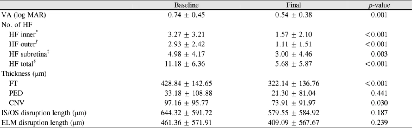
Table 2. Parameters at baseline and final visit
Baseline Final p-value
VA (log MAR) 0.74 ± 0.45 0.54 ± 0.38 0.001
No. of HF
HF inner* 3.27 ± 3.21 1.57 ± 2.10 <0.001
HF outer† 2.93 ± 2.42 1.11 ± 1.51 <0.001
HF subretina‡ 4.98 ± 4.17 3.00 ± 4.46 0.003
HF total§ 11.18 ± 6.36 5.68 ± 5.87 <0.001
Thickness (μm)
FT 428.84 ± 142.65 322.14 ± 136.76 <0.001
PED 33.18 ± 108.88 21.30 ± 81.04 0.441
CNV 97.16 ± 95.77 73.91 ± 91.97 0.030
IS/OS disruption length (μm) 644.32 ± 591.72 579.55 ± 584.92 0.187
ELM disruption length (μm) 461.36 ± 571.91 409.09 ± 567.67 0.239
Values are presented as mean ± SD. All analyses were performed by paired t-test.
VA = visual acuity; log MAR = logarithm of the minimum angle of resolution; HF = hyperreflective foci; FT = foveal thickness; PED = pigment epithelial detachment; CNV = choroidal neovascularization; IS/OS = photoreceptor inner segment and outer segment; ELM = external limiting membrane.
*HF at inner retinal layer; †HF at outer retinal layer; ‡HF at subretinal layer; §HF in total retinal layers.
께가 150 μm 이상 증가할 경우, 형광누출이 증가된 경우에 추가적으로 주입하였다. 최종 방문은 첫 치료 6개월 후로 정의되었고, 최대교정시력, 안저촬영 및 스펙트럼 빛간섭단 층촬영 검사를 시행하였다.
통계분석은 SPSS version 18.0 (SPSS Inc., Chicago, IL, USA)을 이용하였다. 치료 전과 후의 각 인자들의 수치 비 교를 위해 paired t-test를 시행하였다. 치료 전 인자들과 치 료 후 시력의 연관성은 단변량 선형회귀분석을 통해 분석 하였고, 이 중 통계적으로 유의한 인자들(p<0.10)을 대상으로 다변량 선형회귀분석을 후진제거법(backward elimination method)으로 시행하여 치료 후 시력에 대한 유의한 예후인 자를 가려내었다(p<0.05). 두 명의 평가자가 측정한 고반사 점의 개수에 대해 급내상관계수(intraclass correlation co- efficient)를 계산하였다.
결 과
대상 환자는 총 44명 44안이었으며 이 중 남자가 27안, 여자가 17안, 평균 연령은 74.0세였다(Table 1). 평균 주사 횟수는 3.4 ± 0.8회였고, 평균 경과관찰기간은 6.5 ± 1.9개 월이었다. 치료 전 평균 logMAR 시력은 0.74 ± 0.45였다.
치료 전 환자 정보는 Table 1에 요약하였다.
치료 6개월 후 최종 시력은 치료 전에 비해 유의하게 향 상되었다(p=0.001, Table 2). Fig. 1과 Fig. 2에서와 같이 내 망막, 외망막, 망막하 및 전체 고반사점의 개수는 치료 전에 비해 치료 후 유의하게 감소하였다(각각 p<0.001, p<0.001, p=0.003, p<0.001). 고반사점 개수 측정에서 두 평가자 간의 평균 급내상관계수는 0.91로 양호하였다. 중심와 두께 및 맥락막신생혈관 두께는 치료 후 유의하게 감소하였다(각각 p<0.001, p<0.030). 망막색소상피박리 두께는 치료 후 유의 하게 감소되지 않았고(p=0.441), 외경계막과 광수용체 내외 층경계부의 손상 길이는 치료 후 감소하는 경향을 보였으 나 통계적으로 유의하지 않았다(각각 p=0.187, p=0.239).
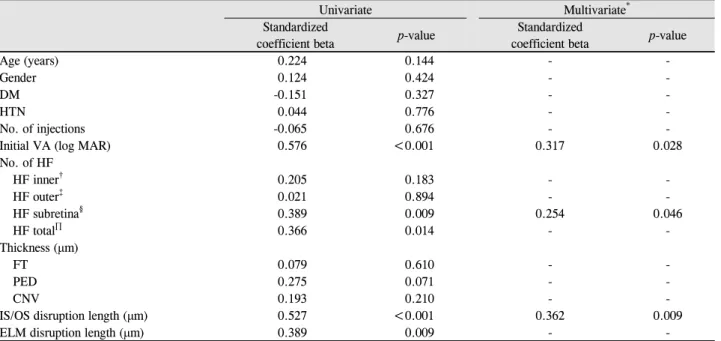
단변량 선형회귀분석을 통해 치료 6개월 후 시력에 영향 을 미치는 인자들을 분석한 결과, 치료 6개월 후 시력은 치 료 전 시력이 나쁠수록, 외경계막과 광수용체 내외층경계 부의 손상이 심할수록, 망막하 고반사점 개수와 전체 고반 사점 개수가 많을수록 유의하게 저하되었다(각각 p<0.001, p=0.009, p<0.001, p=0.009, p=0.014, Table 3). 상기 4개의 치료 전 인자들을 대상으로 다변량 선형회귀분석을 시행한 결과, 치료 6개월 후 시력은 치료 전 시력, 광수용체 내외층 경계부 손상 길이, 망막하 고반사점 개수에 영향을 받는 것 으로 나타났다(각각 p=0.028, p=0.009, p=0.046, Table 3).
다음으로 치료 전 망막하 고반사점이 어떻게 치료 후 시 력에 영향을 미치는지 알기 위해 치료 전 망막하 고반사점
Figure 1. A 66-year-old male presented with neovascular age-
related macular degeneration. (A) Fundus photography at ini- tial visit. Serous elevation is seen at the macula and yellowish fibrinous materials are seen at the parafoveal area. The visual acuity is 20/100. (B) Fundus photography at final visit. Serous elevation is resolved and yellowish fibrinous materials are much decreased. The visual acuity is 20/50. (C) Initial fluo- rescein angiography shows diffuse leakage around macula.(D) Initial indocyanine green angiography shows no definite polypoidal lesions. (E) Horizontal sectional image of optical coherence tomography (OCT) images at initial visit. The OCT shows sparse hyperreflective foci (red arrows) distribution in the inner, outer, and subretinal layers. Subretinal hyper- reflective foci are aggregated at the roof of the subretinal fluid (SRF). (F) Horizontal sectional image of OCT at final visit.
Hyperreflective foci are almost resolved and just small amounts (red arrows) of hyperreflective foci remain at the in- ner retinal layer. SRF is also resolved.
Figure 2. A 76-year-old male presented with neovascular age-
related macular degeneration. (A) Fundus photography at ini- tial visit. Serous elevation at the macula is observed. Baseline visual acuity was 20/60. (B) Fundus photography at final visit.Serous elevation is decreased. (C) Initial fluorescein angiog- raphy shows multiple hyperfluorescene lesions around macula. (D) Initial indocyanine green angiography shows no definite polypoidal lesions. (E) Horizontal sectional image of spectral domain optical coherence tomography (SD-OCT) im- ages at initial visit. The image shows sparse hyperreflective foci (red arrows) distribution at all retinal layers, especially at the subretinal layer. (F) Horizontal sectional image of SD-OCT at final visit. Hyperreflective foci are reduced, but still present around the outer and subretinal layers (red ar- rows). Subretinal fluid has also resolved.
개수와 치료 후 인자들 간의 상관분석을 시행하였다. 치료 전 망막하 고반사점의 개수는 치료 후 중심와 두께 및 맥락 막신생혈관 두께와 유의한 양의 상관관계가 있었다(p=0.002,
p=0.009, Table 4). 또한 상기 두 인자들은 치료 후 시력과 각각 유의한 음의 상관관계가 있었다(p=0.020, p=0.009).
고 찰
본 연구에서 저자들은 삼출성 나이관련황반변성에서 고
A B
C D
E
F
A B
C D
E
F
Table 3. Regression analysis of baseline characteristics for final BCVA (log MAR)
Univariate Multivariate*
Standardized
coefficient beta p-value Standardized
coefficient beta p-value
Age (years) 0.224 0.144 - -
Gender 0.124 0.424 - -
DM -0.151 0.327 - -
HTN 0.044 0.776 - -
No. of injections -0.065 0.676 - -
Initial VA (log MAR) 0.576 <0.001 0.317 0.028
No. of HF
HF inner† 0.205 0.183 - -
HF outer‡ 0.021 0.894 - -
HF subretina§ 0.389 0.009 0.254 0.046
HF total∏ 0.366 0.014 - -
Thickness (μm)
FT 0.079 0.610 - -
PED 0.275 0.071 - -
CNV 0.193 0.210 - -
IS/OS disruption length (μm) 0.527 <0.001 0.362 0.009
ELM disruption length (μm) 0.389 0.009 - -
BCVA = best corrected visual acuity; log MAR = logarithm of the minimum angle of resolution; DM = diabetes mellitus; HTN = hypertension; VA = visual acuity; HF = hyperreflective foci; FT = foveal thickness; PED = pigment epithelial detachment; CNV = choroidal neovascularization; IS/OS = photoreceptor inner segment and outer segment; ELM = external limiting membrane.
*Multivariate linear regression analysis with backward elimination (R2: 0.473); †HF at inner retinal layer; ‡HF at outer retinal layer; §HF at subretinal layer; ∏HF in total retinal layers.
Table 4. Final clinical parameters associated with baseline
number of subretnal hyperreflective focir* p-value
Final VA (log MAR) 0.389 0.009
Thickness (μm)
FT 0.452 0.002
PED 0.023 0.881
CNV 0.390 0.009
IS/OS disruption length (μm) 0.138 0.371
ELM disruption length (μm) 0.149 0.333
VA = visual acuity; log MAR = logarithm of the minimum angle of resolution; FT = foveal thickness; PED = pigment epithelial detachment; CNV = choroidal neovascularization; IS/OS = photoreceptor inner segment and outer segment; ELM = external limiting membrane.
*Pearson's correlation coefficient.
반사점을 포함한 황반부 미세구조와 유리체강내 라니비주 맙 주입술 후 시력 예후와의 연관성을 확인하였다. 기존의 연구 결과를 살펴보면, Akagi-Kurashige et al10은 35명의 삼 출성 나이관련황반변성환자와 61명의 결절맥락망막병증으 로 구성된 환자군에서 치료 후 시력에 대한 치료 전 인자들 의 단변량 선형회귀분석결과, 치료 전 감각신경망막층의 고반사점 개수가 많을수록 치료 후 시력이 저하됨을 발견 하였다. 그러나 다변량 선형회귀분석에서 고반사점은 탈락 되었고, 치료 전 시력과 외경계막의 손상 정도만이 최종 시
력에 대한 예후인자였다. 반면, Coscas et al11은 삼출성 나 이관련황반변성과 결절맥락망막병증으로 구성된 환자군에 서 고반사점은 외망막과 망막색소상피층에 주로 분포하나 그 외의 전 망막층에도 두루 분포하고, 고반사점이 많이 감 소할수록 치료 후 시력예후가 좋았음을 보고한 바 있다.
최근 삼출성 나이관련황반변성과 결절맥락망막병증의 발현에 공통된 유전자들이 관여함이 알려지고, 결절맥락망 막병증을 나이관련황반변성의 아형으로 분류해야 한다는 주장도 있으나,17 결절맥락망막병증의 발병연령이 더 낮고 아시아인에게서 호발하며 출혈성 색소상피박리와 관련성 이 더 높은 점, 광역동학적치료와 항내피세포성장인자에 대한 반응이 삼출성 나이관련황반변성과 다르다는 점을 고 려할 때, 가능한 한 균등한 환자 선정을 위해 라니비주맙 단일 치료를 받은 전형적인 삼출성 나이관련황반변성 환자 들만을 대상으로 하였다.14 한편, 당뇨황반부종에 관한 연구 에서, 치료 전 전망막층에 분포된 고반사점들 중 특히 망막 하액에 위치한 고반사점이 많을수록 최종 시력과 관련 있 음을 발견하여, 고반사점의 수직적 분포가 고려될 필요성 이 제기되었다.16 이에 본 연구에 고반사점을 위치에 따라 내망막, 외망막, 망막하층으로 분류하여 치료 후 시력과의 연관성을 조사하였다.
본 연구 결과, 치료 전에 비해 치료 후 중심와 두께가 유 의하게 감소함은 물론, 전 망막층에서 고반사점의 개수도
유의하게 감소하였다. 시력 역시 치료 후 유의한 향상을 보 였다. 다변량 선형회귀분석결과 치료 후 시력은 치료 전의 망막하액에 위치한 고반사점이 많을수록, 치료 전 시력이 나쁠수록, 광수용체 내외층경계부 손상이 심할수록 유의하 게 저하되었다. 기존의 보고에 따르면 삼출성 나이관련황 반변성에서 최종시력은 치료 전 시력 및 시세포층의 손상 정도와 관련이 있었다.18-20 또한 치료 전 망막하액과 망막색 소상피박리는 치료 후 시력과 상관관계가 없음도 알려져 있다.18 본 연구에서는 기존의 연구 결과와 같이 치료 전 광 수용체 내외층경계부손상을 제외한 망막하액과 망막색소 상피박리는 최종시력과 관련이 없음을 확인할 수 있었고, 망막하액의 고반사점은 시력 저하와 관련된 새로운 예후인 자였다.
고반사점의 기원에 대해 여러 가설들이 제시되고 있으나 정확한 조직학적, 분자적인 구성에 대해 알려지지 않고 있 다. 혈관망막장벽에서 누출된 지단백질, 염증 환경에서 활 성화된 미세아교세포 또는 광수용체 세포의 퇴행과정의 산 물이라는 가설들이 제시되고 있다.6,11,13 또한, 황반변성이 진행하면서 망막색소상피세포가 망막 내측으로 이동한 결 과라고 추측되기도 한다.21,22 본 연구에서 관찰된 망막하 고 반사점은 망막색소상피의 혈관망막장벽이 손상되어 발생 한 것으로 추측된다. 나이관련황반변성에서 조성된 염증반 응은 혈액망막장벽을 손상시켜 혈액에 존재하는 혈장과 지 단백질의 누출을 야기할 것이다. 혈장의 누출은 중심와 두 께의 상승을 유발하고, 지단백질의 누출이 축적되면 빛간 섭단층촬영에서 고반사점으로 나타날 것이다. 본 연구에서 치료 전 증가된 망막하 고반사점은 치료 후 중심와 두께 및 맥락막신생혈관 두께와 양의 상관관계가 있었고, 이 두 인 자들은 치료 후 시력과 음의 상관관계가 있었다. 치료 후 잔여 중심와 두께와 맥락막신생혈관 두께가 크다는 것은 혈관망막장벽의 손상과 맥락막신생혈관의 병적 활성이 더 높은 상태라고 추측할 수 있고, 이들과 망막하 고반사점의 개수가 양의 상관관계가 있음은, 그 기원이 망막상피세포 에서의 이동이나 미세아교세포보다는 혈관외누출임을 더 강하게 시사하는 결과일 것이다.
본 연구의 제한점은 대상 환자 수가 적고 후향적 분석이 었다는 점, 그리고 6개월의 짧은 경과관찰 기간이었다는 점 이다. 또한 SD-OCT상에서 고반사점의 개수를 세는 과정에 서 밝기의 정의가 모호하여 주관이 개입될 가능성이 있음 도 제한점이다. 이는 ImageJ와 같은 소프트웨어를 이용하 여 밝기의 문턱값을 지정하여 고반사점을 표시하는 방법 등으로 보완될 수 있을 것이나, 그 문턱값을 어느 수준으로 정하는 것이 합당할 것인지에 대해서는 추가적인 연구가 필요할 것이다.
REFERENCES
1) Seddon JM, Chen CA. The epidemiology of age-related macular degeneration. Int Ophthalmol Clin 2004;44:17-39.
2) Youm DJ, Oh HS, Yu HG, Song SJ. The prevalence of vitreoretinal diseases in a screened Korean population 50 years and older. J Korean Ophthalmol Soc 2009;50:1645-51.
3) Klein R, Klein BE, Knudtson MD, et al. Fifteen-year cumulative incidence of age-related macular degeneration; the beaver dam eye study. Ophthalmology 2007;114:253-62.
4) Lim LS, Mitchell P, Seddon JM, et al. Age-related macular degeneration. Lancet 2012;379:1728-38.
5) Chen TC, Cense B, Pierce MC, et al. Spectral domain optical co- herence tomography: ultra-high speed, ultra-high resolution oph- thalmic imaging. Arch Ophthalmol 2005;123:1715-20.
6) Bolz M, Schmidt-Erfurth U, Deak G, et al. Optical coherence to- mographic hyperreflective foci: a morphologic sign of lipid ex- travasation in diabetic macular edema. Ophthalmology 2009;116:
914-20.
7) Ogino K, Murakami T, Tsujikawa A, et al. Characteristics of opti- cal coherence tomographic hyperreflective foci in retinal vein occlusion. Retina 2012;32:77-85.
8) Kang JW, Lee H, Chung H, Kim HC. Correlation between optical coherence tomographic hyperreflective foci and visual outcomes after intravitreal bevacizumab for macular edema in branch retinal vein occlusion. Graefes Arch Clin Exp Ophthalmol 2014;252:1413-21.
9) Framme C, Wolf S, Wolf-Schnurrbusch U. Small dense particles in the retina observable by spectral-domain optical coherence tomog- raphy in age-related macular degeneration. Invest Ophthalmol Vis Sci 2010;51:5965-9.
10) Akagi-Kurashige Y, Tsujikawa A, Oishi A, et al. Relationship be- tween retinal morphological findings and visual function in age-re- lated macular degeneration. Graefes Arch Clin Exp Ophthalmol 2012;250:1129-36.
11) Coscas G, De Benedetto U, Coscas F, et al. Hyperreflective dots: a new spectral-domain optical coherence tomography entity for fol- low-up and prognosis in exudative age-related macular degeneration.
Ophthalmologica 2013;229:32-7.
12) Uji A, Murakami T, Nishijima K, et al. Association between hyper- reflective foci in the outer retina, status of photoreceptor layer, and visual acuity in diabetic macular edema. Am J Ophthalmol 2012;153:710-7.
13) Schuman SG, Koreishi AF, Farsiu S, et al. Photoreceptor layer thinning over drusen in eyes with age-related macular degeneration imaged in vivo with spectral-domain optical coherence tomography.
Ophthalmology 2009;116:488-96.
14) Laude A, Cackett PD, Vithana EN, et al. Polypoidal choroidal vas- culopathy and neovascular age-related macular degeneration:
same or different disease? Prog Retin Eye Res 2010;29:19-29.
15) Gomi F, Ohji M, Sayanagi K, et al. One-year outcomes of photo- dynamic therapy in age-related macular degeneration and poly- poidal choroidal vasculopathy in Japanese patients. Ophthalmology 2008;115:141-6.
16) Ota M, Nishijima K, Sakamoto A, et al. Optical coherence tomo- graphic evaluation of foveal hard exudates in patients with diabetic maculopathy accompanying macular detachment. Ophthalmology 2010;117:1996-2002.
= 국문초록 =
나이관련황반변성 환자에서 빛간섭단층촬영의 고반사점 및 치료 전 인자들과 시력과의 연관성
목적: 삼출성 나이관련황반변성 환자의 빛간섭단층촬영에서 나타난 고반사점(hyperreflective foci)과 유리체강내 라니비주맙 주입술 후 시력 예후와의 연관성에 대해 알아보고자 하였다.
대상과 방법: 삼출성 나이관련황반변성 환자 44명 44안을 대상으로 후향적 연구를 시행하였다. 유리체강내 라니비주맙 치료 전 빛간 섭단층촬영에서 나타난 내망막, 외망막, 망막하에 위치한 고반사점의 개수, 중심와 두께, 신생혈관 두께, 외경계막 및 광수용체 내외층 경계부의 상태와 치료 6개월 후 시력과의 연관성을 조사하였다.
결과: 치료 후 고반사점은 망막 전층에서 감소하였다. 치료 후 최대교정시력과 관련 있는 인자는 치료 전 시력, 망막하 고반사점의 개수, 광수용체 내외층경계부의 상태였다(각각 p=0.028, p=0.046, p=0.009). 치료 전 망막하 고반사점은 치료 후 중심와 두께, 맥락 막신생혈관 두께와 유의한 상관관계가 있었다(각각 p=0.002, p=0.009).
결론: 치료 전 망막하 고반사점의 위치 및 개수로 유리체강내 항혈관내피성장인자 주입술 후 시력 예후를 예측할 수 있다.
<대한안과학회지 2015;56(8):1188-1194>
17) Ciardella AP, Donsoff IM, Huang SJ, et al. Polypoidal choroidal vasculopathy. Surv Opthalmol 2004;49:25-37.
18) Simader C, Ritter M, Bolz M, et al. Morphologic parameters rele- vant for visual outcome during anti-angiogenic therapy of neo- vascular age-related macular degeneration. Ophthalmology 2014
;121:1237-45.
19) Kim YM, Kim JH, Koh HJ. Improvement of photoreceptor in- tegrity and associated visual outcome in neovascular age-related macular degeneration. Am J Ophthalmol 2012;154:164-73.
20) Oishi A, Hata M, Shimozono M, et al. The significance of external limiting membrane status for visual acuity in age-related macular degeneration. Am J Ophthalmol 2010;150:27-32.
21) Ho J, Witkin AJ, Liu J, et al. Documentation of intraretinal retinal pigment epithelium migration via high-speed ultrahigh-resolution optical coherence tomography. Ophthalmology 2011;118:687-93.
22) Pieroni CG, Witkin AJ, Ko TH, et al. Ultrahigh resolution optical coherence tomography in non-exudative age related macular degeneration. Br J Ophthalmol 2006;90:191-7.