INTRODUCTION
Neurofibromatosis type 1 (NF1) is a common autosomal dominant genetic disorder with an incidence of approximate- ly 1 in 3,000-3,500 individuals worldwide (1). NF1 is clin- ically characterized by the presence of well established phe- notypic features, including café-au-lait (CAL) spots, neurofi- bromas, freckling of the axillary or inguinal region, Lisch nod- ules, optic nerve gliomas, and bone dysplasias (1). Nearly all NF1 patients have benign dermal neurofibromas, and approx- imately 30% of NF1 patients have benign plexiform neurofi- bromas which can undergo malignant transformation to malignant peripheral nerve sheet tumors (MPNSTs) (2). Malig- nant transformation into MPNSTs was observed in 2-13%
of NF1 patients with plexiform neurofibromas (3) and rep- resents a major cause of mortality in NF1 patients (1).
Haploinsufficiency for neurofibromin has been suggested as the molecular basis of the disease (1, 2). NF1 is caused by mutations in 1 of the 2 alleles of the NF1 gene, located at chromosome 17q11.2, encoding neurofibromin, a GTPase activating protein (GAP). Approximately 5-20% of all NF1 patients have been reported to carry a heterozygous large ~1.5 Mb deletion at chromosome 17q11, where the entirety of the
NF1 gene and several neighboring genes exist, mediated by homologous recombination between the NF1 repetitive sequences (4). It has been reported that a bi-allelic inactiva- tion of NF1, with one allele constitutionally inactivated and the other somatically mutated, is required for neurofibroma and MPNST formation (5), indicating that the tumor progres- sion of NF1 is most likely triggered by the complete loss of the NF1 gene in somatic cells. Somatic loss of heterozygosi- ty (LOH) at the NF1 locus has been described for neurofibro- mas and MPNSTs in previous studies (5).
In a previous study, we reported the case of a 24-yr old male NF1 patient with benign plexiform neurfiboromas and MPN- STs (6). A whole gene expression comparison study was car- ried out using tissue samples obtained by surgical resection and skin biopsy from the patient. Because frequent genomic imbalances in chromosomes 17, 19, and 22q in the neurofi- bromas and MPNSTs have been reported in the patients with NF1 (7), we also tested for genomic alterations and/or chro- mosomal aberrations in the same patient in this study. Com- parison study among normal, benign and malignant tissues and cells was carried out on the chromosomal level. Here, we report a Korean patient with NF1 who had a mosaic Y chromosome loss involved in malignant progression.
804
Seon-Yong Jeong1, Sang-Jin Park2, Su-Jin Lee1, Ho-Jin Park1, and Hyon J. Kim1
Department of Medical Genetics1, School of Medicine, Ajou University, Suwon; MG MED, Inc.2, Seoul, Korea
Address for Correspondence Hyon J. Kim, M.D.
Department of Medical Genetics, Ajou University School of Medicine, 164 Worldcup-ro, Yeougtong-gu, Suwon 443-721, Korea
Tel : +82.31-219-4520, Fax : +82.31-219-4521 E-mail: [email protected]
This work was supported by a grant (KRF-2006-331- E00029) from the Korea Research Foundation (MOEHRD, Basic Research Promotion Fund) and a grant of the Korea Health 21 R&D Project, Ministry of Health & Welfare, Republic of Korea (A050234).
Loss of Y Chromosome in the Malignant Peripheral Nerve Sheet Tumor of a Patient with Neurofibromatosis Type 1
Neurofibromatosis type 1 (NF1) is one of the most commonly inherited autosomal dominant disorders. In order to determine whether genomic alterations and/or chro- mosomal aberrations involved in the malignant progression of NF1 were present in a Korean patient with NF1, molecular and cytogenetic analyses were performed on the pathologically normal, benign, and malignant tissues and primary cells cul- tured from those tissues of the patient. The comparative genomic hybridization (CGH) array revealed a Y chromosome loss in the malignant peripheral nerve sheet tumor (MPNST) tissue. G-banding analysis of 50 metaphase cells showed normal chro- mosomal patterns in the histopathologically normal and benign cultured cells, but a mosaic Y chromosome loss in the malignant cells. The final karyotype for the malig- nant cells from MPNST tissue was 45,X,-Y[28]/46,XY[22]. The data suggest that the somatic Y chromosome loss may be involved in the transformation of benign tumors to MPNSTs.
Key Words : CGH Array; Chromosome Loss; G-banding; Nerve Sheath Neoplasms; Neurofibromatosis 1, Y chromosome
Received : 6 May 2009 Accepted : 11 September 2009
ⓒ 2010 The Korean Academy of Medical Sciences.
This is an Open Access article distributed under the terms of the Creative Commons Attribution Non-Commercial License (http://creativecommons.org/licenses/by-nc/3.0) which permits unrestricted non-commercial use, distribution, and reproduction in any medium, provided the original work is properly cited.
CASE REPORT
A 24-yr old male patient was diagnosed to have NF1 based on standard diagnostic criteria at the Genetic Clinic in Ajou University Hospital. The patient, having a NF1 nonsense mutation (Y2264X) in the NF1 gene, preFsented the clini- cal features of NF1 with café-au-Lait spots, plexiform neurofi- bromas, cutaneous neurofibromas, subcutaneous neurofibro- mas, scoliosis, and neurofibrosarcomas (MPNSTs) (6). After surgical resection of tumors in the patient died at age of 25.
Tumor specimens containing benign and malignant tumors and histopathologically normal tissues were obtained by sur- gical resection and skin biopsy, respectively. Three types of tissues, normal skin fibroblasts, benign plexiform neurofibro- mas, and MPNSTs were prepared and histopathologically eval- uated by routine light microscopy after staining with hema- toxylin and eosin (H&E) as previously described (6).
To determine whether genomic alterations, including mic- rodeletions and gene copy number changes, existed in this male patient, we performed a comparative genomic hybri- dization (CGH) array with high resolution CGH array slides containing 1440 clones, including 356 cancer related genes from BAC libraries at a resolution of 2.3 Mb. Three types of tissues were used; normal skin fibroblasts, benign plexiform neurofibromas, and malignant neurofibrosarcomas (MPNST), all bearing an identical germ line mutation of the Y2264X (c.6792C>G) nonsense mutation on the NF1 gene (6).
DNA was extracted from the tissue samples and used for a CGH array, performed as previously described (8). Com- mercially available CGH array slides (Genomarray; Macro- gen Inc., Korea) were analyzed using the chromofluor image analysis system (Arrayscanner, Array analysis; Macrogen Inc., Korea). The slides contained 1440 clones from BAC libraries.
Each cloned BAC clone DNA was arrayed on the slide in three spots. Non-NF1 male DNA was used as a reference.
The slides were scanned into two 16-bit TIFF image files using GenePix4200A two-color fluorescent scanner (MDS Inc., Canada) and quantitated using GenePix software (MDS Inc., Canada). Green (reference) to red (test) (G/R) ratios were automatically determined for each sample, and the normal- ized G/R ratio was taken to represent the relative average num- ber of copies of the sequence for those spots that were select- ed as controls. Spots with G/R ratios more than the mean plus 2.5 standard deviations (1.25) were considered as ampli- fications or gains of the indicated copy number; less than the mean minus 2.5 deviations (-0.75) were considered as losses of the copy number.
Control hybridization of non-NF1 male DNA was per- formed in four independent experiments and resulted in a mean log2fluorescence ratio of 0.0±0.125 S.D., reflecting the equal copy number in test and reference DNAs (data not shown). Hybridization of non-NF1 male DNA (reference) vs.
DNA from normal tissue of the NF1 patient and further hybridizations of DNA from normal tissue and from benign
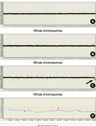
or malignant tissues from the NF1 patient were performed and their results shown in Fig. 1. Multiple tests were per- formed to further narrow down the significant clones dis- cussed in this paper. No genetic alteration, including DNA copy number change across whole chromosomes, was detect- ed in the hybridizations of reference DNA vs. DNA from either normal tissue (Fig. 1A) or benign tissue (Fig. 1B). How- ever, the loss of the whole Y chromosome was detected in the hybridization of the DNA from normal tissue vs. the DNA from MPNST tissue (Fig. 1C). The detailed CGH array pro- file of the Y chromosome for NPNST tissue showed Y chro- mosome-specific genomic loss across the whole region of the Y chromosome (Fig. 1D). Therefore, we focused our study on the Y chromosome aberrations in the MPNST tissue.
To confirm the CGH array findings, G-banding analysis was performed on primary tissue cultured cells. First, primary
Fig. 1. CGH array profiles of the normal, benign and malignant tissues of a patient with NF1. Graphics from the Macrogen’s Mac- Viewer array CGH software show that CGH array profiles for whole chromosomes by (A) hybridization of non-NF1 male DNA (refer- ence) versus test DNA from normal tissue of the NF1 patient, (B) hybridization of DNA from normal tissue versus DNA from benign tissue of the NF1 patient, and (C) hybridization of DNA from nor- mal tissue vs. DNA from MPNST tissue of the NF1 patient. Arrow indicates where loss occurred in the Y chromosome. (D) Graphic shows detailed CGH array profile for the whole region of Y chromo- some in the hybridization of DNAs from normal tissues vs. MPNST tissues of the NF1 patient. Two markers with a letter ‘C’ indicate the position of centromere.
A
B
C
D Whole chromosomes
Whole chromosomes
Whole chromosomes
Y chromosome
tissue culture was performed by primary explant technique from the pathologically normal, benign, and MPNST tissue fragments. The dissected tissues were finely chopped, rinsed with PBS, and the pieces were seeded onto the surface of a culture T24 flask in a small volume (1 mL) of Dulbecco’s mod- ified Eagle’s complete medium (DMEM) supplemented with a high concentration (40%) of heat-treated fetal bovine serum (FBS), 2 mM glutamine, non-essential amino acids, 2.5 mm sodium pyruvate, 100 U/mL penicillin and 100 mg/mL strep- tomycin. After an overnight incubation at 37℃, the medi- um volume was made up to 5 mL and then changed weekly until a substantial outgrowth of cells was observed. Cells were then grown in DMEM media supplemented with 10% heat- treated FBS.
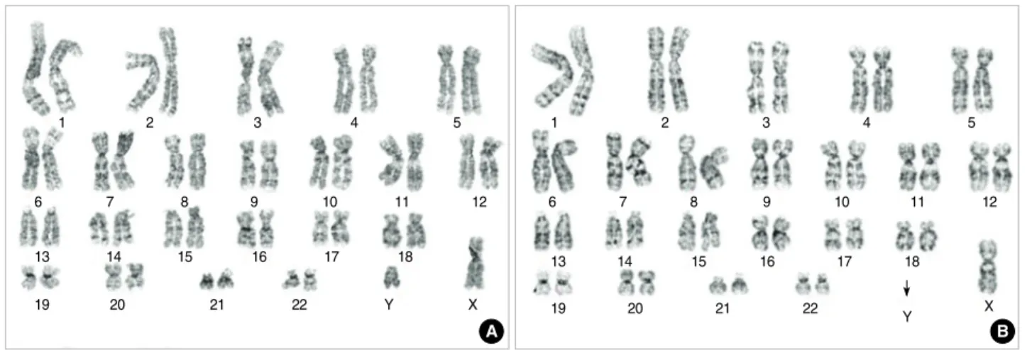
Metaphase cells were harvested and G-banding analyses were carried out using the cultured cells of 5 passages. Cyto- genetic analyses were performed on GTG-banded metaphase spreads prepared from three types of fibroblast cells from the NF1 patient. The karyotypes were described in accordance with ISCN (2009). Chromosome analyses were done in 50 metaphases for each sample with a resolution of 450 bands.
Representative GTG-banded karyotypes of 50 metaphases are shown in Fig. 2. Normal karyotypes were observed in both normal and benign tissue cultured cells (Fig. 2A), but abnor- mal karyotype with a mosaic loss of the Y chromosome was detected in the malignant tissue cultured cells (Fig. 2B). This type of Y chromosome loss was detected in approximately 56%
of the counted cells (28/50). These G-banding study results in primary tissue cultured cells reproduced the CGH array findings in the same tissues. The karyotype for the malignant cells from MPNST tissue revealed 45,X, -Y[28]/46, XY[22].
DISCUSSION
NF1 is notable for the malignant transformation of normal
and/or benign tumor tissues to malignant peripheral nerve sheet tumors (MPNSTs) (3). Three different pathological phe- notypes of tissues, normal, benign and malignant, may exist within one individual despite of an identical germ line muta- tion in the NF1 gene, supporting the two-hit hypothesis that an additional NF1 loss is an independent, somatic event (9).
Studies on the somatic loss of heterozygosity (LOH) in NF1 showed that about 20% of dermal neurofibromas, 40% of plexiform neurofibromas, and 60% of MPNSTs have a LOH either at the NF1 locus or at 17q (5, 10).
The molecular mechanisms potentially required for the full transformation of benign neurofibromas into MPNSTs are poorly understood. Loss of neurofibromin function by bi-allel- ic inactivation of NF1 may be necessary for benign neurofi- broma formation, but is not sufficient for the transformation of neurofibromas to MPNSTs, suggesting that other genetic alterations or epigenetic events must occur in benign tumors for the malignant transformations in NF1 patients. At the gene level, alterations like mutations and/or gene expression changes in other genes including CDKN2A/p14ARF/p15INK4b/p16INK4a, CDKN1B/p27KI11, RB, TP53, PTEN, EGFR, FRAP1 (mTOR), TSC2, TGF-b, HGF-a, TNXB, and TNC, have been identified in MPNSTs (6, 11). In the chromosomal level, several CGH studies of NF1-associated and sporadic MPNSTs reported the existence of gains and/or losses of genetic mate- rials in chromosomes, 4q, 7, 8q, 15q, 17p, 17q, 19, 22q, and X (7, 12, 13). Although no previous studies have reported common patterns of chromosomal aberrations in NF1, the most frequent genomic imbalances in NF-1 associated MPN- STs were detected in chromosome 17q (7).
In our previous study, we reported the results of a gene expression comparison study of a 24-yr old patient with NF1 (6). A whole gene expression comparison study was carried out in normal, benign and malignant tissues and a total of 20 genes were identified showing clear differences in expression patterns among those tissues. Recent studies regarding chro-
Fig. 2. GTG-banded karyotypes of the benign and malignant fibroblast cells from a NF1 patient. (A) Normal karyotype, 46,XY, was detect- ed in metaphases of the benign cells. (B) Abnormal karyotype of loss of Y chromosome, 45,X,-Y, was detected in metaphases of the malignant cells. Arrow indicates where loss occurred in the Y chromosome.
A B
1
6
13
19 20 21 22 Y X
14 15 16 17 18
7 8 9 10 11 12
2 3 4 5 1 2 3 4 5
6 7 8 9 10 11 12
13 14 15 16 17 18
19 20 21 22
Y X
mosomal aberrations in NF1 led to this study in the chromo- somal level of the same patient. Here, we performed molecu- lar cytogenetic analysis using samples of the same normal, benign and malignant tissues from the patient and also per- formed cytogenetic analysis using the primary cultured cells from those tissues. Initially, a comparison study was carried out between tissue types using a CGH array. High-resolution CGH arrays are a powerful tool for the detection of genomic alterations, including gene copy number changes and chro- mosome deletions and duplications (8). Recent studies have identified a gain of a 550 kb segment at 7q, loss of 2.5 Mb at 17q11.2, and duplications in the NF1 locus at 17q11 through CGH array analysis (14). Using a CGH array, we identified the loss of the Y chromosome in the malignant tissue of a patient with NF1. No genetic alterations were detected at the NF1 locus on 17q. This result was supported by G-banding analysis of cultured cells. Karyotype results showed a mosa- ic loss in the Y chromosome of the malignant cells, but not in the normal and benign cells, suggesting that the somatic loss of the Y chromosome in this patient may be associated with the malignant transformation of benign neurofibromas to MPNSTs. In this study, Y chromosome loss was first detect- ed through CGH array in MPNST tissue and confirmed by cytogenetic analysis in cultured cells from MPNST tissue. This ruled out the possibility that the Y chromosome loss occurred during cell culture by chance.
Multiple studies have reported that loss of the Y chromo- some is a normal age-related phenomenon (15). The likelihood of Y chromosome loss increased significantly in normal males after the age of 70. In our case study, the normal age-related Y chromosome loss is less relevant as the NF1 patient is in his mid-twenties. In addition, Y chromosome loss is frequently observed in some types of tumors such as prostate cancer, pan- creatic cancer, squamous cell carcinoma of the head and neck, acute leukemia, hepatocellular carcinoma, and kaposi’s sar- coma (16, 17). A study regarding the significance of Y chro- mosome loss in hematologic diseases reported that the insti- tutional incidence of Y chromosome loss was ~10% of hema- tologic disease groups and the Y loss is frequently seen as mosaicism with a normal (46,XY) clone (18). Our case had a Y chromosome loss with mosaicism in 56% of the count- ed cells. The molecular mechanisms of the loss of Y chromo- some in these tumors remain unclear. Several cancer related genes have been found to be located on the Y chromosome at Yp, TSPY (Y-encoded testis-specific gene), Yq, XKRY (XK- related protein on Y chromosome), PRY (PTPBL-related pro- tein on Y), RRM (RNA recognition motif), YRRM2 (Y chro- mosome RNA recognition motif 2), and CDY1 (Chromod- omain on Y chromosome) (19). Although male predominance has been reported in the NF1 patients with tibial pseudarthro- sis (20), it is not clear whether these genes are associated with malignant transformation of benign neurofibromas to MPN- STs in this case. Further studies are necessary to elucidate the relationship between Y chromosome-specific cancer related
genes and tumor progression of NF1.
To elucidate the mechanisms for the malignant transforma- tion of NF1, we previously performed a comparison study at the gene level in histopathologically different tissues of a Kore- an patient with NF1 (6), and in the present study we carried out comparison study on the genomic and chromosomal levels in the same patient. As a result, we have found the mosaic loss of the Y chromosome in MPNST tissues and cells of the patient. This is the first reported case of Y chromosome loss in NF1. Considering Y chromosome loss has been associat- ed with some types of tumors (16, 17), this result strongly suggests that the somatic loss of the Y chromosome may have caused the transformation of benign tumors to MPNSTs in this patient. Although we are reporting on one case, our data may provide insight into improving the understanding the underlying mechanisms of somatic tumor progression of NF1 on the chromosomal level.
REFERENCES
1. Savar A, Cestari DM. Neurofibromatosis type I: genetics and clini- cal manifestations. Semin Ophthalmol 2008; 23: 45-51.
2. Huson SM. Neurofibromatosis 1: a clinical and genetic overview.
In: Huson, Hughes RAC (eds). The neurofibromatosis. Chapman and Hall Medical, London, 1994; 160-203.
3. Evans DG, Baser ME, McGaughran J, Sharif S, Howard E, Moran A. Malignant peripheral nerve sheath tumours in neurofibromatosis 1. J Med Genet 2002; 39: 311-4.
4. Brunetti-Pierri N, Grange DK, Ou Z, Peiffer DA, Peacock SK, Cooper ML, Eng PA, Lalani SR, Chinault AC, Gunderson KL, Craigen WJ, Cheung SW. Characterization of de novo microdele- tions involving 17q11.2q12 identified through chromosomal com- parative genomic hybridization. Clin Genet 2007; 72: 411-9.
5. Legius E, Marchuk DA, Collins FS, Glover TW. Somatic deletion of the neurofibromatosis type 1 gene in a neurofibrosarcoma sup- ports a tumour suppressor gene hypothesis. Nat Genet 1993; 3:
122-6.
6. Jeong SY, Han JH, Park YY, Kim HJ. Identification of differentially expressed genes related to NF1-associated malignant transforma- tion from a patient with neurofibromatosis type 1. Genes and Genomics 2008; 30: 407-18.
7 Koga T, Iwasaki H, Ishiguro M, Matsuzaki A, Kikuchi M. Frequent genomic imbalances in chromosomes 17, 19, and 22q in peripheral nerve sheath tumours detected by comparative genomic hybridization analysis. J Pathol 2002; 197: 98-107.
8. Yano S, Matsuyama H, Matsuda K, Matsumoto H, Yoshihiro S, Naito K. Accuracy of an array comparative genomic hybridization (CGH) technique in detecting DNA copy number aberrations: com- parison with conventional CGH and loss of heterozygosity analysis in prostate cancer. Cancer Genet Cytogenet 2004; 150: 122-7.
9. Serra E, Puig S, Otero D, Gaona A, Kruyer H, Ars E, Estivill X, Lázaro C. Confirmation of a double-hit model for the NF1 gene in benign neurofibromas. Am J Hum Genet 1997; 61: 512-9.
10. Upadhyaya M, Kluwe L, Spurlock G, Monem B, Majounie E, Mantripragada K, Ruggieri M, Chuzhanova N, Evans DG, Ferner R, Thomas N, Guha A, Mautner V. Germline and somatic NF1 gene mutation spectrum in NF1-associated malignant peripheral nerve sheath tumors (MPNSTs). Hum Mutat 2008; 29: 74-82.
11. Carroll SL, Ratner N. How does the Schwann cell lineage form tumors in NF1? Glia 2008; 56: 1590-605.
12. Lothe RA, Karhu R, Mandahl N, Mertens F, Sauter G, Heim S, Borresen-Dale AL, Kallioneimi. Gain of 17q24-qter detected by com- parative genomic hybridization in malignant tumors from patients with von Recklinghausen’s neurofibromatosis. Cancer Res 1996; 56: 4778- 81.
13. Schmidt H, Taubert H, Meye A, Würl P, Bache M, Bartel F, Holzhausen HJ, Hinze R. Gains in chromosomes 7, 8q, 15q and 17q are characteristic changes in malignant but not in benign peripheral nerve sheath tumors from patients with Recklinghausen’s disease. Cancer Lett 2000; 155: 181-90.
14. Bartsch O, Vlcková Z, Erdogan F, Ullmann R, Novotná D, Spiegel M, Beyer V, Haaf T, Zechner U, Seemanová E. Two independent chromosomal rearrangements, a very small (550 kb) duplication of the 7q subtelomeric region and an atypical 17q11.2 (NF1) microdele- tion, in a girl with neurofibromatosis. Cytogenet Genome Res 2007;
119: 158-64.
15. Wong AK, Fang B, Zhang L, Guo X, Lee S, Schreck R. Loss of the
Y chromosome: an age-related or clonal phenomenon in acute myel- ogenous leukemia/myelodysplastic syndrome? Arch Pathol Lab Med 2008; 132: 1329-32.
16. Wallrapp C, Hahnel S, Boeck W, Soder A, Mincheva A, Lichter P, Leder G, Gansauge F, Sorio C, Scarpa A, Gress TM. Loss of the Y chromosome is a frequent chromosomal imbalance in pancreatic cancer and allows differentiation to chronic pancreatitis. Int J Cancer 2001; 91: 340-4.
17. Kujawski M, Jarmuz M, Rydzanicz M, Szukala K, Wierzbicka M, Grenman R, Golusinski W, Szyfter K. Frequent chromosome Y loss in primary, second primary and metastatic squamous cell carcinomas of the head and neck region. Cancer Lett 2004; 208: 95-101.
18. Wiktor A, Rybicki BA, Piao ZS, Shurafa M, Barthel B, Maeda K, Van Dyke DL. Clinical significance of Y chromosome loss in hemato- logic disease. Genes Chromosomes Cancer 2000; 27: 11-6.
19. Park SJ, Jeong SY, Kim HJ. Y chromosome loss and other genomic alterations in hepatocellular carcinoma cell lines analyzed by CGH and CGH array. Cancer Genet Cytogenet 2006; 166: 56-64.
20. Stevenson DA, Birch PH, Friedman JM, Viskochil DH, Balestrazzi P, Boni S, Buske A, Korf BR, Niimura M, Pivnick EK, Schorry EK, Short MP, Tenconi R, Tonsgard JH, Carey JC. Descriptive anal- ysis of tibial pseudarthrosis in patients with neurofibromatosis 1.
Am J Med Genet 1999; 84: 413-9.