INTRODUCTION
Distant metastases of uterine endometrial stromal sarco- ma (ESS) are uncommon. The most commonly affected site is the lung, with a reported incidence ranging from 7% to 28% (1). Multiple pulmonary nodules are common patterns of metastases of uterine ESS. Unusual presentations included bilateral spontaneous pneumothoraces with predominantly cystic lesions (2-4). Metastatic uterine ESS presenting as a solitary lung nodule is also unusual and only a few cases were documented histologically (1, 5). A case of bilateral reticulo- nodular infiltrates corresponding to a lymphangitic growth pattern was documented (1). We experienced a case of pul- monary metastases of uterine ESS that presented intraalveo- lar micronodules, which is a rare pattern of metastasis not described in English literature.
CASE REPORT
A 37-yr-old woman with increased extent of small random micronodules and ground glass opacities (GGO) in both lungs with dyspnea visited the hospital. She had been diagnosed with ESS in the uterus which had been removed due to post- partum bleeding 4 yr ago. One year later, both of her ovaries and vaginal stump were removed due to the recurred uterine
ESS. At that time, chest computed tomography (CT) showed miliary pattern of micronodules in both lung fields with symp- tom of dyspnea American Thoracic Society (ATS) grade I.
Chemotherapy with Southwest Oncology Group (SWOG) protocol and ifosphamide were performed. During the 3 yr follow-up period, no significant change was found in pul- monary micronodular densities, but occasionally, GGO was found on chest CT with symptoms of dyspnea ATS grade II and fever (Fig. 1). These GGO and her symptoms improved after corticosteroid therapy and the nature of GGO was clini- cally regarded as alveolar hemorrhage or intraalveolar fibroblas- tic polyps. Video assisted thoracotomy biopsy was performed to evaluate the pulmonary micronodules and GGO. Under gross examination, numerous round or ovoid gray white sand- like granular lesions were apparent. Histologic sections showed numerous micronodules in the alveolar spaces (Fig. 2). These nodules consisted of short spindle cells arranged in ill-defined whorls and the neoplastic cells had bland nuclear features and eosinophilic cytoplasm (Fig. 3). Mitotic figures were less than 2 per 10 high power fields. The histologic features of the lung nodules were identical to that of low grade uterine ESS obtained from hysterectomy specimen. Immunohistochem- ically, these neoplastic cells showed strong nuclear staining for estrogen receptor (Novocastra, Newcastle, U.K., 1:100 dilution) (Fig. 3, inset) and progesterone receptor (Novocas- tra, 1:40 dilution), and diffuse cytoplasmic staining for CD10
Gou Young Kim, Chang Ohk Sung, Joungho Han, Joon Oh Park*, Kyung Soo Lee�
Departments of Pathology, Division of Hematology/
Oncology, Departments of Internal Medicine*, and Radiology�, Samsung Medical Center, Sungkyunkwan University School of Medicine, Seoul, Korea
Address for correspondence Joungho Han, M.D.
Department of Pathology, Sungkyunkwan University School of Medicine, Samsung Medical Center, 50 Ilwon-dong, Kangnam-gu, Seoul 135-710, Korea Tel : +82.2-3410-2765, Fax : +82.2-3410-0025 E-mail : [email protected]
901 J Korean Med Sci 2004; 19: 901-3
ISSN 1011-8934
Copyright � The Korean Academy of Medical Sciences
Pulmonary Metastases of Uterine Endometrial Stromal Sarcoma:
Diffuse Micronodular and Ground Glass Opacities : A Case Report
Pulmonary metastases of uterine endometrial stromal sarcoma (ESS) are uncom- mon. The patterns of uterine ESS metastasis to the lung are multiple pulmonary nodules, single nodule, or cystic lesions. Pulmonary intraalveolar micronodular metas- tases of uterine ESS are unusual and have not been reported. We experienced a case of metastatic uterine ESS presenting as pulmonary diffuse micronodules with ground glass opacities on chest computed tomography of a 37-yr-old woman who previously underwent hysterectomy due to low grade ESS of the uterus four years ago. The histologic findings of video assisted thoracotomy biopsy showed numer- ous intraalveolar polypoid micronodules protruding from the alveolar septums. All tumor nodules were composed of short spindle cells arranged in ill-defined whorls, and nuclear feature and sparse cytoplasm were seen in uterine ESS. Immunohis- tochemically, these cells showed strong nuclear staining for estrogen receptor and progesterone receptor, and diffuse cytoplasmic staining for CD10.
Key Words : Sarcoma, Endometrial Stromal; Neoplasm Metastasis; Lung Neoplasms
Received : 11 November 2003 Accepted : 26 December 2003
902 G.Y. Kim, C.O. Sung, J. Han, et al.
(Novocastra, 1:40 dilution). The patient was well and asymp- tomatic when last seen, 8 months post thoracotomy, and the GGO was improved with repeated tamoxifen and ifosphamide therapies.
DISCUSSION
ESS represents approximately 15-25% of all uterine sar- comas and are separated into high and low grades, depending primarily on the mitotic index (more or less than 10 mitoses per 10 high power fields, respectively). Low grade ESS is the most common and has a favorable prognosis. Recurrence is
not uncommon and tends to be limited to the pelvis. Dis- tant metastases may develop after long tumor-free intervals and the lung is the most commonly affected site. The most common growth pattern of pulmonary metastatic ESS, pre- viously described in the literatures, is well circumscribed nodules with entrapped air spaces lined by non-neoplastic respiratory epithelium (1). The unusual growth patterns are solitary nodule, satellite with infiltrative margins, lymphan- gitic pattern and bilateral spontaneous pneumothoraces asso- ciated with predominantly cystic lesions mimicking lym- phangioleiomyomatosis, which can contribute to the diag- nostic dilemma (1-5). This is especially true if clinicians and/
or pathologists are unaware of a prior diagnosis of uterine ESS.
Prior misdiagnosis of a uterine tumor can also be mislead- ing, emphasizing the need to review previous surgical speci- mens, particularly gynecologic specimen in female patients with unusual mesenchymal lung neoplasms (6, 7).
The micronodules of the present case did not show entrap- ped respiratory epithelium and were mainly located in the intraalveolar spaces growing out from alveolar walls suggest- ing hematogeneous spread. This pattern of metastases of uter- ine ESS is interesting and has been not reported in English literatures. We assumed that the lesions were caused by lodg- ing of microtumor emboli through the pulmonary artery.
The metastatic ESS demonstrated the same range of histo- logic features seen in primary uterine ESS.
The differential diagnosis includes benign metastasizing leiomyoma, sclerosing hemangioma, solitary fibrous tumor, hemangiopericytoma, and lymphangioleiomyomatosis in small biopsies. Immunohistochemical stains can be helpful in dif- ferentiation of above diseases. Immunostaining for estrogen receptor and progesterone receptor are positive in nearly all cases of uterine ESS. Immunostaining for keratin and smooth muscle markers are variable. Diffuse immunoreactivity for CD10 in uterine ESS can also be helpful in separating them from uterine smooth muscle neoplasms, which are usually negative.
Fig. 1.Chest computed tomography shows diffuse micronodules and ground glass opacities in both lung fields.
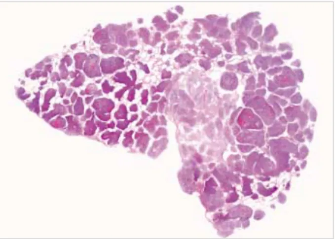
Fig. 2.Histologic section shows numerous micronodules in the alveolar spaces (H&E, ×1).
Fig. 3.Microscopically, nodules consist of short spindle cells arrang- ed in ill-defined whorls. The tumor cells have bland nuclear fea- tures and eosinophilic cytoplasm (H&E, ×200). Inset shows nucle- ar immunoreactivity for estrogen receptor (×400).
Here, we report a case of unusual pulmonary metastatic uterine ESS in a 37-yr-old female with a diffuse micronodule and GGO on chest CT. Pulmonary metastases of low grade ESS may have an excellent prognosis and little effect on sur- vival.
REFERENCES
1. Aubry MC, Myers JL, Colby TV, Leslie KO, Tazelaar HD. Endome- trial stromal sarcoma metastatic to the lung: a detailed analysis of 16 patients. Am J Surg Pathol 2002; 26: 440-9.
2. Mahadeva R, Stewart S, Wallwork J. Metastatic endometrial stromal sarcoma masquerading as pulmonary lymphangioleiomyomatosis. J Clin Pathol 1999; 52: 147-8.
3. Itoh T, Mochizuki M, Kumazaki S, Ishihara T, Fukayama M. Cystic pulmonary metastases of endometrial stromal sarcoma of the uterus,
mimicking lymphangiomyomatosis: a case report with immunohisto- chemistry of HMB45. Pathol Int 1997; 47: 725-9.
4. Dines DE, Cortese DA, Brennan MD, Hahn RG, Payne WS. Malig- nant pulmonary neoplasms predisposing to spontaneous pneumotho- rax. Mayo Clin Proc 1973; 48: 541-4.
5. Satoh Y, Ishikawa Y, Miyoshi T, Mukai H, Okumura S, Nakagawa K. Pulmonary metastases from a low-grade endometrial stromal sar- coma confirmed by chromosome aberration and fluorescence in-situ hybridization approaches: a case of recurrence 13 year after hysterec- tomy. Virchows Arch 2003; 442: 173-8.
6. Abrams J, Talcott J, Corson JM. Pulmonary metastases in patients with low-grade endometrial stromal sarcoma. Clinicopathologic find- ings with immunohistochemical characterization. Am J Surg Pathol 1989; 13: 133-40.
7. Chang KL, Crabtree GS, Lim-Tan SK, Kempson RL, Hendrickson MR. Primary uterine endometrial stromal neoplasms. A clinicopatho- logic study of 117 cases. Am J Surg Pathol 1990; 14: 415-38.
Unusual Pulmonary Metastases of Uterine Endometrial Stromal Sarcoma 903