INTRODUCTION
Urological treatment of urinary calculi has changed much in the past 20 yr. Various endourological treatment modali- ties are available for urinary calculi; ureteroscopic lithotrip- sy, shock wave lithotripsy (SWL), laparoscopic lithotomy, and percutaneous nephrolithotomy. Despite the liberal use of SWL, ureteroscopic lithotripsy is still the preferred treat- ment modality for managing ureter stones at many hospitals and achieves an immediate stone-free state in a high percent of patients. In recent years, the advent of small caliber uretero- scopes and advances in intraureteral lithotripsy have allowed high rates of successful and safe endoscopic treatment of ureter- al calculi (1-3). Currently available semirigid ureteroscopes with a diameter of less than 7Fr and the flexible uretreroscopes can usually be passed up the ureter without ureteral dilation, thus, minimizing morbidity.
Advances in intraureteral lithotripters such as holmium:
YAG laser or Freddy can yield better results. Compared with laser lithotripters, pneumatic lithotripter is old-fashioned and has some limitations of upward migration of stone frag- ments and the lack of fragmentation into small particles. Un- fortunately, pneumatic lithotripter had been the only avail- able tool of ureteroscopic lithotripsy in our hospital for 12 yr. Of course, it is well known that pneumatic lithotripter
has some proven merits of safety and cost-effectiveness. We respectively reviewed our experience of ureteroscopic lithotrip- sy using Swiss Lithoclast.
MATERIALS AND METHODS
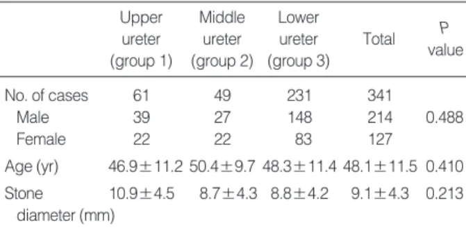
From January 1996 to September 2007, 411 ureteral cal- culi in 392 patients were treated in our institution with ure- teroscopic lithotripsy using Swiss Lithoclast. Medical records of 341 cases in 326 patients were available for use in this ret- rospective study. Exclusion criteria were: radiolucent stone, stone size >2 cm, access failure, failure to apply Swiss Litho- clast. Fifteen patients had bilateral disease. Mean age of 326 patients was 48.1 yr (from 26 to 80).
The 341 cases were divided into three groups according to the stone location. Group 1 consisted of stones located above the pelvic brim (upper), group 2 consisted of stones located over the pelvic brim (mid) and group 3 consisted of stones located below the pelvic brim (lower). The number of cases in each group was 61 in group 1, 49 in group 2, and 231 in group 3. The characteristics of 341 cases are shown on Table 1. There were no significant differences of baseline character- istics including gender, age and stone diameter between the three groups.
Address for correspondence Young Kwon Hong, M.D.
Department of Urology, Bundang CHA Hospital Pochon CHA University, 351 Yatap-dong, Bundang-gu, Seongnam 463-712, Korea
Tel : +82.31-780-5353, Fax : +82.31-780-5323 E-mail : [email protected]
690
Young Kwon Hong and Dong Soo Park
Department of Urology, Bundang CHA Hospital, Pochon CHA University, College of Medicine, Seongnam, Korea
DOI: 10.3346/jkms.2009.24.4.690
Ureteroscopic Lithotripsy Using Swiss Lithoclast for Treatment of Ureteral Calculi: 12-Years Experience
Ureteroscopic lithotripsy using Swiss Lithoclast was performed in 411 cases from January 1996 to September 2007 in a single hospital. Medical records of 341 cases, in which Swiss Lithoclast was successfully applied, were available for this retrospec- tive study. We used 9.5Fr and 10Fr Storz rigid ureteroscopes. A success was defined as being free of stone-related symptoms and residual stones larger than 2 mm. Sixty one stones were located in the upper ureter, 49 stones were in the mid ureter, and 231 stones were in the lower ureter. The overall success rate was 93.5%. The suc- cess rate of upper ureter stone (80.3%) was significantly lower compared with those of mid (93.8%) and lower (96.9%) ureter stones (P=0.001). The higher the calculi was located within the ureter, the more chance of upward migration there was (P<
0.001). The success rate in male patients was lower than in female patients with- out a statistical significance (P=0.068). The success rate decreased as the size of the stone increased (P<0.001), and as the degree of hydronephrosis increased (P=0.03). Perforation rates were 4.9%, 4.1%, and 2.6% from upper to lower ureter stone group. Ureteroscopic lithotripsy using Swiss Lithoclast is a safe and useful treatment modality for ureteral calculi.
Key Words : Ureter; Calculi; Lithotripsy
Received : 10 May 2008 Accepted : 1 September 2008
The parameters used for comparison among the three groups were success rate, stent indwelling, intraoperative ureteral perforation. We also analyzed the success rates according to gender of the patients, stone size, and degree of hydronephro- sis. Our entire equipments for ureteroscopic lithotripsy con- sisted of 9.5Fr, 10Fr Storz rigid ureteroscopes with 5Fr work- ing channel, 5Fr foreign body forceps, stone basket, and Swiss Lithoclast with three probes (1.6, 1, and 0.8 mm). Routine biochemical analysis, blood count, urinalysis and culture of urine were performed preoperatively. Intravenous pyelogram or computed tomogram was taken to confirm the diagnosis and determine the location and the size of stones. Prophy- lactic antibiotics were injected intravenously in all patients.
The procedure was performed with the patients under either general or spinal anesthesia as decided by anesthesiologists.
A safety guide wire was inserted into the ureter as a cysto- scopic procedure. The ureteroscope was introduced via ureter- al orifice with or without ureteral dilation. Ureteral dilation was performed using metal dilator or balloon dilator as nec- essary. Flow of irrigation was controlled by a valve attached to the ureteroscope and accelerated with squeezing pump as needed during operation. Lithoclast probes were passed through the working channel, placed in contact with the calculi, and stones were fragmented down to pieces smaller than 2 mm in diameter under video monitoring and foot control switch.
Fragmented stones were removed out of the ureter as much as possible using basket or forceps.
A JJ stent (6 Fr 22, 24, 26 cm) was placed whenever decid- ed necessary in cases of ureteral edema secondary to an impact-
ed calculus, ureteral injury, and upward migration of stone fragments. A plain radiography of the kidneys, ureters and bladder (KUB) was performed 2 weeks after surgery to assess residual stone fragments. Success was defined as symptom- free and no evidence of residual stones larger than 2 mm in diameter, since stone particles less than 2 mm usually would pass the ureter spontaneously. The outcome of ureteroscopic lithotripsy was compared according to the location of calculi within ureter (Group 1, 2, 3). All variables were expressed as mean±standard deviation. The chi-square test, ANOVA and Fisher’s exact test were used to compare parameters bet- ween the different groups. P<0.05 was considered statisti- cally significant. Statistical analysis was performed with com- puter software (Statistical Package for the Social Science, ver- sion 12.0).
RESULTS Success rate
The overall success rate at 2 weeks postoperative day was 93.5% (Table 2). The success rates were different according to the location of stone. The success rate of group 1 was 80.3%, and those of group 2 and 3 were 93.8% and 96.9%, respec- tively. The success rate of group 1 was significantly lower than other two groups (P=0.001). Success rates of group 2 and group 3 were not significantly different. The higher the cal- culi located within ureter, the more chance of upward migra- tion there was (P<0.001). The success rate in male patients was lower than in female patients (91.6% vs. 96.9%), but there was no statistical significance (P=0.068, Table 3). The success rate decreased as the size of the stone increased (P<
Upper ureter (group 1)
Middle ureter (group 2)
Lower ureter (group 3)
Total P
value
No. of cases 61 49 231 341
Male 39 27 148 214 0.488
Female 22 22 83 127
Age (yr) 46.9±11.2 50.4±9.7 48.3±11.4 48.1±11.5 0.410 Stone 10.9±4.5 8.7±4.3 8.8±4.2 9.1±4.3 0.213
diameter (mm)
Table 1. Baseline data of the subjects
*, group 2, group 3>group 1; �, group 1>gruop 2>group 3.
Upper (group 1)
Parameters Middle (group 2) Lower (group 3) Total P value
No. of cases 61 49 231 341
Success 49 (80.3%) 46 (93.8%) 224 (96.9%) 319 (93.5%) 0.001*
Failure 12 (19.7%) 3 (6.2%) 7 (3.1%) 22 (6.5%)
Migrated stones 10 (16.4%) 4 (8.2%) 4 (1.7%) 18 (5.3%) <0.001�
Perforation 2 (3.3%) 0 (0%) 2 (0.9%) 4 (1.2%) 0.148
Stent indwelling 41 (67.2%) 31 (63.3%) 140 (60.6%) 212 (62.2%) 0.630
Ureteral perforation 3 (4.9%) 2 (4.1%) 6 (2.6%) 11 (3.2%) 0.422
Lasting gross hematuria (>72 hr) 7 (11.4%) 5 (10.2%) 18 (7.8%) 30 (8.8%) 0.453
Pain requiring analgesics 30 (49.1%) 22 (44.9%) 98 (42.4%) 150 (44.0%) 0.633
Table 2. Results and complications according to stone location
Male
Location Female P value
Upper ureter 29/39 (74.4%) 20/22 (90.9%) 0.1821 Middle ureter 25/27 (92.6%) 21/22 (95.5%) 1.0000 Lower ureter 142/148 (96.0%) 82/83 (98.8%) 0.4264 Total 196/214 (91.6%) 123/127 (96.9%) 0.0682 Table 3. Success rates according to gender and stone location
0.001, Table 4), and as the degree of hydronephrosis increased (P=0.03, Table 5). Residual stones larger than 2 mm were noted in 22 cases at 2 weeks after surgery. Eighteen cases of failure was due to upward migration of stones during lithotrip- sy and the other four cases of failure was associated with inci- dental ureteral perforation during lithotripsy. We failed to fragment the stones completely in four cases out of eleven ureteral perforations. Two failed patients were lost to follow- up, and the other 20 failed patients reached stone-free state with auxiliary SWL (15 cases) or medical expulsive therapy (3 cases) or repeated ureteroscopic lithotripsy (2 cases).
Stent indwelling
The rate of stent indwelling was 67.2% (41/61) in group 1, 63.3% (31/49) in group 2 and 60.6% (140/231) in group 3 respectively. The overall rate of stent indwelling was 62.2%
(212/341) (Table 2). The indication of stent indwelling in- cluded ureteral perforation, mucosal avulsion or marked ede- ma, marked bleeding, impacted stone, residual stones, and the surgeon’s preference. Ureteral stent was left in place for 2 to 8 weeks according to postoperative condition of the ureter.
Ureteral perforations were treated with stent indwelling for 4 to 8 weeks without open surgery.
Complications (Table 2)
Ureteral perforation occurred in 11 patients (3.2%), and the incidence was 4.9% in group 1, 4.1% in group 2, and 2.6% in group 3 respectively. The highest incidence was noted in group 1, but the difference was not statistically significant.
Ureteral perforations occurred in eight men and three women who had impacted stones with ureteral narrowing or kink- ing, because guide wires could not pass the obstructed ureter in such cases. Intraoperative ureteral perforation was managed by indwelling a ureteral stent after stopping the procedure as soon as possible. All patients who suffered from ureteral perforations underwent intravenous pyelography 2 weeks after removal of ureteral stent. There was no evidence of ureteral stricture or extravasation of contrast material in any patient with ureteral perforation. The most common postoperative complications were pain requiring analgesics and gross hema- turia lasting for more than 72 hr. In postoperative complica- tions there was no difference of incidence between the three groups.
DISCUSSION
Means of ureteroscopic lithotripsy include ultrasound, elec- trohydraulic, pneumatic, and laser. These instruments are passed through the working channel of the ureteroscope to fragment stones into extractable pieces. In choosing a spe- cific lithotripter operators should take into account not only the characteristics of the stone but also the potential adverse events of the specific lithotripsy technique (4). Every device has its advantages and limitations. Electrohydraulic lithotrip- sy (EHL), the first intracorporeal option, was developed in the 1950s (5). Stone fragmentation is achieved via an electri- cal discharge through a fluid medium, causing a hydraulic shockwave. Although some authors reported fragment rates reaching up to 100% related to EHL, tissue trauma were the main problem (6-9). Stone free rates of EHL for ureteral calculi ranges from 85.3% to 100% and complication rates ranges from below 10% to 45%. Compared with other ure- teroscopic lithotripters, EHL has more complications includ- ing ureteral perforation.
Ultrasonic lithotripsy (USL) relies on rapid vibration of the probe tip, which grinds the stone into fragments. USL requires large-diameter instruments with a straight operating chan- nel; therefore preoperative dilation of the intramural ureter in necessary. This process prolongs the time required for the procedure and increases the amount of ionizing radiation to the patient. The reported stone-free rate following a single treatment with USL ranges from 73.3% in a large scale study (10) to 89.4% in a small study (8). According to a large scale study, ureteral perforation requiring surgical correction oc- curred in 0.65% (10). USL could be a safe and effective treat- ment option for ureteral stones; however, other therapeutic strategies should also be considered in patients with current- ly identified risk factors associated with treatment failure fol- lowing a single USL procedure.
Holmium:YAG laser lithotripsy differs from prior genera- tion lasers such as alexandrite, pulsed dye, and Q-switched lasers. Older lithotripters had short pulse durations that de- posited laser energy quickly, causing a high-energy vapor bub- ble. This bubble subsequently collapsed, thereby fragment- ing calculi through a ‘photoacoustic effect’ (11). In contrast, the holmium:YAG laser has long pulse duration with a pear- shaped bubble and fragmentation occurs through a ‘photother- mal mechanism’ (11). The net result of this modality is small- er fragmentation, and thereby less efficient/slower lithotrip-
<5 mm 5-10 mm >10 mm P value
Upper 2/2 (100.0%) 34/38 (89.5%) 13/21 (61.9%) 0.0323 Middle 9/9 (100.0%) 26/27 (96.3%) 11/13 (84.6%) 0.2507 Lower 56/57 (98.32%) 138/141 (97.9%) 30/33 (90.9%) 0.0981 Total 66/68 (97.1%) 198/206 (96.1%) 55/67 (82.1%) 0.0005 Table 4. Success rates according to stone size and location
Mild HN Moderate HN Severe HN P value
Upper 27/32 (84.4%) 14/17 (82.4%) 8/12 (66.7%) 0.4296 Middle 28/29 (96.6%) 11/12 (91.7%) 7/8 (87.5%) 0.3606 Lower 126/128 (98.4%) 72/75 (96.0%) 26/28 (92.9%) 0.1747 Total 181/189 (95.8%) 97/104 (93.3%) 41/48 (85.4%) 0.0349 Table 5. Success rates according to degree of hydronephrosis (HN) and stone location
Location
sy. However, the overriding major advantage is the holmium’s ability to fragment all stone compositions (11).
The Swiss Lithoclast (a pneumatic lithotripter), originally developed at the University Teaching Hospital in Lausanne, Switzerland, is based on a jackhammer principle (12). A pro- jectile in the handpiece is propelled by compressed air through the probe. The compressed air originates from a small gen- erator that is connected to a dry, clean air supply. The ballis- tic energy produced is conveyed to the probe base at a rate of 12 Hz (13). Continued impaction of the probe tip against the stone results in stone breakage once the tensile forces of the calculus are overcome. The metallic probe rods are avail- able in five diameters: 0.8 mm, 1.0 mm, 1.6 mm, 2.0 mm, and 3.5 mm. Pneumatic lithotripsy has the benefit of better stone targeting and visualization than is possible with the laser. Rapid flashes of light emanating from the laser and visu- ally obscuring protective eyewear may interfere with target- ing. Nevertheless, according to recent studies comparing holmium:YAG laser with Swiss Lithoclast, holmium:YAG laser has higher stone-free rate or fragmentation rate and less complications (14-16). Stone free rate of holmium:YAG laser ranged from 92% to 97% and complication rate was as low as below 4%. On the other hand, stone free rate of pneumatic lithotripsy ranged from 82% to 86% and complication rate was 8% to 14% in these studies. Swiss Lithoclast had been the only available tool of ureteroscopic lithotripsy in our hos- pital for 12 yr, so we did not have a chance to compare it with other treatment modalities including holmium:YAG laser.
It is well established that pneumatic lithotripter has merits of safety and cost-effectiveness. Pneumatic lithotripter is very effective on every stone composition including calcium oxalate monohydrate and cystine stones (13), and it is rarely traumat- ic to tissue and has a low complication rates (17-19). The rate of successful fragmentation of ueteral calculi has wide spec- trum from 70.7% to 96.8%, showing a trend of higher suc- cess rate as the number of patient increases in each study and as the follow-up time increases from the day of operation (14- 19). Our results are comparable with those of other studies about pneumatic lithotripsy. The only appreciable disadvan- tages of pneumatic lithotripsy are the limitation of probe rigid- ity and the potential for proximal stone migration during treatment. The overall rate of stone migration in this study was 5.28%, and 16.4% of upper ureter stone cases failed due to upward migration. The use of a suction device (Lithovac) in conjunction with the Lithoclast or of occlusion devices (basket, occlusion balloon catheter, Stone Cone) or occlusion material (lidocaine jelly) decreases the migration rate (20-22).
We used a basket only in some indicated cases of upper ureter stone during pneumatic lithotripsy. We did not have a flex- ible ureteroscope, so we used SWL for migrated stones left in renal collecting system. The reported rate of ureteral per- foration and avulsion during ureteroscope is 0-4% (23), and the rate of ureteral stricture was reported as 0.5% (24). The rates of ureteral perforation (3.2%) and stricture formation
(0%) in this study are similar to or lower than these values.
Ureteral stenting after ureteroscopic lithotripsy is a com- mon practice to prevent postoperative complications such as ureteral obstruction. Some investigator noted that uncompli- cated ureteroscopy can be performed without routine stent- ing with minimal patient discomfort and a low incidence of postoperative complications (25, 26). Denstedt et al. report- ed that patients, in whom a stent was not inserted, were not at increased risk for complications and postoperative symp- toms including flank pain after ureteroscopy compared with those with a stent, and ureteral stenting after uncomplicat- ed ureteroscopic stone fragmentation was no longer absolutely necessary in all cases (27). We placed ureteral stent even when ureteral injury was not remarkable to prevent postoperative complications such as ureteral stricture. We believe that the liberal use of stent in this study could lead a good result against ureteral stricture.
In conclusion, pneumatic lithotripsy with Swiss Lithoclast is an effective and safe treatment modality for ureter stones.
Its efficacy is reduced in case of large sized upper ureter stones with marked hydronephrosis because of higher chance of stone fragment migration during lithotripsy.
REFERENCES
1. Mugiya S, Nagata M, Un-No T, Takayama T, Suzuki K, Fujita K.
Endoscopic management of impacted ureteral stones using a small caliber ureteroscope and a laser lithotriptor. J Urol 2000; 164: 329-31.
2. Dubosq F, Pasqui F, Girard F, Beley S, Lesaux N, Gattegno B, Thi- bault P, Traxer O. Endoscopic lithotripsy and the FREDDY laser:
initial experience. J Endourol 2006; 20: 296-9.
3. Marks AJ, Teichman JM. Lasers in clinical urology: state of the art and new horizons. World J Urol 2007; 25: 227-33.
4. Piergiovanni M, Desgrandchamps F, Cochand-Priollet B, Janssen T, Colomer S, Teillac P, Le Duc A. Ureteral and bladder lesions after ballistic, ultrasonic, electrohydraulic, or laser lithotripsy. J Endourol 1994; 8: 293-9.
5. Denstedt JD. Intracorporeal lithotriptors. In: Smith A, Badlani GH, Bagley DH, et al., eds. Smith’s Textbook of Endourology. St. Louis, Mo: Quality Medical Publishing, 1996; 47-63.
6. Willscher MK, Conway JF Jr, Babayan RK, Morrisseau P, Sant GR, Bertagnoll A. Safety and efficacy of electrohydraulic lithotripsy by ureteroscopy. J Urol 1988; 140: 957-8.
7. Denstedt JD, Clayman RV. Electrohydraulic lithotripsy of renal and ureteral calculi. J Urol 1990; 143: 13-7.
8. Biri H, Ku_peli B, Isen K, Sinik Z, Karaoglan U, Bozkirli I. Treatment of lower ureteral stones: extracorporeal shockwave lithotripsy or intracorporeal lithotripsy? J Endourol 1999; 13: 77-81.
9. Hofbauer J, Ho_barth K, Marberger M. Electrohydraulic versus pneu- matic disintegration in the treatment of ureteral stones: a random- ized, prospective trial. J Urol 1995; 153: 623-5.
10. Kurahashi T, Miyake H, Oka N, Shinozaki M, Takenaka A, Hara I, Fujisawa M. Clinical outcome of ureteroscopic lithotripsy for 2,129
̆
̆
patients with ureteral stones. Urol Res 2007; 35: 149-53.
11. Teichman JM. The use of holmium:YAG laser in urology. AUA Up- date Series 2001; 20: 154-8.
12. Denstedt JD, Eberwein PM, Singh RR. The Swiss Lithoclast: a new device for intracorporeal lithotripsy. J Urol 1992; 148: 1088-90.
13. Teh CL, Zhong P, Preminger GM. Laboratory and clinical assess- ment of pneumatically driven intracorporeal lithotripsy. J Endourol 1998; 12: 163-9.
14. Jeon SS, Hyun JH, Lee KS. A comparison of holmium:YAG laser with Lithoclast lithotripsy in ureteral calculi fragmentation. Int J Urol 2005; 12: 544-7.
15. Tipu SA, Malik HA, Mohhayuddin N, Sultan G, Hussain M, Hashmi A, Naqvi SA, Rizvi SA. Treatment of ureteric calculi--use of Holmi- um: YAG laser lithotripsy versus pneumatic lithoclast. J Pak Med Assoc 2007; 57: 440-3.
16. Bapat SS, Pai KV, Purnapatre SS, Yadav PB, Padye AS. Compari- son of holmium laser and pneumatic lithotripsy in managing upper- ureteral stones. J Endourol 2007; 21: 1425-7.
17. So_zen S, Ku_peli B, Tunc L, Senocak C, Alkibay T, Karaoglan U, Bozkirli I. Management of ureteral stones with pneumatic lithotrip- sy: report of 500 patients. J Endourol 2003; 17: 721-4.
18. Yinghao S, Linhui W, Songxi Q, Guoqiang L, Chuanliang X, Xu G, Yongjiang M. Treatment of urinary calculi with ureteroscopy and Swiss lithoclast pneumatic lithotripter: report of 150 cases. J Endourol 2000; 14: 281-3.
19. Tan PK, Tan SM, Consigliere D. Ureteroscopic lithoclast lithotrip- sy: a cost-effective option. J Endourol 1998; 12: 341-4.
20. Dirim A, Tekin MI, Aytekin C, Peskircioglu L, Boyvat F, Ozkardes H. Ureteroscopic treatment of proximal ureter stones with the aid of an antegrade occlusion balloon catheter. Acta Radiol 2006; 47: 103-6.
21. Maislos SD, Volpe M, Albert PS, Raboy A. Efficacy of the stone cone for treatment of proximal ureteral stones. J Endourol 2004;
18: 862-4.
22. Mohseni MG, Arasteh S, Alizadeh F. Preventing retrograde stone displacement during pneumatic lithotripsy for ureteral calculi using lidocaine jelly. Urology 2006; 68: 505-7.
23. Schuster TG, Hollenbeck BK, Faerber GJ, Wolf JS Jr. Complications of ureteroscopy: analysis of predictive factors. J Urol 2001; 166:
538-40.
24. Harmon WJ, Sershon PD, Blute ML, Patterson DE, Segura JW. Ure- teroscopy: current practice and long-term complications. J Urol 1997; 157: 28-32.
25. Hosking DH, McColm SE, Smith WE. Is stenting following uretero- scopy for removal of distal ureteral calculi necessary? J Urol 1999;
161: 48-50.
26. Rane A, Cahill D, Larner T, Saleemi A, Tiptaft R. To stent or not to stent? That is still the question. J Endourol 2000; 14: 479-81.
27. Denstedt JD, Wollin TA, Sofer M, Nott L, Weir M, D’A Honey RJ.
A prospective randomized controlled trial comparing nonstented ver- sus stented ureteroscopic lithotripsy. J Urol 2001; 165: 1419-22.
̆
̆