• 검색 결과가 없습니다.

Effect of Trunk Inclination Angles on Trunk Muscle Activity and Subjective Difficulties During Supine Bridge Exercise with a Suspension Device

N/A
N/A
Protected

Academic year: 2021

Share "Effect of Trunk Inclination Angles on Trunk Muscle Activity and Subjective Difficulties During Supine Bridge Exercise with a Suspension Device"

Copied!
7
0
0

로드 중.... (전체 텍스트 보기)

전체 글

(1)

| Abstract |

Purpose: Recent studies have indicated that applying different inclination angles and suspension devices could be a useful way of performing exercises that include the co-activation of the trunk muscles. Present study was to examine the influences of changes in the inclination angle during trunk muscle activity while engaging in a bridge exercise with a suspension device.

Methods: 18 healthy, physically active male volunteers completed three trunk inclination angles (15°, 30°, and 45°) for bridge exercise variations. The surface electromyography responses of the rectus abdominis, internal oblique (IO), erector spinae (ES), and rectus femoris (RF), as well as the subjective difficulty (Borg RPE score), were investigated during these bridge exercises.

Results: The bridge with a 45° inclination angle suspension significantly increased the muscular activities of the RA and RF and increased the Borg RPE scores (p <0.05). The bridge with a 15° suspension significantly elevated the ES activities when compared to the other conditions.

Conclusion: The present study demonstrated that a higher inclination angle could not activate the overall trunk muscles during the bridge exercise. The RA and RF produced greater activation during the bridge exercise with the higher inclination angle. On the other hand, the activities of the erector spine were greater during the bridge exercise with the lower inclination angle. The present study suggests that applying a low trunk inclination angle for the supine bridge exercise is suitable for activating the erector spine muscles.

Key Words: Abdominal strengthening, Electromyography, Supine bridge, Training

†Corresponding Author : Se-Yeon Park ([email protected])

Original Article Open Access

Effect of Trunk Inclination Angles on Trunk Muscle Activity and

Subjective Difficulties During Supine Bridge Exercise with a Suspension Device

Jwa-Jun Kim, P.T., Ph.D.⋅Se-Yeon Park, P.T., Ph.D.

1† 2)

Department of Physical Therapy, Choonhae College of Health Sciences

1

Department of Physical Therapy, Kaya University Received: April 19, 2020 / Revised: May 7, 2020 / Accepted: May 13, 2020

ⓒ 2020 Journal of Korea Proprioceptive Neuromuscular Facilitation Association

This is an Open Access article distributed under the terms of the Creative Commons Attribution Non-Commercial License

(http://creativecommons.org/licenses/by-nc/3.0) which permits unrestricted non-commercial use, distribution, and reproduction

in any medium, provided the original work is properly cited.

(2)

Ⅰ. INTRODUCTION

In the field of rehabilitation, enhancing abdominal muscle strength and appropriate order of activation of the abdominal muscles are major issues for improving performance and functional movement, as well as for reducing incidence of the musculoskeletal problem (Cho

& Park, 2019; Choi et al., 2019; Dafkou et al., 2020;

Yoon et al., 2018). Various exercises are used to improve the abdominal muscle function, including curl-ups, sit-ups, bridge exercises, and exercises using suspension or unstable surfaces (Cho & Park, 2019; Dafkou et al., 2020;

Yoon et al., 2018). Available evidence suggests that multi-joint full body exercises, such as bridge with suspension, may be useful for rehabilitation training (Cho

& Park, 2019). The exercise characterized as low-intensity exercise, and those that demand minimal movement of the body, so that recommended for rehabilitation (Cho

& Park, 2019; Rutkowska-Kucharska & Szpala, 2017).

Among the bridge types of exercises, a supine bridge is a general low-intensity abdominal exercise that granted minimal stress on the lumbar spine (Axler & McGill, 1997). Previous researchers have suggested variations to the supine bridge exercise to maximize its strengthening effects (Escamilla et al., 2016). It was previously demonstrated that abduction of the lower extremity and lifting one leg had positives with rectus abdominis (RA) activation during supine bridge exercises (Yoon et al., 2018). Choi and Kang (2013) reported that supine bridge performed with the feet suspended are advantageous for activating abdominal muscles.

Although previous findings reported additional hip movement, elevated feet position have positive effects for activating abdominal muscles during bridge form of exercise, additional exercise component not always lead to mechanical advantage for recruiting muscles. A study, in which muscle activities during supine bridge performed

at hip position were examined, bilateral movements induced more activity of the multifidus, while a reduced activation of the global muscles (Park et al., 2014). In addition, it is questionable to maximize the muscular recruitment through increases of exercise intensity, especially regarding that the supine bridge exercise is low-intensity exercise.

The supine bridge exercise using suspension device is generally used by physical therapist and athletic trainer, to enhance the trunk muscles. Recently, the supine bridge with suspension device which the feet elevation could be controlled by the height adjustable sling has been suggested (Cho & Park, 2019; Choi & Kang. 2013). The magnitude of feet elevation could change the inclination angle of the trunk and lower extremity, which affects the subjective difficulties and muscular activations around the trunk. However, there were lack of study, which has evaluated trunk muscle activities and subjective difficulties according to the inclination angles.

Therefore, the major purpose of the study was to compare inclination angle variations with respect to trunk muscle activation during supine bridge exercises. The secondary purpose was to find out whether difficulty level could be changed according to exercise conditions.

Ⅱ. MATERIALS AND METHOD

1. PARTICIPANTS

Asymptomatic individuals participated in this study, who were recruited from a local university. The inclusion criterion for participants were determined as excluding musculoskeletal problems during recent 6 months.

Participants with body mass index (BMI) ≥25 were

excluded for obviating the latent influence of fatty tissue

on surface electromyographic (EMG) investigation. A

(3)

final sample were 19 male subjects and demographics of sample participants were 20–24 years old (21.26 ± 1.41, mean ± SD) with height and weight of 171.27 ± 4.90cm and 62.58 ± 7.17kg, respectively. The average BMI was 21.26 ± 1.41. All participants recieved informed written consent following the protocol approved by Kaya University Faculty of Health Science Human Ethics Committee (Kaya IRB-274).

2. INSTRUMENTATION

Four channel of the surface electromyography (LXM3204, LAXTHA, Korea) was used to correct the muscular activation. The sampling rate of the sEMG signals were as 1000Hz frequency. The data were processed with the acquisition software (Telescan, LAXTHA, Korea). Four surface electrodes were embedded parallel to the muscle fibers on the right side as follows:

on the rectus abdominis (RA) muscle at approximately 3 cm lateral to the umbilicus; on the internal oblique (IO) muscle at approximately 1 cm medial to the anterior superior iliac spine; on the erector spine of L3 (ES) muscle at the a approximately 2 cm form the vertebral spine over the muscle mass; and on the rectus femoris (RF) muscle at approximately the midpoint at the line between anterior superior iliac spine and patella (Cram et al., 1998).

The skin was prepared for EMG measurement by cleaning the electrode site with alcohol.

3. PROCEDURES

Five minutes of practice and rest time is given to participant to practice the exercise variations, each subject has performed three different supine bridge exercises with suspension device. Subjects were in the supine position with neutral hip and knee extension. With making a line between the axis of suspension and ankle joint to vertical

to the ground, level of the ankle strap was determined by the wedge under the buttocks. Inclination of wedge was measured with digital inclinometer as 15°, 30°, and 45°, then the wedge was removed during exercise procedures. During exercises, distal portions of both legs placed in the sling suspension system (Redcord Trainer AS, Norway).

A neutral lumbar spine position was ensured by the examiner (anterior and posterior iliac spines in line) during the exercise performance, and the subject was instructed to maintain this position for 10 seconds controlled by metronome. The first and last three seconds were excluded, and the activity from middle of four second was used for further analysis. The participants were allowed to rest three minute between three trials in each condition. After collecting sEMG data during exercise variations, each subject was asked to perform two trials at maximal voluntary muscle contraction (MVIC) for the included muscles against manual resistance (Kendall et al., 2005).

Although the bridge exercise is known as low-intensity exercise, psychological and subjective difficulties were not well identified, especially for bridge exercise using suspension device. To identify the subjective difficulties of each exercise, therefore, the Borg rating for perceived exertion score, which ranged from six to twenty were investigated after performing each exercise. It was designed the rating of perceived exertion (RPE) scale, which is widely believed to be one of the best indicators of degree of physical strain. Score of 7 represents “very, very light”, 9 represents “very light”, 11 represents “fairly light”, and 13 represents “Moderately light” (Scherr et al., 2013).

4. STATISTICAL ANALYSIS

The sEMG signal was calculated with root mean square

(4)

formula and set widow length at 0.125 second. The data were averaged and demonstrated as the %MVIC relative to normalized data. The maximum value between the two MVIC trials was used for the normalization procedure.

The normalized values of RA, IO, ES, and RF were presented as %MVIC. The values including normalized surface EMG, the Borg RPE scale values were used for statistical analysis. PASW Statistics (version 18.0; SPSS, Chicago, IL, USA) was used to determine the significance of differences in %MVIC values between exercise conditions. For investigating the normal distribution of data, the test of the Kolmogorov-Smirnov has been used before the statistical analysis. One-way repeated-measure ANOVA was performed to test for differences in

normalized muscle activity and subjective difficulties. The Bonferroni correction was performed for pairwise multiple comparisons to identify specific differences between exercise variations. In all analyses, significant level was set as 0.05.

Ⅲ. RESULTS

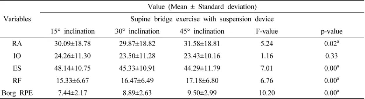
The mean values of normalized EMG data and Borg RPE score were demonstrated in table 1. The factor of inclination angle significantly affected the muscular activities of RA, ES, RF, and Borg RPE score (p < 0.05).

The supine bridge with 15° inclination significantly

Variables

Value (Mean ± Standard deviation) Supine bridge exercise with suspension device

15° inclination 30° inclination 45° inclination F-value p-value

RA 30.09±18.78 29.87±18.82 31.58±18.81 5.24 0.02

a

IO 24.26±11.30 23.50±11.28 23.43±10.16 1.16 0.33

ES 48.14±10.75 45.33±10.91 44.29±11.79 7.01 0.00

a

RF 15.33±6.67 16.47±6.49 17.18±6.80 6.76 0.00

a

Borg RPE 7.44±2.17 8.89±2.63 9.50±2.99 10.20 0.00

a

a

: significant difference among exercise conditions (p <0.05)

MVIC: maximum voluntary isometric contraction, RA: rectus abdominis, IO: internal oblique abdominis, ES: erector spinae, RF: rectus femoris, Borg RPE: Borg rating of perceived exertion

Table 1. Descriptive statistics of normalized EMG data (%MVIC) of the four muscles and Borg RPE

(rating for perceived exertion) score during supine bridge exercise conditions with suspension device

* Significant difference between conditions (p < 0.05)

Fig. 1. The normalized EMG data (%MVC) of the RA, IO, and ES during exercise variations.

(5)

elevated the muscular activity of the ES, compared to the exercise with 30° and 45° inclination (F

2.16

= 7.09, observed power = 0.87, p < 0.05). On the other hand, the normalized sEMG data for the RA and RF differed significantly among exercise variations, which were greater in supine bridge with 45° inclination, compare with the other exercise conditions (RA: F

2.16

= 5.24, observed power = 0.75, RF: F

2.16

= 6.76, observed power

= 0.86) (p < 0.05). (Fig. 1 & 2). The subjective difficulty measured with the Borg RPE score was serially increased with the increases of the inclination angle (F

2.16

= 10.20, observed power = 0.96) (p < 0.05).

Ⅳ. DISCUSSION

This study was to identify the effects of different inclination angle on the activation of the trunk muscles, and subjective difficulties during supine bridge exercises with suspension device. The supine bridge exercise with 45° inclination elicited more RA and RF activations and was considered more difficult. A previous study also investigated the muscular activation during supine bridge exercise with sling device (Lee et al., 2015): supine bridge

with high sling position could elicited overall abdominal muscular activities, which is similar results to present result of RA. However, IO activity did not differ significantly by inclination angle in present study. These results are not same with those of previous finding demonstrating overall trunk muscle were more activated with high sling position. This difference might be caused by the methodological changes. The previous study included adduction and abduction of the hip joint as well as sling height. On the other hand, present study only included the neutral hip position. Presence and absence of the additional muscle recruitment might affect the difference between the present and previous results.

The RF activity also increased with the high inclination angle, but this result might not be positively interpreted.

Because the increased activation of the RF could rotated the pelvis anteriorly, which contributes lumbar lordosis.

Considering results of the Borg RPE score and RF activities, supine bridge with high inclination angle might be unsuitable for the subject with weak abdominal muscle or lumbar pathologies. Although exercise types are different, previous finding also reported the similar suggestions about increased activation of the RF (Escamilla et al., 2006).

* Significant difference between conditions (p < 0.05)

Fig. 2. The normalized EMG data (%MVC) of the RF and Borg RPE (rating for perceived exertion) score

during exercise variations.

(6)

The major finding of present study is that supine bridge exercise with low inclination angle decreased the subjective difficulties, and showed relatively higher activation of the erector spine. Atkins et al. (2015) also reported that increased exercise intensity compromised by the unstable surface improved activation of the anterior core musculature rather than lateral or posterior muscles.

Based on previous studies demonstrating activities of the trunk musculature during curl up, prone bridge (plank), bilateral leg raise, and one leg hip extension, supine bridge is not appropriate exercise for activating the abdominal trunk muscles (Czaprowski et al., 2014; Escamilla et al., 2016; Yoon et al., 2018).

According to a previous study classifying muscle activity, it was considered low muscle activity was ranged 0–20% maximum voluntary contraction (MVC), while 21–

40% was moderate, 41–60% was high, and greater than 60% was very high (Escamilla et al., 2010). In our results demonstrated that the low level of muscular activities from 10% to 30% except the erector spines. This manifest that supine bridge exercise is suitable for activating posterior trunk muscles, but not for the anterior trunk muscles.

Considering the supine bridge exercise is low-intensity exercise which is examined with present results of Borg RPE score, application of the low inclination angle is sufficient for activating trunk extensor muscles in clinical setting.

Several limitations were remained in present study.

First, there was a lack of electromyography data on the other hip muscles, such as the biceps femoris, which may also be influenced by the inclination angle during exercise.

Second, our results of RA could not reach to 80% of power, so that it is hard to have clinical meaning. Lastly, kinematic considerations in the pelvis could not be measured. Further investigations addressing these limitations are warranted.

Ⅴ. CONCLUSION

Supine bridge exercise with three inclination angles were examined in the present study: 15° inclination, 30°

inclination, and 45° inclination. These inclination angles significantly affected muscular activities during supine bridge exercises, as well as Borg rating of perceived exertion (RPE) scale scores. Although 45° inclination angle of the exercise significantly elevated the muscular activities of the rectus femoris and rectus abdominis, present study did not recommend adopting high inclination angle on bridge exercise with suspension device considering subjective difficulties and low mean value of muscular activities. Applying low inclination angle on supine bridge exercise is suitable for activating erector spine muscles.

ACKNOWLEDGEMENTS

This research was supported by the National Research Foundation of Korea (NRF) grant funded by Ministry of Science and ICT (No. 2019R1G1A1007809).

References

Atkins SJ, Bentley I, Brooks D, et al. Electromyographic response of global abdominal stabilizers in response to stable- and unstable-base isometric exercise. The Journal of Strength & Conditioning Research.

2015;29(6):1609-1615.

Axler CT, McGill SM. Low back loads over a variety of abdominal exercises: searching for the safest abdominal challenge. Medicine & Science in Sports

& Exercise. 1997;29(6):804-811.

Cho SH, Park SY. Immediate effects of isometric trunk

(7)

stabilization exercises with suspension device on flexion extension ratio and strength in chronic low back pain patients. Journal of Back and Musculoskeletal Rehabilitation. 2019;32(3):431-436.

Choi CW, Koo JW, Jeong YG. Comparison of trunk muscle activity during modified side-bridge exercises and traditional side bridge exercise. Journal of Sport Rehabilitation. 2019;8:1-7. [Epub ahead of print].

Choi Y, Kang H. The effects of sling exercise using vibration on trunk muscle activities of healthy adults. Journal of Physical Therapy Science. 2013;25(10):1291-1294.

Cram JR, Kasman GS, Holtz J. Introduction to surface electromyography. Gaithersburg. Aspen. 1998.

Czaprowski D, Afeltowicz A, Gębicka A, et al. Abdominal muscle EMG-activity during bridge exercises on stable and unstable surfaces. Physical Therapy in Sport.

2014;15(3):162-168.

Dafkou K, Kellis E, Ellinoudis A, et al. The effect of additional external resistance on inter-set changes in abdominal muscle thickness during bridging exercise. Journal of Sports Science and Medicine. 2020;19(1):102-111.

Ekstrom RA, Donatelli RA, Carp KC. Electromyographic analysis of core trunk, hip, and thigh muscles during 9 rehabilitation exercises. Journal of Orthopaedic

& Sports Physical Therapy. 2007;37(12):754-762.

Escamilla RF, Babb E, DeWitt R, et al. Electromyographic analysis of traditional and nontraditional abdominal exercises: implications for rehabilitation and training.

Physical Therapy. 2006;86(5):656-671.

Escamilla RF, Lewis C, Bell D, et al. Core muscle activation during Swiss ball and traditional abdominal exercises.

Journal of Orthopaedic & Sports Physical Therapy.

2010;40(5):265-276.

Escamilla RF, Lewis C, Pecson A, et al. Muscle activation

among supine, prone, and side position exercises with and without a swiss ball. Sports Health. 2016;8 (4):372-379.

Kim HD, Jeon DM, Bae HW, et al. Changes in activation of abdominal muscles at selected angles during trunk exercise by using ultrasonography. Annals of Physical and Rehabilitation Medicine. 2015;39(6):950-956.

Lee D, Park J, Lee S. Effects of bridge exercise on trunk core muscle activity with respect to sling height and hip joint abduction and adduction. Journal of Physical Therapy Science. 2015;27(6):1997-1999.

Yoon JO, Kang MH, Kim JS, et al. Effect of modified bridge exercise on trunk muscle activity in healthy adults:

a cross sectional study. Brazilian Journal of Physical Therapy. 2018;22(2):161-167.

Park HJ, Oh DW, Kim SY. Effects of integrating hip movements into bridge exercises on electromyographic activities of selected trunk muscles in healthy individuals.

Manual Therapy. 2014;19(3):246-255.

Rutkowska-Kucharska A, Szpala A. The use of electromyography and magnetic resonance imaging to evaluate a core strengthening exercise programme.

Journal of Back and Musculoskeletal Rehabilitation.

2018;31(2):355-362.

Scherr J, Wolfarth B, Christle JW, et al. Associations between Borg’s rating of perceived exertion and physiological measures of exercise intensity. European Journal of Applied Physiology. 2013;113(1):147-155.

Schoenfeld BJ, Contreras B, Tiryaki-Sonmez G, et al. An electromyographic comparison of a modified version of the plank with a long lever and posterior tilt versus the traditional plank exercise. Sports Biomechanics.

2014;13(3):296-306.

수치

Table  1.  Descriptive  statistics  of  normalized  EMG  data  (%MVIC)  of  the  four  muscles  and  Borg  RPE
Fig. 2. The normalized EMG data (%MVC) of the RF and Borg RPE (rating for perceived exertion) score during  exercise  variations.

참조

관련 문서

Based on the above results, the 12-week complex type correction program had a positive effect on the shoulder angle, trunk rotation angle, and body shape

This study investigates the effect of complex exercise program activities using Motion-based games on high school students with developmental disabilities on

Wrestler anaerobic exercise on body composition and function by analyzing the impact of physical man wrestler necessary for the efficient exercise

In the health-related fitness of middle-aged women who participated in the Zumba fitness exercise, muscle strength, muscular endurance, cardiopulmonary

Changes in the level of physical fitness of obese women who performed complex exercise were significantly different in muscle endurance,

The purpose and necessity of this study was to investigate the effect of the 8-week GX complex exercise program on the stress index, blood lipid and APG

12 weeks of combined exercise (body strength exercise and aerobic exercise) was applied to measure health fitness (muscle strength, muscular endurance,

First, the degree of exercise participation of weight lifting hobbyists did not differ according to gender among socio-demographic characteristics, but the