J Korean Skull Base Society 4 : 39~42, 2009
3 9
수술적 감압술 후 회복된 외상성 위눈확틈새 증후군
종설1 원저1 증례1 증례2 증례3 증례4 증례5
순천향대학교부천병원 신경외과학교실 여동규, 황선철, 신동성, 김범태 증례6
Department of Neurosurgery, Soonchunhyang University Bucheon Hospital
Dong Kyu Yeo, M.D., Sun Chul Hwang, M.D., Dong Sung Shin, M.D., Bum Tae Kim, M.D.
수술적 감압술 후 회복된 외상성 위눈확틈새 증후군
Early Surgical Decompression in Traumatic Superior Orbital Fissure Syndrome after Failed Steroid Therapy : A Case Report
The traumatic superior orbital fissure (SOF) syndrome is a rare complication of craniofacial trauma. The treatment modalities remain still in controversy. A 35 year-old man came to emergency room after the impact of his face to other player’s knee during football game. His left eye had ptosis, a dilated fixed pupil and complete paralysis of ocular movements and he also complained of sensory loss of the left forehead and cheek. His visual acuity was preserved. On the facial bone CT, a spicule of fractured sphenoid bone was impinged into the left SOF. Although methylprednisolone was administered for 4 days, his eye symptoms were not improved at all. So the patient was managed by complete resection of fractured bony segment. His symptoms were getting better gradually and completely recovered at the postoperative 3rd month. Surgical decompression may help improve the neurological deficits when the corticosteroid is not effective and the fractured bony segment is displaced into the SOF.
논문 접수일 : 2009년 5월 20일 심사 완료일 : 2009년 6월 5일 주소 : 경기도 부천시 원미구 중동 1174
순천향대학교부천병원 신경외과학교실 전화 : 032-621-5291
전송 : 032-621-5016 이메일 : [email protected]
황 선 철
교신저자
Superior orbital fissure, Ptosis, Ophthalmoplegia, Decompression
Key Words
4 0 JOURNAL OF KOREAN SKULL BASE SOCIETY JUNE |Vol. 4 |No. 1
▒
서 론위눈확틈새(superior orbital fissure ; SOF)는 뇌신경 중에서 눈돌림신경, 도로래신경, 가로돌림신경, 삼차신경의 눈신경이 통 과한다. SOF에 병변으로 인해 안구의 운동장애, 눈꺼풀처침 (ptosis)와 빛반사(light reflex)의 이상을 초래하는 것은 위눈확틈 새 증후군(superior orbital fissure syndrome ; SOFS)이라고 한다1. SOFS는 안면부 외상, 안면골 수술 후, 감염, 종양 등에 의 해 드물게 발생한다2-4, 6. 외상으로 발생한 SOFS는 고용량의 스테 로이드 투여1와 수술적 감압술10을 고려할 수 있다. 본 논문은 골절 편에 의한 직접적으로 SOF의 압박으로 발생한 SOFS를 골절편 (fractured bony spicule) 제거를 시행한 수술적 감압술로 좋은 결과를 얻은 증례보고와 문헌고찰을 통해 조기수술에 대한 적절성 을 고찰해 보고자 한다.
▒
증 례35세 남자가 축구 경기를 하던 중에 상대편 무릎으로 얼굴을 가격 당한 후에 좌측 안와 주위의 부종과 두통으로 응급실로 내원하였다.
안와 주위의 부종과 멍 이외에는 외상은 관찰되지 않았고, 환자는 자발적으로 눈을 뜰 수 없는 눈꺼풀처짐이 관찰되었다. 신경학적 검 사에서 의식은 명료하였고, 좌측 동공은 7mm로 산대되어 있었고, 직접과 간접 빛반사는 소실되었다. 좌측 안구는 중앙에 고정이 되어 있었고, 안구 운동은 모든 방향에서 전혀 움직임이 없었다(Fig. 1).
시력은 외상 전과 비교하여 변함이 없이 유지되었다. 좌측 이마와
광대뼈 주위로 감각이 저하되어 있었다. 얼굴 표정근의 움직임은 정 상적이었다.
응급실에서 촬영한 두부 전산화단층촬영에서 좌측 안와를 이루는 위턱뼈(maxillary bone), 벌집뼈(ethomoid bone), 나비뼈 (sphenoid bone)의 골절과 좌측 측두골의 골절이 관찰되었다(Fig.
2A). 전산화단층촬영의 관상면에서 SOF가 골절편에 의해 좁아져 있었다(Fig. 2B). 골절편에 의한 SOF의 손상에 따른 외상성 위눈확 틈새 증후군을 진단할 수 있었다. 초기치료로 methylprednisolone (Solu-Medrol ; Pharmacia & Upjohn, NJ) 30mg/kg을 부하용량 (loading dose)으로 투여하였고, 그 후에 500mg 을 8시간 간격으로 정주하였다. 외상 3일째까지 약물치료에서 안구운동과 빛반사의 호 전은 전혀 관찰되지 않았다.
Photographs for the eye movements after trauma. Ptosis and full dilated pupil on the left eye is shown with the periorbital contusion.
His left eye doesn’t move in all directions.
Fig. 1
Preoperative and postoperative CT scan with bone algorithm.
A. The axial image shows multiple fractures (arrow head) involving the ethmoid, sphenoid, and temporal bones.
B. The left superior orbital foramen (SOF) is compressed by the fractured bony segment (black arrow) on the coronal CT scan.
C. The postoperative CT scan shows the fracture bony segment is removed and the SOF is well decompressed (white arrow).
A B C
Fig. 2
4 1
수술적 감압술 후 회복된 외상성 위눈확틈새 증후군
외상 4일째 SOF의 수술적 감압술을 계획하였다. 전신마취하에서 좌측 전두-측두 개두술을 시행하였고, 나비뼈의 외측을 드릴로 제 거하여 SOF를 노출하였다. 측두부의 경막을 측두골에서 박리하여 원형구멍(foramen rotundum)을 노출하였다. SOF의 후외측을 구 성하고 있는 나비뼈의 작은날개(lesser wing)의 골절이 있었으며, 드릴을 이용하여 조심스럽게 SOF를 외측에서 내측으로 원형구멍을 향하면서 골편을 제거하였다. 원형구멍의 전상방의 내측 SOF에 골 편이 함입되어 압박되는 소견이 보여 이를 제거하였고, 신경 주행은 유지되는 것을 확인하였다. 수술 직후 안구운동과 빛반사는 호전을 보이지 않았다. 수술 후 스테로이드는 10일간 유지하고 중지하였다.
수술 후 전산화단층촬영의 관상면에서 SOF를 압박하는 골편은 제 거되어 감압이 잘 되었음이 확인되었다(Fig. 2C).
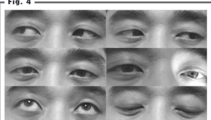
외래 추적관찰 중에 빛반사의 회복이 가장 먼저 관찰되었고, 수 술 45일째에 눈꺼풀처짐은 완전히 호전되어 자발적인 개안은 가능 하였다. 안구운동은 회복되고 있었으나 양안으로 주시할 때 복시가 남아 불편함이 있었다. 빛반사도 회복되어 동공의 크기는 정상적이 었으나 반응속도는 늦었다(Fig. 3). 수술 3개월째에는 안구운동이 완전히 회복되어 복시는 없어졌으나 어두운 곳에서 밝은 곳은 이동 할 때 눈부심이 오래 지속되는 조절부전마비(accommodation paresis)를 호소하였으나 심한 불편함을 보이지는 않았다(Fig. 4).
이러한 눈부심은 점차 호전되기는 하였으나 수술 2년째에도 불편함 이 남아 있었다.
▒
고 찰SOFS는 눈꺼풀처짐, 눈근육마비, 동공확대와 빛반사의 소실을 특징으로 하고, 눈확꼭지증후군(orbital apex syndrome)은 SOFS 의 증상과 더불어 시신경의 손상을 동반한 경우이다1. 외상으로 인 한 SOFS는 관련된 신경의 부종, 좌상, 압박, 혈종에 의해서 발생하
며, 관상영상(coronal image)를 포함한 전산화단층촬영이 좋은 진 단 도구이다2, 8. 만일 골절이 동반되어 있지 않은 경우에는 자기공명 영상촬영, 혈관조영술 등이 필요하다.
외상성 SOFS이 치료방법에는 경과관찰, 스테로이드의 투여, 수 술적 감압술을 고려할 수 있다. Acarturk 등1은 5명의 외상성 SOFS 환자에서 고용량의 스테로이드(methylprednisolone 30mg/kg을 부하용량으로 15분간 정주하고, 시간당 5.4mg/kg을 48시간 투여)를 투여하였고, 48시간 이내에 증상의 호전이 관찰되 었고, 6개월째에는 증상이 완전히 회복되었다고 하였다. 그러나 이 들의 증례에서 골절편이 SOF를 직접 전위되어 압박하는 경우는 없 었다.
외상성 SOFS에 대한 수술적 치료에 대한 보고는 매우 드물어 명 확한 수술 적응증이나 수술방법은 확립되어 있지 않다. 하지만, 외 상성 SOFS에서 안와와 안면골의 골절은 대부분 고정술을 시행하 고, SOF의 감압 여부는 골절편에 의한 직접적인 압박이나 혈종이 있는 경우, 그리고 스테로이드에도 증상이 호전을 보이 않는 경우라 고 알려져 있다1, 2, 9, 10, 12
. 골절편이 전위되지 않은 경우에는 수술 중
General photographs for the eye movements at the postoperative 3rd month. His eye movement, ptosis and light reflex are completely recovered.
Fig. 4
Summary of clinical course. He recovers from ptosis and ophthalmoplegia in the early period, but the subjective discomfort of papillary accommodation is remained in the late period.
Fig. 3
Ptosis (+) Ophthalmoplegia
LR (-)
Ptosis (-) Diplopia (-) Accommodation (±) Op
Time course
Trauma POD #3 mos
Ptosis (-) Diplopia (+)
LR (+) POD #1½ mos
4 2 JOURNAL OF KOREAN SKULL BASE SOCIETY JUNE |Vol. 4 |No. 1
신경손상을 악화시킬 수 있다고 하였다2. 본 증례에서는 내원 직후 에 고용량의 스테로이드를 투여하였으나 외상 4일째까지 증상의 호 전이 전혀 보이지 않았고, 골절편으로 SOF가 압박되어 수술적인 감 압술을 시행하였다. 수술 방법은 고전적인 개두술에 의한 방법과 비 강을 통해 내시경을 이용한 골편제거8, 안와경유(transorbital) 감압 술9이 있다. 개두술은 동반된 안와의 골절을 동시에 고정할 수 있는 장점이 있고, SOF의 외측에 골절편이 위치한 경우에 접근이 가능하 다. 본 증례에서는 SOF의 외측으로 전위되어 있던 골절편의 제거하 였다. 비강을 통한 내시경 수술의 경우에는 SOF의 내측의 접형동의 골절편 제거에서 유용할 것이다.
대부분의 보고에서 SOFS의 예후는 대개 양호하다고 보고되고 있 다. 먼저, 중심 시력에서 복시가 소실되기 시작하며 대부분 3~6개월 내에 복시와 눈꺼풀처짐은 완전히 호전되었다. Chung 등3의 증례 에서 3개월 후에 약간의 동공반사지연을 제외하고 모두 회복되었다 고 하였다. 본 증례에서도 안구운동은 조기에 완전히 회복되었으나 수술 3개월 이후에도 조절부전마비를 호소하였고 2년 후에도 약간 의 불편함이 남아 있었다. 이는 눈돌림신경의 바깥쪽에 부교감신경 이 위치하고 있어 외상에 더 많이 노출되었기 때문으로 생각된다.
뇌신경마비는 경과관찰과 약물치료가 주요하다. 뇌신경마비의 치 료 방침에 대한 연구가 가장 많은 연구가 된 분야가 안면신경 마비 인 벨마비(Bell’s palsy)이다. 벨마비는 약 80~85%가 자연적으로 치유되나 나머지는 후유증상을 가지게 된다5. 후유증상은 마비의 지 속, 구축(contracture), 안면경련, 연합운동(synkinesis), 미각눈물 (crocodile tear) 등이다. 특히, 마비의 등급이 심한 경우에 불완전한 회복과 후유증상의 발생이 높다. 눈돌림신경 마비에서도 이상재생 (aberrant regeneration)에 대해서 잘 알려져 있다11. 이상재생으로 안구운동의 연합운동이 나타나게 된다. 벨마비(Bell’s palsy)에서 예 후가 불량할 것으로 예상되는 경우에 수술을 통한 신경절의 감압술 은 증상의 호전과 후유증의 완화에 효과적이며, 증상 발생 2주 이내 에 수술을 시행하여야 한다고 하였다7. 그러므로 눈돌림신경의 마비 의 정도가 심한 경우는 회복 후에도 이상운동으로 인한 후유증이 예 상이 될 수 있다. 이러한 것을 근거로 할 때 SOFS에서 감압술은 가 능한 조기에 시행하는 것이 좋을 것이다.
▒
결 론외상성 SOFS는 스테로이드에 반응이 없는 경우 수술적 감압술이 유용할 것이며, 특히 전위된 골절편에 의해 압박되는 경우에는 가능 한 조기에 수술을 시행하는 것이 타당할 것이다.
References
1. Acarturk S, Sekucoglu T, Kesiktas E. Mega dose corticosteroid treatment for traumatic superior orbital fissure and orbital apex syndromes. Ann Plast Surg 53:60-64, 2004
2. Bun RJ, Vissink A, Bos RR. Traumatic superior orbital fissure syndrome: report of two cases. J Oral Maxillofac Surg 54:758-761, 1996
3. Chung JW, Kim SJ, Lee SY, Lee EH. A case of Superior Orbital Fissure Syndrome. J Korean Ophthalmol Soc 42:654-657, 2001 4. de Keizer RJ, Padberg GW, de Wolff-Rouendaal D. Superior orbital
fissure syndrome caused by intraorbital spread of a cutaneous squamous cell carcinoma and not detected on computed tomography and magnetic resonance imaging. Jpn J Ophthalmol 41:104-110, 1997 5. Finsterer J. Management of peripheral facial nerve palsy. Eur Arch
Otorhinolaryngol 265:743-752, 2008
6. Fujiwara T, Matsuda K, Kubo T, Tomita K, Yano K, Hosokawa K.
Superior orbital fissure syndrome after repair of maxillary and naso- orbito-ethmoid fractures: a case study. J Plast Reconstr Aesthet Surg 62:e565-569, 2009
7. Gantz BJ, Rubinstein JT, Gidley P, Woodworth GG. Surgical management of Bell's palsy. Laryngoscope 109:1177-1188, 1999 8. Gasco J, Hooten K, Ridley RW, Rangel-Castilla L, Adewumi A, Nauta
HJ, et al. Neuronavigation-guided endoscopic decompression of superior orbital fissure fracture: case report and literature review. Skull Base 19:241-246, 2009
9. Giaoui L, Lockhart R, Lafitte F, Girard B, Chikani L, Fleuridas G, et al.
Traumatic superior orbital fissure syndrome: report of 4 cases and review of literature. J Fr Ophtalmol 24:295-302, 2001
10. Rohrich RJ, Hackney FL, Parikh RS. Superior orbital fissure syndrome:
current management concepts. J Craniomaxillofac Trauma 1:44-48, 1995
11. Weber ED, Newman SA. Aberrant regeneration of the oculomotor nerve: implications for neurosurgeons. Neurosurg Focus 23: E14, 2007 12. Zachariades N, Vairaktaris E, Papavassiliou D, Papademetriou I, Mezitis M, Triantafyllou D. The superior orbital fissure syndrome. J Maxillofac Surg 13:125-128, 1985