The author reports a case of orbital Sarcoidosis in a 70-year-old female that initially presented as diffuse swelling of the lower eyelid
3
0
0
전체 글
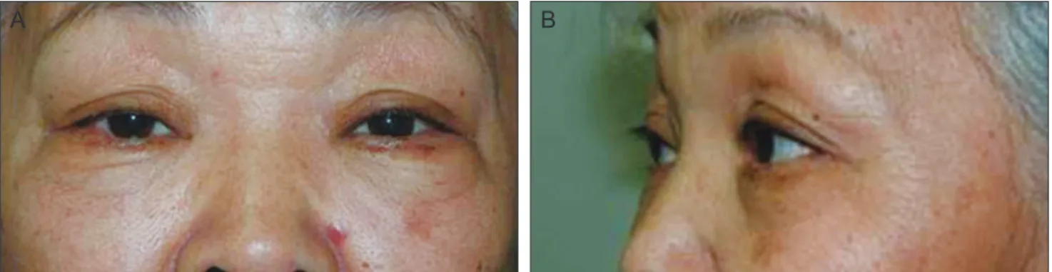
(2) JK Lee, et al. Orbital Sarcoidosis in the Lower Lid. A. B. C. Fig. 1. (A) Photograph of a 70-year-old female with diffuse swelling of the left lower eyelid. Computed tomographic scans, (B) axial and (C) coronal, showed no evidence of a mass lesion or soft tissue infiltration.. A. Fig. 2. Chest radiograph shows multiple confluent irregular opacities (arrow) in right upper lobe. There are no remarkable findings in hilar region with shadow of aortic arch (arrowhead).. on prednisolone (10 mg/day) in consideration of the significant improvement in eyelid swelling after surgical debulking (Fig. 4). The symptoms completely resolved within one month, and she has been followed for 12 months without a recurrence.. B. Discussion The distinctive feature of our case is the initial presentation of diffuse eyelid swelling without mass lesions, even on CT examination. Despite the suspicion of sarcoidosis with a slightly elevated ACE level, inconclusive laboratory and radiologic results necessitated a biopsy to verify the diagnosis. In our patient with diffuse inflammatory lesions without any signs of a mass effect, surgical debulking of the lower lid was considered diagnostic and curative. The histologic examination showed non-caseating granulomatous infiltration in the orbital fat, which was consistent with sarcoidosis, and infectious etiologies were ruled out, as well as foreign body granulomatous disease. Subsequent investigations revealed hilar lymphadenopathy on a chest CT. A prompt response to systemic steroids also supported the diagnosis of sarcoidosis.. Fig. 3. (A) There are multifocal granulomas with lymphocytic infiltration in the fat (H&E, ×40). (B) High magnification view shows a well-formed, non-caseating granuloma composed of epithelioid histiocytes with surrounding lymphocytes (H&E, ×400).. 53.
(3) Korean J Ophthalmol Vol.27, No.1, 2013. A. B. Fig. 4. (A) The patient showed significant improvement of the left lower lid swelling after surgery. (B) Left lower lid swelling was completely resolved after low dose steroid treatment.. Systemic corticosteroids are known to be effective in the treatment of orbital sarcoidosis. Although an initial dose of 1 mg/kg of body weight of oral steroids and gradual tapering is recommended [5], our patient was initially treated with 10 mg of oral prednisolone in consideration of the improvement in eyelid swelling after surgical debulking, and she responded promptly without a recurrence. Surgical debulking might reduce the required dose of steroids by eliminating the amount of inflammatory tissues and enhancing drug diffusion. To the best of our knowledge, the case reported here is the first report of orbital fat infiltration of sarcoidosis without palpable mass in lower lid. A few examples of soft tissue involvement in orbital sarcoid have been reported, but most of them occurred within cutaneous or connective tissue, and isolated orbital fat infiltration has rarely been documented [5-7]. A case of retrobulbar fat infiltration of sarcoid mimicking a superior orbital fissure syndrome has been reported [8], our case is unique in that only the lower eyelid was involved, and there were no remarkable findings on the orbital CT scan. In conclusion, unexplained chronic eyelid swelling without clinical evidence of an orbital mass should be considered as a possible ophthalmic manifestation of orbital sarcoidosis.. Conflict of Interest No potential conflict of interest relevant to this article was reported.. 54. References 1. Prabhakaran VC, Saeed P, Esmaeli B, et al. Orbital and adnexal sarcoidosis. Arch Ophthalmol 2007;125:1657-62. 2. Simon EM, Zoarski GH, Rothman MI, et al. Systemic sarcoidosis with bilateral orbital involvement: MR findings. AJNR Am J Neuroradiol 1998;19:336-7. 3. Biswas J, Krishnakumar S, Raghavendran R, Mahesh L. Lid swelling and diplopia as presenting features of orbital sarcoid. Indian J Ophthalmol 2000;48:231-3. 4. Pollock JM, Greiner FG, Crowder JB, et al. Neurosarcoidosis mimicking a malignant optic glioma. J Neuroophthalmol 2008;28:214-6. 5. Mavrikakis I, Rootman J. Diverse clinical presentations of orbital sarcoid. Am J Ophthalmol 2007;144:769-75. 6. Moin M, Kersten RC, Bernardini F, Kulwin DR. Destructive eyelid lesions in sarcoidosis. Ophthal Plast Reconstr Surg 2001;17:123-5. 7. Marchell RM, Judson MA. Cutaneous sarcoidosis. Semin Respir Crit Care Med 2010;31:442-51. 8. Shaikh ZA, Bakshi R, Greenberg SJ, et al. Orbital involvement as the initial manifestation of sarcoidosis: magnetic resonance imaging findings. J Neuroimaging 2000;10:1803..
(4)
수치
관련 문서