Copyright ⓒ 2019 by Korean Society for Surgery of the Hand, Korean Society for Microsurgery, and Korean Society for Surgery of the Peripheral Nerve. All Rights reserved.
This is an Open Access article distributed under the terms of the Creative Commons Attribution Non-Commercial License (http://creativecommons.org/licenses/by-nc/4.0/) which permits unrestricted non-commercial use, distribution, and reproduction in any medium, provided the original work is properly cited.
Ganglion cyst is a small fluid filled sac that forms over a joint or tendon sheath and can develop almost in any joint space of the body. Most cases present as a slow growing (usually over months) solitary lesion with no fur- ther symptoms. But in some patients, numbness or pain may occur and one of its mechanism can be understood as nerve compression especially in zones of anatomic nerve entrapment. Although they are benign tumors, gan- glion cysts are prone to expand and cause scarring, which can devastate neighboring articular space. This would be synergistic with the confined and limited compliance
of joint, thereby accelerating symptoms of compression and ischemia. When it comes to the ankle joint, one can represent it as a tarsal tunnel syndrome originated from a ganglion cyst1. Tarsal tunnel is located posterior to the medial malleolus and many structures pass through this fibro-osseous space2. Posterior tibial nerve is one of them and covered by the flexor retinaculum whose role is sta- bilizing its understructures. Tarsal tunnel syndrome is an entrapment neuropathy of the tibial nerve or its branches in the tarsal tunnel. Decompression with complete surgi- cal excision has been reported as effective treatment for Hand and
Microsurgery
신경절 낭종과 관련된 족근 터널 증후군의 수술적 감압술: 증례 보고
홍우택ㆍ정윤규
연세대학교 원주의과대학 원주세브란스기독병원 성형외과학교실
Surgical Decompression of Tarsal Tunnel Syndrome Associated with Ganglion Cyst: A Case Report
Woo Taik Hong, Yoon Kyu Chung
Department of Plastic and Reconstructive Surgery, Wonju Severance Christian Hospital, Yonsei University Wonju College of Medicine, Wonju, Korea
Ganglion cyst which originates from joint space can affect nearby nerves through an articular branch. Despite known as benign mucinous lesion, ganglion cyst can show significant fascicular invasion and expansion, resulting in compression neuropathy. Number of pathogenesis have been insisted but the most widely acknowledged one is synovial herniation hypothesis. It describes a capsular defect in joint and discharged fluid to aggregate as cyst along the epineurium of articu- lar branch. Tarsal tunnel syndrome is an entrapment neuropathy of the tibial nerve or its branches in the tarsal tunnel of ankle. In this case report, we describe a case of ganglion cyst associated tarsal tunnel syndrome and its surgical treatment.
Key Words: Ganglion cysts, Tarsal tunnel syndrome
Received August 1, 2019, Accepted August 20, 2019 Corresponding author: Yoon Kyu Chung
Department of Plastic and Reconstructive Surgery, Wonju Severance Christian Hospital, Yonsei University Wonju College of Medicine, 20 Ilsan- ro, Wonju 26465, Korea
TEL: +82-33-741-0611, FAX: +82-33-732-4022, E-mail: [email protected], ORCID: https://orcid.org/0000-0002-0401-3912
Case Report
tion of tumor. As is widely known, incomplete excision may lead to recurrence.
CASE REPORT
A 56-year-old male presented to our department for paresthesia and tingling sensation on the left foot.
This symptom occurred spontaneously and persisted 6 months according to the patient (July 2003). He also had no significant medical or trauma history but had some
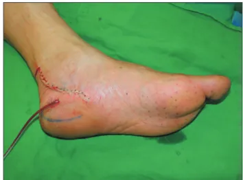
plantar nerve distribution area. We commissioned the Department of Rehabilitation Medicine to conduct an electromyography test to further clarify the sensory de- terioration of this particular area4. On examination, the patient was suspected of having tarsal tunnel syndrome (Fig. 1). We concluded that release of flexor retinaculum and elimination of the occurrence factor would be the fundamental treatments. In order to directly identify what is causing the compression on the medial plantar nerve and to remove it, we decided to undergo surgical treat- ment with general anesthesia. On the surgical field (Au- gust 7, 2003), we noticed that there was severe medial plantar nerve compression by 3×1×1 cm3 size fusiform shaped white cyst and thickened perineural fibrous band (Fig. 2). The cyst was multinodular and had no direct connection with the posterior tibial nerve. Release of flexor retinaculum was done with surgical excision and elaborated fibrous adipose tissue dissection (Fig. 3). Both protection of the nerve branch and complete elimination of cyst were successfully carried out. The dissected cyst was presumed to be ganglion cyst, but we referred the pathology department for further confirmation (Fig. 4).
They revealed it as a ganglion cyst after 4 days. After the surgery, the patient recovered immediately and no longer complained of sensory abnormality or numbness in the
Fig. 2. Intraoperation 1: fusiform shaped white cyst and Fig. 1. Preoperation: demonstrating posterior tibial nerve and artery under flexor retinaculum.
same area (Fig. 5).
DISCUSSION
Intrinsic or extrinsic compression of peripheral nerve can cause several symptoms like paresthesia or tingling sensation. Tarsal tunnel syndrome is entrapment neu- ropathy associated with the posterior tibial nerve or its branches at the tarsal tunnel5. Various factors like bony deformity of talus or calcaneus, hypertrophy of the ab- ductor hallucis, osteochondroma, posttraumatic fibrosis or even ganglion cyst can be the cause of tarsal tunnel syndrome6,7. Precise cause of neural ganglia formation is still on controversy. Two different pathogenesis have been discussed considering the origin of the ganglion.
One presumes that ganglion occurs in the nerve itself and the other one seeks its origin outside of the nerve. The first hypothesis insists cystic degeneration of the con- nective tissue of the epineurium. Ganglion presenting in restricted space may lead to compression of the epineu- rium and the increased pressure would induce degenera- tion of the epineurium8. This degeneration may form a ganglion cyst. On the other hand, the second hypothesis takes charge of invasion of ganglion cyst from the joint or surrounding tissue. However, the mechanism how the ganglion cyst penetrated into the nerve have not been thoroughly revealed.
Neural ganglia occurring in the foot and ankle are rela-
tively rare and epineural ganglion has been reported even lesser than a ganglion cyst originated from intra-fascic- ular location8. For physicians, the location of ganglion is critical for treatment decision. If the ganglion is located within the nerve, complete resection of the cyst without causing damage to the nerve branch may be impossible.
If the ganglion involves only the epineurium and doesn’t have direct connection with the nerve fibers, excsion can be done safely.
In our case, the ganglion cyst of the posterior tibial nerve was multinodular and had no direct connection with the nerve. The patient was farmer and had history of repeated ankle sprains. It suggests that repeated minor injuries may induce cause an epineural ganglion cyst as second hypothesis mentioned above.
Though treatment of tarsal tunnel syndrome depends on the cause or physician’s preference, surgical decom- pression is widely accepted as effective treatment nowa- days9,10. If there is a space-occupying lesion in the tarsal tunnel, Surgical excision would be the best choice of treatment. Total resection of the lesion could be associ- ated with sacrificing the nerve fibers, causing permanent neurologic deficit. Fortunately, the epineural ganglion cysts were not directly connected to the nerve fascicles, surgery could be performed without injury to the nerve fibers in this case. Thorough resection of the involved epineurium was necessary to reduce the recurrence rate because the cyst wall originate from the epineurium.
Fig. 4. Excised ganglion cyst. Fig. 5. Postoperation.
REFERENCES
1. Singh SK, Wilson MG, Chiodo CP. The surgical treatment of tarsal tunnel syndrome. Foot. 2005;15:212-6.
2. De Prado M, Cuervas-Mons M, Golanó P, Rabat E, Va- quero J. The tarsal tunnel syndrome. Fuß Sprunggelenk.
2015;13:227-36.
3. Jerosch J, Schunck J, Khoja A. Results of surgical treatment of tarsal tunnel syndrome. Foot Ankle Surg.
2006;12:205-8.
4. Cancilleri F, Ippolito M, Amato C, Denaro V. Tarsal tun- nel syndrome: four uncommon cases. Foot Ankle Surg.
2007;13:214-7.
5. Lee MF, Chan PT, Chau LF, Yu KS. Tarsal tunnel syn- drome caused by talocalcaneal coalition. Clin Imaging.
Ankle Surg. 2002;8:41-4.
7. Suranigi S, Rengasamy K, Najimudeen S, Gnanadoss J.
Extensive osteochondroma of talus presenting as tarsal tunnel syndrome: report of a case and literature review.
Arch Bone Jt Surg. 2016;4:269-72.
8. Fujita I, Matsumoto K, Minami T, Kizaki T, Akisue T, Yamamoto T. Tarsal tunnel syndrome caused by epineural ganglion of the posterior tibial nerve: report of 2 cases and review of the literature. J Foot Ankle Surg. 2004;43:185- 90.
9. Mullick T, Dellon AL. Results of decompression of four medial ankle tunnels in the treatment of tarsal tunnels syn- drome. J Reconstr Microsurg. 2008;24:119-26.
10. Ahmad M, Tsang K, Mackenney PJ, Adedapo AO. Tarsal tunnel syndrome: a literature review. Foot Ankle Surg.
2012;18:149-52.
신경절 낭종과 관련된 족근 터널 증후군의 수술적 감압술: 증례 보고
홍우택ㆍ정윤규
연세대학교 원주의과대학 원주세브란스기독병원 성형외과학 교실
관절강에 생긴 신경절 낭종은 해당 부위를 지나가는 신경에 영향을 줄 수 있다. 신경절 낭종은 양성 점액성 병변이 지만 내막 침범이나 부피 팽창을 통해 압박성 신경 병증을 유발할 수 있기 때문이다. 병인으로 몇 가지 이론이 거론 되지만, 가장 널리 인정되고 있는 것은 관절낭의 결손을 통해 배출된 체액이 신경의 피복을 따라 응집돼 낭종을 형 성한다는 활막 탈출 가설이다. 이것이 발목 관절에서 발생할 경우 후경골 신경 또는 후경골 신경의 발목 터널 내 분 지의 압박에 의해 발목 터널 증후군이 발생할 수 있다. 이 증례 보고에서는 신경절 낭종에 의한 발목 터널 증후군 1 예와 그 수술적 치료에 대해 기술한다.
색인단어: 결절종, 족근관증후군
접수일 2019년 8월 1일 게재확정일 2019년 8월 20일 교신저자 정윤규
26465, 원주시 일산로 20, 연세대학교 원주의과대학 원주세브란스기독병원 성형외과학교실 TEL 033-741-0611 FAX 033-732-4022 E-mail [email protected]
ORCID https://orcid.org/0000-0002-0401-3912