Partial Purification of OsCPK11 from Rice Seedlings and Its Biochemical Characterization
Jae-Hwa Shin
1and Sung-Ha Kim
2*
1Gyeonggi Science High School, 135, Sooil-ro, Jangan-gu, Suwon-si, Gyeonggi-do 16297, Korea
2Department of Biology Education, Korea National University of Education, 250 Taesungtabyeon-ro, Gangnae-myeon, Heungdeok-gu, Cheongju-si, Chungbuk 28173, Korea
Received February 3, 2020 /Revised February 21, 2020 /Accepted February 22, 2020
Calcium is one of the important secondary signaling molecules in plant cells. Calcium-dependent pro- tein kinases (CDPK)—the sensor proteins of Ca
2+and phosphorylating enzymes—are the most abun- dant serine/threonine kinases in plant cells. They convert and transmit signals in response to various stimuli, resulting in specific responses in plants. In rice, 31 CDPK gene families have been identified, which are mainly involved in plant growth and development and are known to play roles in response to various stress conditions. However, little is known about the biochemical characteristics of CDPK proteins. In this study, OsCPK11—a CDPK in rice—was partially purified, and its biochemical charac- teristics were found. Partially purified OsCPK11 from rice seedlings was obtained by three-step col- umn chromatography that involved anion exchange chromatography consisting of DEAE, hydrophobic interaction chromatography consisting of phenyl-Sepharose, and gel filtration chromatography consist- ing of Sephacryl-200HR. An in vitro kinase assay using partially purified OsCPK11 was also performed.
This partially purified OsCPK11 had a molecular weight of 54 kDa and showed a strong hydrophobic interaction with the hydrophobic resin. In vitro kinase assay showed that the OsCPK11 also had Ca
2+-dependent autophosphorylation activity. The OsCPK11 phosphorylated histone III-S, and the op- timum pH for its kinase activity was found to be 7.5~8.0. The native OsCPK11 shared several bio- chemical characteristics with recombinant OsCPK11 studied previously, and both had Ca
2+-dependent autophosphorylation activity and favored histone III-S as a substrate for kinase activity, which also had a Ca
2+-dependence.
Key words :
Autophosphorylation, Ca
2+-mediated signaling, CDPK, column chromatography, OsCPK11, rice, transphosphorylation
*Corresponding author
*Tel : +82-43-230-3738, Fax : +82-43-232-7176
*E-mail : [email protected]
This is an Open-Access article distributed under the terms of the Creative Commons Attribution Non-Commercial License (http://creativecommons.org/licenses/by-nc/3.0) which permits unrestricted non-commercial use, distribution, and reproduction in any medium, provided the original work is properly cited.
Journal of Life Science 2020 Vol. 30. No. 2. 137~146 DOI : https://doi.org/10.5352/JLS.2020.30.2.137
Introduction
Among the molecules used in the signaling pathways of the plant cell, there is no molecule that represents the re- sponse to a variety of stimuli than the free Ca
2+in the cyto- plasm [46]. Salinity/drought [28], hypo-osmotic stress [50], cold [27], heat shock [18], and NOD factors [15] have been found to cause the change of free [Ca
2+cyt]. The ability of a single molecule to transmit information about various stimuli is due to Ca
2+signal magnitude, duration, frequency, location, and interactions with cellular structures and signal- ing pathways [36]. The unique signature of stimulus - specif-
ic Ca
2+signal is called the calcium signature [35]. Since Ca
2+is not metabolized in the cells, [Ca
2+cyt] should be thoroughly controlled through various Ca
2+-binding proteins. For this, Ca
2+-binding proteins have the optimal structure to bind Ca
2+, and it plays a role in lowering [Ca
2+cyt] or initiating a secondary signaling pathway [11].
In order for calcium to act as a secondary signaling mole- cule in the intracellular signal transduction, the dormant [Ca
2+cyt] should be kept at a very low level. Conversely, giv- en a particular stimulus, the [Ca
2+cyt] is elevated at a rapid rate to initiate a specific response. It should also be con- sumed very quickly to go back to the idle state again for the next signal transfer [23]. Temporally increased recog- nition of free Ca
2+in the cytoplasm is associated with a pri- mary Ca
2+sensor or a specific substrate on the signaling pathway [46].
Ca
2+-dependent protein kinases (CDPKs) are primary Ca
2+sensors that bind to high [Ca
2+cyt] and are a unique class
of kinases that transmit signals by transferring phosphoryl
groups to cellular substrate proteins [23]. They are also the most abundant serine/threonine kinases in plants [48].
CDPKs are found in vascular plants, non-vascular plants, green algae and some protozoa [21]. CDPKs are encoded by the multi-gene family [52], and Arabidopsis (34) [24], rice (29)[6], and maize (35)[32] carry the CDPK genes. CDPKs also exist in a variety of cellular locations, indicating that they may be involved in a variety of signaling pathways [34]. Harmon et al. [19] suggested that CDPKs are involved in the potential regulation of gene expression, metabolism and signal transduction, migration of various ions and water through the membrane, and the dynamic movement of the cytoskeleton.
Indeed, as a Ca
2+sensor and a protein kinase, CDPKs reg- ulate the diverse functions of plants, and the biological func- tion of each CDPK isoform has been demonstrated in several plants. CDPKs are known as modulators of plant growth and developmental processes. Expression of pollen-specific CDPK genes in corn [16] and in Arabidopsis [38] are examples. CDPKs are also involved in hormone delivery and biotic and abiotic stress signaling [47]. In connection with the signaling of abscisic acid (ABA), several CDPKs in
Arabidopsis play a central role in environmental stress re-sponses, including drought, salinity, and cold [4]. CDPKs are also involved in the signal transduction pathways re- quired to defend against infectious and herbal feeding [43].
They regulate various metabolic enzymes [49] and attenuate Ca
2+signal [14]. Although various functions performed by these cells have been revealed, but much still remains unknown.
Asano et al. [6] showed 29 CDPK genes in rice by a ge- nome-wide analysis. Ray et al. [41] confirmed that 31 rice CDPK genes were identified by adding two new genes, and that each CDPK was involved in specific organ development and specific developmental stages. CDPK isoforms have been shown to be tissue-specific [6, 52]. Especially, OsCPK8 and OsCPK19 (OsCDPK2) were found in the panicle, while OsCPK10, OsCPK24 and OsCPK29 were mainly expressed in roots, and they were related to signal transmission in the panicle and root, respectively [52]. Each CDPK isoform has a different function in the cell, which can be broadly sum- marized as being involved in the growth and development of rice and in the signaling pathway of response to various stresses.
It is almost certain that rice CDPKs are involved in the growth and development. First, in the study using OsCPK2
and OsCPK11 [currently OsCPK7 based on Asano et al. [6]
mRNA, it was confirmed that mRNA amount of OsCPK2 in white light and anaerobic treatment was much lower than that of OsCPK11 [8]. In addition, in a study by Frattini et
al. [17] OsCDPK2 protein is expressed at a low level in theearly stage of seed development, but later increased in amount and OsCDPK11 shows the opposite pattern.
OsCDPK11 transcript and protein in leaves are not affected by light. While OsCDPK2 protein is almost absent upon ex- posure to light, it increases rapidly at night. It suggested that two CDPK isoforms perform different functions in re- sponse to seed development and light. This suggests a sim- ilar conclusion in Morello et al. [37] where the stability of the OsCDPK2 protein is regulated by light and is also in- volved in the seed formation. In the study of the location of CDPKs in the cells, OsCPK2 was found to be associated with the membrane by the myristoylation and palmitoyla- tion [34].
There are many studies that rice CDPKs are involved in various stress responses. Wan et al. [52] reported that the promoter region of OsCPK1~OsCPK29 genes contained mul- tiple stress-responsive cis-elements and that CDPK genes were extensively involved in the stress responses. OsCDPK7 belongs to stress-inducible CDPKs, and their transcripts in- crease by a low temperature and salt stress conditions [45].
Under the low temperature stress condition, the role of ABA in controlling the activity of 45 kDa CDPK in rice was determined. In addition to OsCDPK13, which was found to be stress-inducible [1], OsCPK6, OsCPK17, and OsCPK25 were also found to be very important in stress tolerance of rice [52]. OsCPK21 and OsCPK12 are positive regulators of the salt stress signaling pathway [5]. Like the CDPKs of oth- er plant species, many of the unique biological functions of CDPKs in rice remain largely unknown.
The first study to purify and characterize plant CDPKs was performed by Harmon et al. [20] They used CDPKs from soybean to validate the key properties of CDPKs that require calcium for enzyme activity, but do not require calmodulin.
This is due to structural features with a calmodulin-like do-
main that can bind directly to calcium at the C-terminal of
CDPKs [21]. In rice, it was the first obtained by a partial
purification of CDPK in leaves and its activity was con-
firmed to be calcium dependent [25]. The activity of CDPK
purified from sandalwood [3], and beetroot cell membranes
[30] depends on calcium precisely. In addition, CDPKs had
a unique biochemical characteristics in autophophorylation
and transphosphorylation of specific substrates. In the kin- ase assay using CDPK1 of chickbean (Cicer arietium) ex- pressed in E. coli, calcium-dependent autophophorylation and transphosphorylation were confirmed [13]. Cho [10]
studied the biochemical characteristics of recombinant OsCPK11 expressed in E. coli. In this study, a biochemical characteristics of native OsCPK11 obtained from rice leaves was investigated. And the similarities and differences be- tween native OsCPK11 and recombinant OsCPK11 will be discussed.
Materials and Methods
Materials
Rice (Nipponbare) seeds were sown on MS medium and grown for 2-3 weeks. At this time, a chamber having a con- stant temperature of 30℃, and a photoperiod of 16 hr light/8 hr dark was used. Leaves were harvested and used for the purification experiment. Most chemicals used in this study are from Sigma Aldrich (USA) as follows; 2-amino-2-(hy- droxymethyl)-1,3-propanediol (Trizma base), sodium chlor- ide, magnesium chloride, calcium chloride, ammonium sul- fate, sodium phosphate monobasic, potassium phosphate monobasic, glycine, polyethylene glycol sorbitan mono- laurate (Tween-20), 4-(2-hydroxyethyl)piperazine-1-ethane- sulfonic acid (HEPES), 2-(N-morpholino)ethanesulfonic acid hydrate (MES), adenosine 5’-triphosphate (ATP), sodium or- thovanadate (Na
3VO
4), ethylene glycol-bis(2-aminoethylether)- N,N,N,N’-tetraacetic acid (EGTA), phenylmethanesulfonyl fluoride (PMSF), ethylenedinitrilo tetraacetic acid (EDTA), 1,4-dithiothreitol (DTT), histone Ⅲ-S, myelin basic protein and casein. Methanol is the product from Emsure. [γ
32-P]
Adenosine 5’-triphosphate (6,000 Ci/mmol) was from Perkin Elmer (Boston, USA).
Purification of OsCPK11
The rice leaves grown for 2-3 weeks were harvested and their proteins were extracted. Rice leaves was measured and at least 1 ml of protein extraction buffer (50 mM Tris, 150 mM NaCl, 10 mM MgCl
2,1 μM approtinin pH 7.5) per 100 mg of the fresh weight was added and ground it finely with a grinder. Crude extract was filtered through gauze and miracloth (Millipore, USA), then centrifuged at 11,000× g for 20 min at 4℃ using centrifuge (Model Avanti JXN-30, Beckman). The supernatant was collected and used for the purification.
Column chromatography
Chromatography was carried out using a biologic LP sys- tem (Bio-Rad, USA) in a 4℃ chamber. First, gel filtration chromatography was performed to remove low molecular substances including salts contained in the crude extract. At this time, 30 ml of crude extract was desalted at once using Bio-Gel P-6 desalting cartridge (Bio-Rad, USA, 50 ml) and 25 mM Tris buffer (pH 8.0). Desalted crude extract was col- lected and proceeded to the anion exchange chromatography.
5 ml of DEAE Sepharose (GE Healthcare, USA) was packed in an open column (1×10 cm, Bio-Rad, USA). Start Buffer was 25 mM Tris buffer (pH 8.0) and Binding Buffer was 25 mM Tris buffer (pH 8.0) containing 1 M NaCl. Equili- brium was performed for 200 min with Start Buffer. After 25 min, sample was loaded instead of the Start Buffer. In the gradient, the proteins attached to the resin were eluted by increasing the salt concentration from 0% to 100% by mix- ing the Start Buffer and the Binding Buffer for 80 min. And eluted proteins were collected in each 1 ml fractions. After elution of all the proteins attached to the resin with Binding Buffer for 25 min, re-equilibrium was performed with Start Buffer for 50 min. The flow rate was kept constant at 1 ml/min. Through buffer exchange with Start Buffer (50 mM sodium phosphate buffer containing 1.5 M ammonium sul- fate, pH 7.5) for the next purification step using Centriprep 10K (Miliipore, Germany), a final sample was obtained to carry out the next step.
In order to perform hydrophobic interaction chromatog- raphy, 3 ml of Phenyl-Sepharose (GE Healthcare, USA) was packed in an open column (1×10 cm, Bio-Rad, USA). Start Buffer was 50 mM sodium phosphate buffer (pH 7.5) con- taining 1.5 M ammonium sulfate and the Binding Buffer was 50 mM sodium phosphate buffer (pH 7.5). Equilibrium was performed for 75 min with Start Buffer. After 15 min, sample was loaded instead of the Start Buffer. In the gradient, the proteins attached to the resin were eluted by decreasing the salt concentration from 100% to 0% by mixing the Start Buffer and the Binding Buffer for 60 min. And eluted pro- teins were collected in each 2 ml fractions. After elution of all the proteins attached to the resin with Binding Buffer for 15 min, re-equilibrium was performed with Start Buffer for 30 min. The flow rate was kept constant at 1 ml/min.
As in the previous step, fractions which contained target proteins were combined and concentrated with Centriprep 10K (Miliipore, Germany).
As a final step, gel filtration chromatography using
Sephacryl-200HR (Sigma Aldrich, USA) was performed.
Forty-two milliliter of Sephacryl-200HR was used to pack the open column (1×30 cm, Bio-Rad, USA) and PBS buffer (137 mM NaCl, 2.7 mM KCl, 10 mM Na
2HPO
4, 2 mM KH
2PO
4) was used for the process, and the flow rate was maintained at 0.4 ml/min. After loading the sample on the packed resin, PBS buffer was flowed for 100 min. Finally, fractions containing target proteins were combined and con- centrated using Centriprep 10K (Miliipore, Germany). In or- der to confirm the efficient the purification process, several confirmation steps were carried out. Total protein obtained from each purification step was measured. Samples were as follows: ⅰ) desalted crude extract, ⅱ) sample eluted from anion exchange chromatography, ⅲ) sample eluted from hy- drophobic interaction chromatography and ⅳ) fractions from gel filtration chromatography. Total protein content was measured using the BCA Protein Assay kit (Sigma Aldrich, USA) [31].
SDS-PAGE
SDS-PAGE was performed using a stepwise sample to confirm protein pattern in each purification process. Protein separation was performed using SDS-PAGE mini kit (Major Science, South Korea). The sample was loaded into each well of 10% SDS-PAGE gel and protein separation was performed.
Gel obtained after electrophoresis was stained with Coomas- sie Blue solution and protein bands were observed.
Western Blot
Fractions containing the target proteins in each purifica- tion step were confirmed by western blot using OsCPK11 antibody. Purified OsCPK11 antibody was prepared and used. Synthetic peptide was made based on known amino acid sequence of OsCPK11, and it was injected into the rab- bits to induce antibody formation. Serum from the blood collection was purified and OsCPK11 antibody with high affinity was obtained. Western blotting was performed on all fractions obtained from the gradient section of DEAE chromatography and Phenyl-Sepharose chromatography, and the fractions centered on the peak of Sephacryl-200HR chromatography. After selecting the samples to be analyzed for each purification step, SDS-PAGE was performed. Gel was transferred to PVDF membrane (Immobilon-P, Milli- pore, Germany). Transfer from the gel to the PVDF mem- brane was carried out in a Transfer Buffer (25 mM Tris, 192 mM glycine, 20% methanol) for 90 min at 4℃ and 100V
(Electro Blot System, Major Science, South Korea). Blocking was done at room temperature for 3 hr in Blocking Solution (5% skim milk in 0.1% PBST). It was treated with OsCPK11 antibody (1:2,000 dilution) in 2.5% skim milk, and the mem- brane was reacted at room temperature for 3 hr, and then the wash step was initiated. It was washed with 2.5% skim milk in 0.1% PBST twice for 15 min, and with 0.1% TBST for 15 min once. And reaction was done at room temperature for 1 hr in 2.5% skim milk containing secondary antibody (anti-rabbit IgG AP conjugate, Promega, USA; 1:5,000 dilu- tion). After washing with 0.1% TBST twice for 15 min and once for 15 min with TBS, color development was performed in alkaline phosphate buffer (100 mM Tris, 150 mM NaCl, 1 mM MgCl
2, pH 9.0) containing NBT and BCIP (Promega, USA) to confirm the final band pattern.
Autophosphorylation assay
Autophosphorylation was performed in vitro using a par- tially purified OsCPK11 as described before. 4X Kinase Reaction Buffer (20 mM HEPES, 200 nM ATP, 1 mM Na
3VO
4, 2 mM DTT, 0.5 mM PMSF, 2 mM EDTA, pH 7.4) was prepared. 0 or 100 μM of MgCl
2, 0 to 1 mM CaCl
2, 1 mM of EGTA, 1 μg of OsCPK11 were added to each Reaction Buffer, and the final volume was adjusted to 12 μl with 20 mM HEPES (pH 7.4). Then, 10 UCi [γ
32-P]ATP diluted 10 fold with 20 mM HEPES was added and incubated for 0.5~
1 hr at 30℃. Four microliter of 4X Sample Buffer was added and boiled for 5 min at 100℃. Sample was loaded into each well of 12% SDS-PAGE gel and protein separation was performed. Gel was dried using gel dryer (Bio-Rad, Model 583) for 30 min at 80℃, and exposed to BAS plate for 2~3 days at -80℃. The film was developed and examined. The intensity of raidioactivity was calculated by Image J software (National Institutes of Health, USA).
Transphosphorylation assay
In order to examine the transphosphorylation properties
of partially purified OsCPK11, following experiments were
conducted. First, three different substrates of histone Ⅲ-S,
myelin basic protein and casein were used to find the best
substrate for a partially purified OsCPK11 in vitro. One mi-
crogram of each substrate, 1 μM of CaCl
2, 100 μM of MgCl
2,
1 mM of EGTA and 1 μg of OsCPK11 were added to the
4X Kinase Reaction Buffer. After this, rest of experimental
procedure was the same as one described in autophosphor-
ylation experiment.
A
B
C
Fig. 1. A. Elution profile of DEAE column chromatography. It showed the gradient range between 200 and 280 min in the entire DEAE chromatography. 1 M of NaCl con- centration was gradually increased from 0 to 100%, and the proteins bound to the resin were eluted. The interval indicated by (↑) mark was the interval over which OsCPK11 was eluted, which corresponds to 0.175 M to 0.55 M NaCl. B. Elution profile of Phenyl-Sepharose col- umn chromatography. It showed the gradient range be- tween 75 and 135 min in the entire Phenyl-Sepharose chromatography. Proteins bound to the resins were elut- ed by reducing the concentration of 1.5 M ammonium sulfate gradually from 100% to 0%. The interval in- dicated by (↑) mark was the interval over which OsCPK11 was eluted, which corresponds to 0.4 M to 0 M (NH4)2SO4. C. Elution profile of Sephacryl-200HR col- umn chromatography. The time in between two mark (↑) at the bottom indicates when OsCPK11 was eluted.
Table 1. Total protein content during purification step
Purification step Total protein
(mg) Crude extract
DEAE-Sepharose column chromatography Phenyl-Sepharose column chromatography Sephacryl-200HR column chromatography
1,187.3 67.1
9.6 0.2
In order to find the optimal pH for the transphosphor- ylation activity of partially purified OsCPK11, 1 μM CaCl
2, 100 μM MgCl
2, 1 μg of OsCPK11 and histone Ⅲ-S were add- ed to 4X Kinase Reaction Buffer of pH 6 to pH 9. A series of different 20 mM buffer solution from pH 6 to pH 9 was added to each to make a final volume to 12 μl. After this, rest of experimental procedure was the same as one de- scribed in autophosphorylation experiment.
Results
Partial purification of OsCPK11
From DEAE chromatography, the OsCPK11 was eluted in the 17.5%~55% NaCl gradient section (0.175 M to 0.55 M) as shown in Fig. 1A, which was confirmed by the west- ern blot analysis. At this step, OsCPK11 was presumed to have a weak anionic amino acid on its surface. From Phenyl-Sepharose chromatography, OsCPK11 was eluted in the 27%~0% (NH
4)
2SO
4gradient section (0.4 M to 0 M) as shown in Fig. 1B, which was confirmed by the western blot analysis. This indicated that OsCPK11 has a strong hydro- phobic binding Phenyl-Sepharose resin. Most CDPKs were characterized by their ability to bind strongly to hydro- phobic resins such as Phenyl-Sepharose [42]. CDPKs of soy- bean, maize and silver beet were known to have hydro- phobic binding sites [7, 40] and potato protein kinase was eluted by 4 M urea [33], and the salt-resistant CDPKs of
Dunaliella tertiolecta were eluted with 50% of ethylene glycol[54]. This was also confirmed by the refining process of this study. From Sephacryl-200HR chromatography, fractions # 27 and #28 contained partially purified OsCPK11 (Fig. 1C) and this was confirmed by western blotting.
Several confirmation procedure was performed to de-
termine if the purification level proceeded effectively during
the purification steps. The total protein content of each sam-
ple during purification steps was measured and result was
shown in Table 1. During purification steps, fractions con-
taining the target protein was recovered and the next step
was carried out. At each step, Western blot analysis was per-
Fig. 2. Western blot analysis of the fractions from Sephacryl-200 HR column chromatography. Eluted samples from Sephacryl-200HR column chromatography were deter- mined by Western blot analysis using OsCPK11 antibody.
OsCPK11 was present in the fractions #27~#28. Its mo- lecular weight was estimated to be 54 kDa.
A
B C
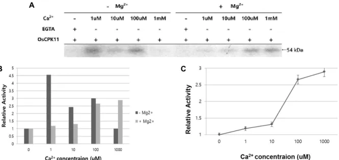
Fig. 3. Effects of Mg2+ and Ca2+ on the autophosphorylation activity of OsCPK11. A. Partially purified OsCPK11 showed a strong autophosphorylation activity at 1 μM Ca2+ in the absence of Mg2+. In the presence of 100 μM Mg2+, the autophosphorylation activity tended to increase as Ca2+ concentration increased, and the strongest autophosphorylation activity was observed at 1 mM Ca2+. B. This showed the autophosphorylase activity of partially purified OsCPK11 with or without Mg2+ under different Ca2+ concentration. When Ca2+ was not present, no autophosphorylation activity was observed regardless of the presence or absence of Mg2+. C. This showed the change of autophosphorylation activity of partially purified OsCPK11 as Ca2+ concentration increased in the presence of 100 μM Mg2+. The higher the Ca2+ concentration, the greater the autophosphor- ylation activity of partially purified OsCPK11, and it had the highest activity at 1 mM Ca2+.
formed to find OsCPK11, and results were shown in Fig.
2. The molecular weight of OsCPK11 was estimated to be 54 kDa, which was confirmed by western blotting as well as autoradiography (Fig. 3). This is similar to the study by Cheng et al., [9] that CDPK is a monomer and has a molec-
ular weight in the range of 40~90 kDa. The molecular weights of known CDPKs indicate 52 kDa from soybean [20, 40], 53 kDa from groundnut [12] and 45 kDa from barley [26].
In vitro kinase assay
Ca
2+and Mg
2+influence the activity of autophosphor- ylation of OsCPK11 (Fig. 3). In the absence of Mg
2+, auto- phosphorylation of OsCPK11 was activated, but when Ca
2+was absent, no activity of autophosphorylation was ob- served (Fig. 3), suggesting that Ca
2+is essential for the activa- tion of autophosphorylation of partially purified OsCPK11.
In the absence of Mg
2+, autophosphorylation activity was ob-
served even in the presence of lower Ca
2+, while the activity
of autophosphorylation was decreased when Ca
2+become
higher (Fig. 3). Even when Ca
2+was at 1 mM, autophosphor-
ylation was not activated at all. Conversely, when 100 μM
Mg
2+was present, the autophosphorylation activity of parti-
ally purified OsCPK11 gradually increased as Ca
2+increased
(Fig. 3). As a result, the strongest autophosphorylation activ-
ity was observed when 1 μM Ca
2+was present without Mg
2+,
and the strongest autophosphorylation activity was ob-
served when 1 mM Ca
2+was present with 100 μM Mg
2+(Fig.
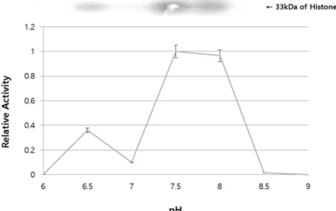
Fig. 5. Optimal pH for the transphosphorylation activity of OsCPK11. Partially purified OsCPK11 and histone III-S were reacted at the different pH from pH 6 to pH 9, respectively. Each buffer contained 1 mM Ca2+ and 100 μM Mg2+ as well. It showed the strongest activity at a slightly alkaline pH 7.5~8.0.
Fig. 4. Searching for the substrate for OsCPK11. One microgram of partially purified OsCPK11 was reacted with each 1 μg of three different substrates of histone Ⅲ-S, MBP and casein. Among them, histone Ⅲ-S seemed to be the best substrate for the partially purified OsCPK11 phosphor- ylase activity was the most strongly observed in histone III-S. It had a stronger kinase activity in the presence of 1 μM Ca2+, while it did not phosphorylate either MBP or casein.
3). Therefore, the partially purified OsCPK11 in this study has a Ca
2+-dependent autophosphorylation function. Num- erous studies supported the autophosphorylation of ser- ine/threonine residues in CDPKs is Ca
2+-dependent and in- dependent of calmodulin [20, 22]. Conversely, winged bean CDPK has an autophosphorylation activity independent of calcium [44]. Little is known about the effects of autophos- phorylation on CDPK function in vitro, and almost nothing is known about its physiological or mechanistic role in vivo [22]. Therefore, more extensive studies will be needed to ex- amine the effect of Ca
2+on autophosphorylation of CDPKs.
Partially purified OsCPK11 in this study was tested with three different substrates: histone Ⅲ-S, MBP, and casein with or without 1 μM Ca
2+. Result showed that partially purified OsCPK11 strongly phosphorylated histone III-S, which was Ca
2+-dependent (Fig. 4). That is, kinase activity was stronger in the presence of Ca
2+. On the other hand, MBP and casein were almost not phosphorylated, as shown in Fig. 3. In addi- tion, partially purified OsCPK11 and histone III-S were re- acted in the presence of 1 mM Ca
2+and 100 μM of Mg
2+, and the pH of the reaction buffer varied from pH 6 to pH 9. Result indicated that partially purified OsCPK11 showed the strongest activity at a slightly alkaline pH 7.5~8.0 (Fig.
5). It was similar to the optimum pH range of the phosphor- ylation activity of CDPKs of other species [30]. As a special case, three types of soybean CDPKs have been known to have optimal pH 6-9 for the phosphorylation of syntide-2 [29].
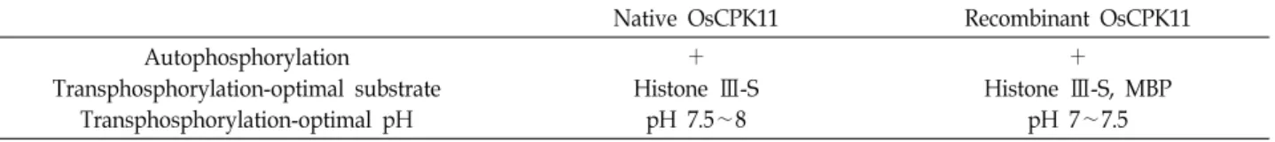
Comparison of native OsCPK11 and recombinant OsCPK11
Biochemical characteristics of OsCPK11 identified in this study could be compared in some aspects with the character- istics of recombinant OsCPK11 identified studied by Cho [10]. Results were shown in Table 2.
Native OsCPK11 and recombinant OsCPK11 all had Ca
2+-dependent autophosphorylation activity. However, there are some differences. Native OsCPK11 showed the au- tophosphorylation activity at 1 μM Ca
2+in the absence of Mg
2+, whereas recombinant OsCPK11 did not show any au- tophosphorylation activity.
In the presence of Mg
2+, native OsCPK11 showed an in- crease in autophosphorylation activity as Ca
2+concentration increased. However, recombinant OsCPK11 showed auto- phosphorylation activity was almost constant when the Ca
2+concentration was increased. This difference needs to be studied further.
Both native OsCPK11 and recombinant OsCPK11 pre- ferred histone Ⅲ-S as a substrate for the kinase activity in
vitro, and that kinase activity was dependent on Ca2+. This appears to be due to the structural similarity of the substrate binding sites and calcium binding sites of both proteins.
Partially purified native OsCPK11 and recombinant OsCPK11 phosphorylated histone III-S, but further studies are needed to determine if OsCPK11 has a function associated with his- tone III-S in plant cells.
Both the native OsCPK11 and the recombinant OsCPK11
were similar in terms of optimal pH range of the kinase ac-
tivity in vitro. There was a little bit of difference between
native OsCPK11 in the range of pH 7.5~8 and the recombi-
Table 2. Comparison of native OsCPK11 and recombinant OsCPK11
Native OsCPK11 Recombinant OsCPK11
Autophosphorylation
Transphosphorylation-optimal substrate Transphosphorylation-optimal pH
+ Histone Ⅲ-S
pH 7.5∼8
+ Histone Ⅲ-S, MBP
pH 7∼7.5
nant OsCPK11 in the range of pH 7~7.5. There are many studies that showed native and recombinant proteins are functionally almost identical [39], and further study will be needed on the detailed differences between them.
There were some limitations in purifying OsCPK11 and revealing its biochemical characteristics. OsCPK11 was found to be a very small amount in plant cells and it was difficult to purify with a higher purity. Due to the non-spe- cific binding characteristics of the OsCPK11 antibody used in western blot, it was sometimes difficult to differentiate and separate OsCPK11 band.
To confirm a correct biological function of autophosphor- ylation and transphosphorylation of CDPKs, experiments should be needed in vivo. And it will provide a more ex- tended knowledge of the role of OsCPK11 in signaling path- ways in plant cells. This study will provide a basic knowl- edge in making such an extended study.
Discussion
In this study, OsCPK11, one of CDPKs from rice, was par- tially purified from rice seedlings. The partial purification steps included anion exchange chromatography, hydro- phobic interaction chromatography and gel-filtration chro- matography. OsCPK11 seems to have a weak anion on the surface and a strong hydrophobic bond with the hydro- phobic resin (Fig. 1). It was found that molecular weight of the subunit was estimated to be 54 kDa. Its in vitro kinase assays also showed that it had a Ca
2+-dependent autophos- phorylation activity and was influenced by Mg
2+(Fig. 3).
Especially, it was found that autophosphorylation activity increases with increasing concentration of Ca
2+in the pres- ence of 100 μM of Mg
2+(Fig. 3). In addition, partially purified OsCPK11 was found to phosphorylate histone III-S strongly (Fig. 4), and it was found that the optimum pH range was pH 7.5-8.0 (Fig. 5). The biochemical characteristics of this native OsCPK11 were compared with those of recombinant OsCPK11 [10]. Both OsCPK11 have Ca
2+-dependent auto- phosphorylation activity and histone Ⅲ-S is preferred as a substrate for the kinase activity, and the optimum pH is
about 7.5(Fig. 3). These results should help understanding the biochemical characteristics of OsCPK11. If we extend the experiments in vivo, we will be able to find the physiological function of OsCPK11 in rice.
The Conflict of Interest Statement
The authors declare that they have no conflicts of interest with the contents of this article.
References
1. Abbasi, F., Onodera, H., Toki, S., Tanaka, H. and Komatsu, S. 2004. OsCDPK13, a calcium-dependent protein kinase gene from rice, is induced by cold and gibberellin in rice leaf sheath. Plant Mol. Biol. 55, 541-552.
2. Abo-El-Saad, M. and Wu, R. 1995. A rice membrane cal- cium-dependent protein kinase is induced by gibberellin.
Plant Physiol. 108, 787-793.
3. Anil, V. S. and Rao, K. S. 2001. Purification and character- ization of a Ca2+-dependent protein kinase from sandalwood (Santalum album L.): evidence for Ca2+-induced conforma- tional changes. Phytochemistry 58, 203-212.
4. Asano, T., Hayashi, N., Kikuchi, S. and Ohsugi, R. 2012.
CDPK-mediated abiotic stress signaling. Plant Signal. Behav.
7, 817-821.
5. Asano, T., Hayashi, N., Kobayashi, M., Aoki, N., Miyao, A., Mitsuhara, I. and Ohsugi, R. 2012. A rice calcium dependent protein kinase OsCPK12 oppositely modulates salt‐stress tolerance and blast disease resistance. Plant J. 69, 26-36.
6. Asano, T., Tanaka, N., Yang, G., Hayashi, N. and Komatsu, S. 2005. Genome-wide identification of the rice calcium-de- pendent protein kinase and its closely related kinase gene families: comprehensive analysis of the CDPKs gene family in rice. Plant Cell Physiol. 46, 356-366.
7. Battey, N. H. 1990. Calcium-activated protein kinase from soluble and membrane fractions of maize coleoptiles.
Biochem. Biophy. Res. Comm. 170, 17-22.
8. Breviario, D., Morello, L. and Gianì, S. 1995. Molecular clon- ing of two novel rice cDNA sequences encoding putative calcium-dependent protein kinases. Plant Mol. Biol. 27, 953- 967.
9. Cheng, S. H., Willmann, M. R., Chen, H. C. and Sheen, J. 2002.
Calcium signaling through protein kinases. The Arabidopsis calcium-dependent protein kinase gene family. Plant Physiol.
129, 469-485.
10. Cho, I. S., Lee, S. H., Park, C. M. and Kim, S. H. 2017.
Phosphorylation properties of recombinant OsCPK11, a cal- cium-dependent protein kinase from rice. J. Life Sci. 27, 1393-1402.
11. Clapham, D. E. 1995. Calcium signaling. Cell 80, 259-268.
12. DasGupta, M. 1994. Characterization of a calcium-depend- ent protein kinase from Arachis hypogea (groundnut) seeds.
Plant Physiol. 104, 961-969.
13. Dixit, A. K. and Chelliah, J. 2013. Molecular cloning, over- expression, and characterization of autophosphorylation in calcium-dependent protein kinase 1 (CDPK1) from Cicer arietinum. Appl. Microbiol. Biotechnol. 97, 3429-3439.
14. Dodd, A. N., Kudla, J. and Sanders, D. 2010. The language of calcium signaling. Annu. Rev. Plant Biol. 61, 593-620.
15. Ehrhardt, D. W., Wais, R. and Long, S. R. 1996. Calcium spiking in plant root hairs responding to Rhizobium nod- ulation signals. Cell 85, 673-681.
16. Estruch, J. J., Kadwell, S., Merlin, E. and Crossland, L. 1994.
Cloning and characterization of a maize pollen-specific cal- cium-dependent calmodulin-independent protein kinase.
Proc. Natl. Acad. Sci. 91, 8837-8841.
17. Frattini, M., Morello, L. and Breviario, D. 1999. Rice cal- cium-dependent protein kinase isoforms OsCDPK2 and OsCDPK11 show different responses to light and different expression patterns during seed development. Plant Mol.
Biol. 41, 753-764.
18. Gong, M., Van de Luit, A. H., Knight, M. R. and Trewavas, A. J. 1998. Heat-shock-induced changes in intracellular Ca2+
level in tobacco seedlings in relation to thermotolerance.
Plant Physiol. 116, 429-437.
19. Harmon, A. C., Gribskov, M. and Harper, J. F. 2000. CDPKs- a kinase for every Ca2+ signal? Trends Plant Sci. 5, 154-159.
20. Harmon, A. C., Putnam-Evans, C. and Cormier, M. J. 1987.
A calcium-dependent but calmodulin-independent protein kinase from soybean. Plant Physiol. 83, 830-837.
21. Harper, J. F., Sussman, M. R., Schaller, G. E., Putnam-Evans, C., Charbonneau, H. and Harmon, A. C. 1991. A cal- cium-dependent protein kinase with a regulatory domain similar to calmodulin. Science 252, 951-954.
22. Hegeman, A. D., Rodriguez, M., Han, B. W., Uno, Y., Phillips, G. N., Hrabak, E. M. and Sussman, M. R. 2006. A phylopro- teomic characterization of in vitro autophosphorylation in calcium-dependent protein kinases. Proteomics 6, 3649- 3664.
23. Hrabak, E. M. 2000. Calcium-dependent protein kinases and their relatives. Adv. Bot. Res. 32, 185-223.
24. Hrabak, E. M., Chan, C. W., Gribskov, M., Harper, J. F., Choi, J. H., Halford, N. and Thomas, M. 2003. The Arabid- opsis CDPK-SnRK superfamily of protein kinases. Plant Physiol. 132, 666-680.
25. Karibe, H., Komatsu, S. and Hirano, H. 1996. Partial purifi- cation and characterization of a calcium-dependent protein kinase in rice leaves. Phytochemistry 41, 1459-1464.
26. Klimczak, L. J. and Hind, G. 1990. Biochemical similarities between soluble and membrane-bound calcium-dependent protein kinases of barley. Plant Physiol. 92, 919-923.
27. Knight, H., Trewavas, A. J. and Knight, M. R. 1996. Cold
calcium signaling in Arabidopsis involves two cellular pools and a change in calcium signature after acclimation. Plant Cell 8, 489-503.
28. Knight, H., Trewavas, A. J. and Knight, M. R. 1997. Calcium signaling in Arabidopsis thaliana responding to drought and salinity. Plant J. 12, 1067-1078.
29. Lee, J. Y., Yoo, B. C. and Harmon, A. C. 1998. Kinetic and calcium-binding properties of three calcium-dependent pro- tein kinase isoenzymes from soybean. Biochemistry 37, 6801- 6809.
30. Lino, B., Carrillo-Rayas, M. T., Chagolla, A. and de la Vara, L. E. G. 2006. Purification and characterization of a cal- cium-dependent protein kinase from beetroot plasma mem- branes. Planta 225, 255-268.
31. Lowry, O. H. 1951. The Lowry protein assay. J. Biol. Chem.
193, 265-275.
32. Ma, P., Liu, J., Yang, X. and Ma, R. 2013. Genome-wide iden- tification of the maize calcium-dependent protein kinase gene family. Appl. Biochem. Biotechnol. 169, 2111-2125.
33. MacIntosh, G. C., Ulloa, R. M., Raíces, M. and Téllez-Iñón, M. T. 1996. Changes in calcium-dependent protein kinase activity during in vitro tuberization in potato. Plant Physiol.
112, 1541-1550.
34. Martín, M. L. and Busconi, L. 2000. Membrane localization of a rice calcium‐dependent protein kinase (CDPK) is mediated by myristoylation and palmitoylation. Plant J. 24, 429-435.
35. McAinsh, M. R. and Pittman, J. K. 2009. Shaping the calcium signature. New Phytol. 181, 275-294.
36. McAinsh, M. R., Brownlee, C. and Hetherington, A. M. 1997.
Calcium ions as second messengers in guard cell signal transduction. Physiol. Plant 100, 16-29.
37. Morello, L., Frattini, M., Gianì, S., Christou, P. and Breviario, D. 2000. Overexpression of the calcium-dependent protein kinase OsCDPK2 in transgenic rice is repressed by light in leaves and disrupts seed development. Transgenic Res. 9, 453-462.
38. Myers, C., Romanowsky, S. M., Barron, Y. D., Garg, S., Azuse, C. L., Curran, A. and Harper, J. F. 2009. Calcium- dependent protein kinases regulate polarized tip grow thin pollen tubes. Plant J. 59, 528-539.
39. Pitsin, S. M., D'andrea, R. J., Vandeleur, L., Moretti, P. A., Pu, X. I. A., Gamble, J. R. and Wattenberg, B. W. 2000.
Human sphingosine kinase: purification, molecular cloning and characterization of the native and recombinant enzymes.
Biochem. J. 350, 429-441.
40. Putnam-Evans, C. L., Harmon, A. C. and Cormier, M. J. 1990.
Purification and characterization of a novel calcium-depend- ent protein kinase from soybean. Biochemistry 29, 2488-2495.
41. Ray, S., Agarwal, P., Arora, R., Kapoor, S. and Tyagi, A.
K. 2007. Expression analysis of calcium-dependent protein kinase gene family during reproductive development and abiotic stress conditions in rice (Oryza sativa L. ssp. indica).
Mol. Genet. Genomics 278, 493-505.
42. Roberts, D. M. and Harmon, A. C. 1992. Calcium-modulated proteins: targets of intracellular calcium signals in higher
초록:벼 유식물에서 OsCPK11의 부분 정제 및 생화학적 특성 규명
신재화
1․김성하
2*
(1경기과학고, 2한국교원대 생물교육과)
식물에서 Ca
2+는 세포의 중요한 2차 신호 전달 분자 중 하나이다. Ca
2+및 인산화 효소의 센서 단백질인 칼슘-의
존성 단백질 카이네즈(CDPKs)는 식물 세포에서 가장 풍부한 세린/트레오닌 키나아제이다. 이들은 다양한 자극에 대한 신호를 변환하여 식물에서 특정 반응을 일으킨다. 벼에는 31개의 CDPK 유전자 족이 확인되었다. 그들은 주로 식물의 생장과 발달에 관여하며 다양한 스트레스 조건에 반응하여 기능을 하는 것으로 알려져 있다. 그러나 CDPK 단백질의 생화학적 특성에 대해서는 알려진 바가 별로 없다. 이 연구에서는 벼의 CDPK 중 하나인 OsCPK11을 부분 정제하여 그 생화학적 특성을 조사하고자 하였다. 벼 유식물에서 3단계 칼럼 크로마토그래피 과정을 거쳐 부분 정제된 OsCPK11을 얻었다. 정제 과정에는 DEAE를 사용한 음이온 교환 크로마토그래피, Phenyl-Sepharose를 사용한 소수성 상호작용 크로마토그래피 및 Sephacryl-200HR를 사용한 겔 여과 크로마토그 래피를 포함하였다. 부분 정제된 OsCPK11은 분자량이 54kDa이며 소수성 수지와 강한 소수성 상호작용을 보였 다. 부분 정제된 OsCPK11으로 in vitro kinase assay를 실시한 결과, OsCPK11은 Ca
2+-의존성 자가인산화 활성을 가짐을 보여 주었다. OsCPK11은 histone III-S를 인산화 하였으며, 카이네즈 활성의 최적 pH는 7.5-8.0이었다.
Native OsCPK11은 이전에 연구된 재조합 OsCPK11과 몇 가지 생화학적 특징을 공유하였는데, 둘 다 Ca
2+-의존성
자가인산화 활성을 나타냈다. 또한, 둘 모두 카이네즈 활성을 위한 기질로서 histone III-S를 선호하였으며, Ca
2+의존성을 보여 주었다.
plants. Annu. Rev. Plant Biol. 43, 375-414.
43. Romeis, T. and Herde, M. 2014. From local to global: CDPKs in systemic defense signaling upon microbial and herbivore attack. Curr. Opin. Plant Biol. 20, 1-10.
44. Saha, P. and Singh, M. 1995. Characterization of a winged bean (Psophocarpus tetragonolobus) protein kinase with calmodulin-like domain: regulation by autophosphorylation.
Biochem. J. 305, 205-210.
45. Saijo, Y., Hata, S., Kyozuka, J., Shimamoto, K. and Izui, K.
2000. Overexpression of a single Ca2+ dependent protein kinase confers both cold and salt/drought tolerance on rice plants. Plant J. 23, 319-327.
46. Sanders, D., Brownlee, C. and Harper, J. F. 1999. Communi- cating with calcium. The Plant Cell 11, 691-706.
47. Schulz, P., Herde, M. and Romeis, T. 2013. Calcium-depend- ent protein kinases: hubs in plant stress signaling and development. Plant Physiol. 163, 523-530.
48. Sheen, J. 1996. Ca2+-dependent protein kinases and stress signal transduction in plants. Science 274, 1900.
49. Simeunovic, A., Mair, A., Wurzinger, B. and Teige, M. 2016.
Know where your clients are: subcellular localization and
targets of calcium-dependent protein kinases. J. Exp. Bot. 67, 3855-3872.
50. Taylor, A. R., Manison, N. F. H., Fernandez, C., Wood, J.
W. and Brownlee, C. 1996. Spatial organization of calcium signaling involved in cell volume control in the Fucus rhizoid. Plant Cell 8, 2015-s2031.
51. Venter, J. C., Adams, M. D., Myers, E. W., Li, P. W., Mural, R. J., Sutton, G. G. and Gocayne, J. D. 2001. The sequence of the human genome. Science 291, 1304-1351.
52. Wan, B., Lin, Y. and Mou, T. 2007. Expression of rice Ca2+
dependent protein kinases (CDPKs) genes under different environmental stresses. FEBS Lett. 581, 1179-1189.
53. Yang, G., Shen, S., Yang, S. and Komatsu, S. 2003. OsCDPK13, a calcium-dependent protein kinase gene from rice, is in- duced in response to cold and gibberellin. Plant Physiol.
Biochem. 41, 369-374.
54. Yuasa, T. and Muto, S. 1992. Ca2+-dependent protein kinase from the halotolerant green alga Dunaliella tertiolecta: parti- al purification and Ca2+-dependent association of the en- zyme to the microsomes. Arch. Biochem. Biophys. 296, 175- 182.