http://crossmark.crossref.org/dialog/?doi=10.14474/ptrs.2019.8.2.86&domain=pdf&date_stamp=2019-6-25
Received: 31 May, 2019 Revised: 25 June, 2019 Accepted: 25 June, 2019 Corresponding author: Yijung Chung (ORCID https://orcid.org/0000-0002-2431-8895)
Department of Physical Therapy, College of Health Science and Social Welfare, Sahmyook University, 815 Hwarang-ro, Nowon-gu, Seoul 01795, Republic of Korea
Tel: 82-2-3399-1637 Fax: 82-2-3399-1639 E-mail: [email protected]
This is an Open-Access article distributed under the terms of the Creative Commons Attribution Non-Commercial License (http://creativecommons.org/licenses/
by-nc/4.0) which permits unrestricted non-commercial use, distribution, and reproduction in any medium, provided the original work is properly cited.
Copyright © 2019 Korean Academy of Physical Therapy Rehabilitation Science
https://doi.org/10.14474/ptrs.2019.8.2.86 pISSN 2287-7576
eISSN 2287-7584
Phys Ther Rehabil Sci 2019, 8 (2), 86-92 www.jptrs.org
A comparison of trunk and lower extremity muscle activity during the performance of squats and kneeling squats in persons with stroke: a preliminary study
Suyoung Shim a , Yijung Chung b
a
Department of Physical Therapy, IM Convalescent Hospital, Uijeongbu, Republic of Korea
b
Department of Physical Therapy, College of Health Science and Social Welfare, Sahmyook University, Seoul, Republic of Korea
Objective: The purpose of this study was to compare the effects of performing squats and kneeling squats on trunk and lower ex- tremity muscle activity in persons with stroke.
Design: Cross-sectional study.
Methods: Ten persons with stroke (3 male and 7 female) were recruited. The subjects were instructed to randomly perform the 4 different squat conditions: squat with 30 degrees of knee flexion, squat with 60 degrees of knee flexion, squat with 90 degree of knee flexion, and the kneeling squat. During the squat performance, surface electromyograms (sEMG) was used to assess muscle activity of the erector spinae (ES), gluteus maximus (Gmax), gluteus medius (Gmed), and biceps femoris (BF) muscles.
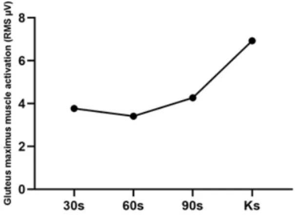
Results: Muscle activation of the ES and BF were significantly increased with the kneeling squats compared to the general squats with 30 degrees and 60 degrees of knee flexion (p<0.05), and muscle activation of the Gmax and Gmed were significantly in- creased with the kneeling squats compared to all other squat conditions (p<0.05).
Conclusions: The results suggest that the kneeling squat is an effective exercise to strengthen the proximal muscles of the lower extremities. Rather than applying a difficult general squat to the stroke population, the kneeling squat may be applied as a safer method for training the proximal muscles.
Key Words: Electromyography, Lower extremity, Stroke
Introduction
The majority of persons with stroke experience muscle weakness and movement disorders due to lesions in the de- scending motor pathway [1] and decreases in skeletal mus- cle activation [2]. As a result, due to asymmetric posture, body imbalances, and reduced weight shifting abilities, it is difficult for stroke survivors to perform functional activities [3].
In general, many stroke survivors tend to shift their center of gravity towards the paralysis side in order to compensate for limited movement and muscle weakness.
Many stroke survivors tend to have a lack of ability to pro- duce adequate voluntary muscle contractions and are unable to perform exercises normally or maintain balance due to the inability to coordinate the timing and the intensity of muscle activity during contractions [4]. Ineffective weight shifting onto the affected side leads to sustained weakness [5]. Thus, strength training performed on the affected side can improve the functional movement of persons affected with stroke [6].
Hwang and Kim [7] lower limb muscle strength training on
the paralyzed side and non-paralyzed side increases weight
support rate in stroke patients. Park and Chung [8] lower
limb muscle Strength training improves balance in stroke
Left 5 (50.0)
MMSE-K 26.20 (1.95)
Values are presented as n (%) or mean (SD).
MMSE-K: Korean version of the Mini-Mental State Examination.
is a complex exercise in which not only the femoral muscle, but also the whole muscles of the trunk and lower limbs.
There have been recent studies involving squat move- ments and the stroke population.
A study by Gray et al. [10] included squatting at a high speed and it was suggested that the squat movement should be accompanied by the kinetic energy and speed, which are components of the force. Choi et al. [13] compared the mus- cle activity between the affected and unaffected sides of stroke survivors and found that squats performed at higher speeds exhibited increased muscle activity of the rectus femoris. Ki et al. [14] did a study on stroke survivors and found that modifying the squat by changing the inclination of the ankle joint of one foot to 15° showed greater muscle activity of the vastus lateralis and vastus medialis when compared to performing the squat movement in the neutral state of the ankle joint and when the foot was flexed.
As shown in previous studies, research based on squat movements for the stroke population are actively being im- plemented, however, research on modified squat move- ments are insufficient. In particular, there are no studies per- formed that includes the squats performed while standing on the knees.
Therefore, this study was designed to investigate the ef- fect of performing squats while standing on the knees and squats according to various knee joint angles, as well as to determine whether the modified squat movement is effec- tive on the body and leg muscle activity. In such cases where squatting is difficult for the stroke survivor, the existing squat exercise can be modified so that it can be more easily applied within the clinical setting. This study is also an at- tempt to provide basic data in introducing an effective ex- ercise method.
Methods Subject
This study is a cross-sectional study design. A total of 10 persons admitted at the IM Convalescent Hospital in Gyeonggido who were diagnosed with stroke and hemi- plegia, were receiving physical therapy treatment, had met the study conditions and had agreed to participate in the study were included. The inclusion criteria for the partic- ipants were presence of hemiplegia due to stroke, the ability to walk more than 10 m independently without assistance, a score of 24 points or more on the Korean version of the Mini-Mental State Examination, and no visual defects or ab- normalities in the vestibular system. Those who did not have orthopedic disease in the trunk and bilateral legs were se- lected, and those who experienced falls or trauma or pain during the past 6 months were excluded. The characteristics of the participants are as follows (Table 1).
The experiment was implemented after the experimental conditions and procedures of the study were fully explained and after the agreement to participate was obtained by the subjects. This study was approved by the research Sahm- yook University Life Science ethics committee (2-7001793- AB-N-012019020HS) and informed consent was obtained.
Procedures
With electrodes applied onto the erector spinae, gluteus
maximus, gluteus medius, and biceps femoris muscles, the
peak muscle activation values were assessed while the sub-
jects performed squats with three different knee flexion an-
gles as well as while standing on the knees. Prior to the ex-
periment, the researchers explained about the squat move-
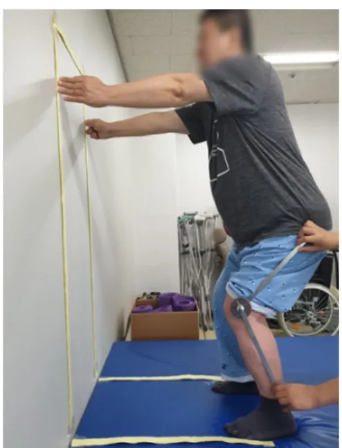
Figure 1. Guide line. Figure 3. Squat posture.
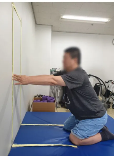
Figure 2. Kneeling position.
ment using three different knee flexion angles as well as in the standing posture of the knee, and the subjects were al- lowed to practice the squats more than 3 times in order to en- able them to fully understand the experimental method. The squat movement was performed in four ways as stated above. In order to prevent the influence of order that the squats were performed, each type of squat was performed three times each, in random order.
The descriptions of peforming the squat movement while standing on the knees and with various knee flexion angles
are as follows.
The squats with use of various knee flexion angles and while standing on the knees were performed while following the guidelines that have been attached vertically onto the wall to enable the upper body to move down along with the knee flexion angles and in order to control the movement of the torso (Figure 1).
When performing the squats with use of various knee an- gles and while standing on the knees, the distance of the squat postures from the wall was defined as the distance from the palm and extended fingers of the unaffected side running parallel along the acromion process. During the squat movement, the feet were placed shoulder-width apart and were positioned so that the acromion process was placed vertically along the lateral malleolus.
During the performance of the squats while standing on the knees, the knees were positioned so that the acromion process and the femur were placed along a vertical line, and the legs were placed in parallel position with the knee and ankle joints aligned (Figure 2).
The knee flexion angle of the squat was set at 30°, 60°, and 90° using a goniometer (Baseline FEI Baseline Ss 360 Degree Goniometer; Fabrication Enterprises, Inc., White Plains, NY, USA).
The stationary arm of the goniometer was positioned par-
allel along the longitudinal axis of the femur while the mov-
ing arm was placed parallel along the long axis of the fibula
(Figure 3).
Table 2. Comparison of muscle activity during squat knee angles and kneeling squat (N=10)
Condition 30S 60S 90S KS χ² (p)
ES ( μV) 13.50 (7.03) 15.21 (9.54) 19.15 (9.28)
a,b20.06 (8.33)
a,b22.280 (<0.001)
GMx ( μV) 3.77 (2.62) 3.41 (1.80) 4.27 (2.01) 6.93 (2.21)
a,b.c21.240 (<0.001)
Gme ( μV) 2.48 (2.51) 2.23 (1.62) 2.42 (1.10) 4.31 (1.39)
a,b.c15.960 (0.001)
BF ( μV) 3.86 (1.41) 4.58 (1.49)
a6.25 (1.60)
a,b6.47 (2.78)
a,b20.520 (<0.001) Values are presented as peak ( μV) or mean (SD).
30S: knee flexion 30° squat, 60S: knee flexion 60° squat, 90S: knee flexion 90° squat, KS: kneeling squat, ES: elector spinae, GMx: gluteus maximus, GMe: gluteus medius, BF: bicep femoris.
a