Cordycepin Induced Apoptosis via Intracellular Ca
2+Modulation and Mitochondrial Dysfunction in Human Prostate Cancer PC-3 Cells
Dong-Min Kang
1, Kwang-Youn Kim
1, Sun-Nyoung Yu
1, Young-Rang Jin
1, Hyun-Joo Jeon
1, Sang-Hun Kim
1, Sung-Sik Chun
2, Hack-Ryong Ko
3and Soon-Cheol Ahn
1,4*
1
Department of Microbiology and Immunology, Pusan National University School of Medicine, Yangsan 626-870, Korea
2
Division of Food Science, International University of Korea, Jinju 660-759, Korea
3
Bio21 Center, Jinju 660-844, Korea
4
Medical Research Institute, Pusan National University, Yangsan 626-870, Korea
Received March 5, 2011 /Accepted March 12, 2011Cordycepin (3'-deoxyadenosine), a nucleoside derivative isolated from Cordyceps militaris, is reported to have antitumor effects. However, neither its molecular mechanism nor its molecular targets are well understood. In the present study, molecular mechanisms for the anti-tumor effects of cordycepin were investigated in human prostate cancer PC-3 cells. The MTT assay was used to detect cell viability.
Annexin V/FITC assay, reactive oxygen species (ROS) production, mitochondrial membrane potential (MMP), and Ca
2+flux were used to assess for the presence of apoptosis. Western blot analysis was used to detect protein expression. Treatment of cordycepin resulted in significantly decreased cell via- bility of PC-3 cells in a dose- and time-dependent manner. A dose-dependent apoptotic cell death was also measured by flow cytometery analysis. Molecular mechanistic studies of apoptosis unraveled cor- dycepin treatment resulted in significant mitochondrial dysfunction, ROS production, and elevation of Ca
2+concentrations. These phenomena were followed activation of caspase-3, subsequently leading to PARP cleavage and cell apoptosis. Taken together, cordycepin induces apoptosis in PC-3 cells through regulation of a mitochondrial mediated pathway.
Key words : Cordycepin, apoptosis, Ca
2+flux, mitochondrial membrane potential (MMP)
*Corresponding author
*Tel:+82-51-510-8092, Fax:+82-55-382-8090
*E-mail : [email protected]
서 론
전립선암은 북미나 서구 유럽 등에서 가장 흔한 암으로 , 미 국 암 협회 (American Cancer Society)의 보고에 따르면 2010 년 기준으로 전체 암환자 중 발병률 1위, 암 관련 사망률 2위를 차지하고 있다 [11]. 우리나라의 경우도 서구화 된 식습관과 생 활습관의 변화 및 고령화로 인해 발생빈도가 빠르게 증가하고 있는 추세이며 , 현재 국내 암 발병률 5위, 증가율 1위를 차지하 고 있다 [3]. 전립선암은 나이, 인종, 국가, 가족력, 유전자, 식이, 비만 및 전립선에의 염증 및 감염 등으로 인해 발병할 수 있다 고 알려져 있으나 정확한 메커니즘은 밝혀져 있지 않다 [1]. 발 병 초기 전립선암 치료를 위해 androgen을 제거하는 방법을 기초로 하여 화학 , 방사선 및 외과적 요법을 병행 하지만 부작 용이 따를 뿐만 아니라 androgen 비의존성 암 환자의 경우 치료하기가 아주 힘들기 때문에 근본적인 치료책으로는 사용 되지 못하고 있다 . 이에 최근에는 천연물을 이용한 생물학적 치료를 통해 암의 세포사멸을 유도하는 연구가 많이 이루어지 고 있다 [21,25].
동충하초 (genus Cordyceps)는 자낭균아문, 맥각균강, 맥각
균목 , 맥각균과에 속하고, 일반적으로 곤충의 몸을 숙주로 사 용하여 내생균핵을 만든 다음 밖으로 자실체를 형성하는 것으 로 알려져 있다 [19,26]. 이러한 동충하초는 전통 한약제로서 면역력 증강 , 항산화 효과, 호흡기질환 그리고 자양강장 등에 사용되었으며 , 최근 연구에 따르면 동충하초의 한 종류인 C.
militaris와 C. sinensis로부터 항암 및 항염증 효과, 면역 조절 및 항산화 효과 그리고 혈중 콜레스테롤 수치의 감소에 관한 효과에 대한 연구가 수행되었다 [6,8,14,15,20,24]. 또한 동충하 초에는 cordycepin, ergosterol, myriocin, polysaccharide 및 glycoprotein 등 다양한 생리활성 물질들이 함유되어 있다. 특 히 cordycepin (3’-deoxyadenosine)은 동충하초에 함유된 주 요 생리활성 물질로서 , 아데노신 리보오스 잔기의 3번째 탄소 에 산소가 결핍된 구조로 (Fig. 1A), 다양한 암세포에서 세포사 멸 유도에 관한 활성이 보고되어 있다 [4,12,18,23].
본 연구에서는 cordycepin이 인간 전립선암 세포주인 PC-3
와 LNCaP 세포에 대하여 세포독성을 확인하고, 호르몬 의존
성 세포주보다 항암 치료가 힘들다고 알려진 호르몬 비의존성
PC-3 세포에 대하여 세포사멸 기전을 조사하여 얻어진 결과를
보고하고자 한다 .
Fig. 1. Chemical structure of cordycepin and its inhibition on proliferation in prostate cancer cells. A. The structure of cordycepin (3’-deoxyadenosine), B, PC-3 cells; C, LNCaP (androgen -); D, LNCaP (androgen +). Cell viability was determined using the MTT assay as described in Materials and Methods. Data are presented as mean±SD (n=3 in each group).#
p
<0.05,¶p
<0.01,*
p
<0.001 vs. the control group.재료 및 방법
사용시약
3-(4,5-Dimethylthiazol-2-yl)-2,5-diphenyltetrazolium bro- mide (MTT), propidium iodide (PI), 2',7'-dichlorodihydro- fluorescein diacetate (DCFH-DA), 3,3-dihexylox- acarbocyanine (DiOC
6), Fluo-3/AM 그리고 N-acetyl-
L-cys- tein (NAC)은 Sigma Chemical Co. (St. Louis, MO, USA) 에서 구입하였다 . 1,2-bis-(ο-Aminophenoxy) ethane-tetra- acetic acid tetra-(acetoxymethyl) ester (BAPTA/AM)는 Calbiochem (Merck, Darmstadt, Germany)에서 구입하였으 며 , Annexin-V-Fluo Staining Kits는 BD Bioscience (San Jose, CA, USA)에서 구입하여 사용하였다. Western blot- ting에 사용한 antibody로 Bax, Caspase-3, PARP 그리고 β -Actin은 Santa Cruz Biotechnology (Santa Cruz, Ca, USA) 에서 구입하였고 , Bcl-2는 Cell Signaling (Beverly, MS, USA)에서 구입하였다.
본 실험에서 사용한 cordycepin은 Sigma Chemical Co. (St.
Louis, MO, USA)에서 구입하여, methanol로 50 mM의 농도
로 녹여 -20℃에서 보관하여 실험에 사용하였다.
세포의 배양
인간 전립선 암세포주인 PC-3와 LNCaP cell은 각각 American Type Culture Collection (ATCC, Manassas, VA, USA)과 Korea Cell Line Bank (KCLB, Seoul, Korea)로부터 분양 받아 10% fetal bovine serum (FBS) (WelGene Inc., Korea)과 100 U/ml penicillin, 100 μg/ml streptomycin (WelGene Inc., Korea)이 함유된 Dulbecco’s modified Eagle’s medium (DMEM)과 RPMI1640 (WelGene Inc., Korea) 배지에 서 5%의 CO
2가 포함된 37℃ 배양기에서 배양하였다. 모든 실 험에서 항산화제인 NAC와 Ca
2+chelator인 BAPTA/AM은 cordycepin을 처리하기 1시간 전에 전처리하였다.
MTT assay를 이용한 세포독성의 측정
세포 배양용 48-well plate를 이용하여 1×10
4cells/well의
농도로 전립선 암세포주인 PC-3와 LNCaP 세포를 분주하고
24시간 동안 안정화시킨 후, cordycepin을 다양한 농도로 24
또는 48 시간 동안 처리하였다. 이후 0.5 mg/ml 농도가 되게
MTT 시약을 처리하여 37℃에서 4 시간 반응하였다. 생성된 보라색의 formazan을 dimethyl sulfoxide (DMSO)로 녹인 후 ELISA reader (VERSA
MAXmicroplate reader, Molecular Devices, Toronto, Canada)로 570 nm에서 흡광도를 측정하였 다 [10]. 이 흡광도는 MTT가 세포에 의해서 환원된 양으로, 각 well에 존재하는 세포의 생존 수와 비례한다.
Annexin V / PI assay를 이용한 apoptosis 유도 세포 의 측정
PC-3 세포에서 cordycepin의 apoptosis 유도 여부를 확인 하기 위하여 FITC Annexin-V Apoptosis Detection kit를 이용 하여 측정하였다 [22]. PC-3 세포를 1×10
5이 되도록 6-well plate에서 24 시간 동안 안정화시킨 후, cordycepin을 적정 농 도로 24 시간 동안 처리한 후, 세포를 분리하여 phosphate buf- fered saline (PBS, pH 7.4)으로 세척하고, cell pellet을 Annexin-V/PI 시약과 1× binding buffer를 이용하여 조심스 럽게 현탁 후 , 상온에서 15 분간 염색하였다. 염색된 세포는 flow cytometry (FACS Calibur, BD Biosciences)를 이용하여 측정하였고 , Cell Quest software (Becton Dickinson Co.)를 이용하여 apoptosis가 유도된 세포를 분석하였다.
세포 내 Ca
2+농도의 측정
Cordycepin에 의한 Ca
2+이온 농도 변화를 Fluo-3/AM 형 광 염색법을 이용하여 측정하였다 [9]. PC-3 세포를 1×10
5이 되도록 6-well plate에서 24 시간 동안 안정화시킨 후, cordyce- pin을 200 μM의 농도로 6 시간 동안 처리하였다. 세포를 분리 하여 PBS (pH 7.4)로 세척한 뒤, 5 μM Fluo-3/AM을 처리하여 37℃에서 30 분간 염색하였다. 염색된 세포는 flow cytometry 를 이용하여 측정하였고 , Cell Quest software를 이용하여 Ca
2+농도의 변화를 분석하였다 .
세포 내 reactive oxygen species (ROS) 생성 측정 Cordycepin에 의한 세포 내 ROS의 생성을 DCFH-DA 형광 염색법을 이용하여 측정하였다 [2]. PC-3 세포를 1×10
5이 되도 록 6-well plate에서 24 시간 동안 안정화시킨 후, cordycepin 을 200 μM의 농도로 다양한 시간 동안 처리하였다. 세포를 분리하여 PBS (pH 7.4)로 세척한 뒤, 10 μM의 DCFH-DA를 처리하여 37℃에서 30 분간 염색하였다. 염색된 세포는 flow cytometry를 이용하여 측정하였고, Cell Quest software를 이 용하여 ROS 생성을 분석하였다.
Mitochondrial membrane potential (MMP)의 측정
Cordycepin에 의한 MMP의 변화를 DiOC
6형광 염색법을 이용하여 측정하였다 [7]. PC-3 세포를 1×10
5이 되도록 6-well plate에서 24 시간 동안 안정화시킨 후, cordycepin을 200 μM 의 농도로 다양한 시간 동안 처리하였다 . 세포를 분리하여
PBS (pH 7.4)로 세척한 뒤, 100 nM의 DiOC
6를 처리하여 37℃
에서 30 분간 염색하였다. 염색된 세포는 flow cytometry를 이용하여 측정하였고 , Cell Quest software를 이용하여 MMP 의 변화를 분석하였다 .
Western blotting을 이용한 단백질의 분석
Cordycepin에 의한 PC-3의 apoptosis 유도 기전을 알아보 기 위하여 western blotting을 이용하여 단백질의 발현변화를 관찰하였다 . PC-3 세포를 1×10
5이 되도록 6-well plate에서 24 시간 안정화시킨 후 , cordycepin을 적정 농도로 24 시간 동안 처리하였다 . 세포를 분리하여 PBS (pH 7.4)로 세척하였고 ly- sis buffer [150 mM NaCl, 10 mM Tris (pH 7.4), 5 mM EDTA (pH 8.0), 1%-Triton X-100, 1mM PMSF, 20 mg/ml aprotinin, 50 μg/ml leupeptin, 1 mM benzaidine, 1 mg/ml pepstatin]를 이용하여 분해하였다 . Bradford 법을 이용하여 정량 한 뒤, 40 μg의 protein은 12-15%의 sodium dodecyl sulfate – poly- acrylamide gel electrophoresis (SDS-PAGE)를 이용하여 분리 하였다 . 분리된 protein을 polyvinylidence fluofide (PVDF) membrane으로 transfer 하여 5% skim milk가 함유 된 TBS-T buffer [20 mM Tris (pH 7.4), 150 mM NaCl, 0.1% Tween-20]
를 이용하여 실온에서 1 시간 동안 blocking 하였고 primary antibody와 horseradish peroxidase (HRP)가 결합 된 secon- dary antibody를 이용하여 target protein에 결합하였다. 이후, Enhanced Chemiluminescence (ECL) western blotting de- tection 시약을 이용하여 발색하고, fluorescence scanner (LAS-3000, Fuji Film, Tokyo, Japan)을 이용하여 확인 하였으 며 , Multi Gauge V3.0 software를 이용하여 분석하였다.
통계처리
모든 실험의 표시된 결과는 3번 수행하였으며, 통계분석은 mean±S.D로 표시하였고, ANOVA에 의해 분석하였다. 통계 적 유의성은 p≤0.05로 판정하였다.
결과 및 고찰
PC-3 세포에 대한 cordycepin의 세포독성 효과
사람 전립선암 세포주인 PC-3와 LNCaP 세포에 대한 cor-
dycepin의 세포독성을 알아보기 위하여 시간 및 농도별로
cordycepin을 처리하여 MTT assay를 실시하였다. 그 결과,
농도 및 시간 의존적으로 cordycepin에 의해 유의적으로 세포
독성이 증가하였으며 , IC
50(50% 저해농도) 값은 200 μM로 관
찰되었다 (Fig. 1B). 또한, 호르몬 의존성 세포주인 LNCaP의
경우 , dihydrotestosterone (DHT)를 첨가하지 않고 배양한
LNCaP 세포가 DHT를 첨가하여 배양한 세포보다 높은 세포
독성을 나타내었다 (Fig. 1C, D). 게다가 호르몬 비의존성 PC-3
세포와 호르몬 의존성 LNCaP 세포의 생존율을 비교하였을
때 , PC-3 세포가 LNCaP 세포보다 cordycepin에 대한 저항성 이 높음을 알 수 있었다 (Fig. 1B, C, D). 따라서 cordycepin이 전립선 암세포주인 PC-3와 LNCaP 세포의 증식을 억제하는 것을 확인하였고 , 세포증식에서 호르몬이 영향을 미치는 것을 확인하였다 . 이후 실험에서는 호르몬 비의존성 세포인 PC-3 세포를 사용하여 다양한 apoptosis 유도 기전에 대해 탐색을 하였다 .
PC-3 세포에 대한 cordycepin의 apoptosis 유발
Cordycepin의 PC-3 세포에 대한 세포독성이 apoptosis 유 도에 의한 것임을 Annexin-V/PI 염색을 통해 확인하였다.
Cordycepin을 50-400 μM의 농도로 24 시간 처리한 뒤, FITC Annexin-V Apoptosis Detection kit를 이용하여 apoptosis를 정량화 하였다 . Annexin-V가 염색된 부분인 초기 apoptosis와 annexin-V와 PI가 함께 염색된 부분인 후기 apoptosis 세포가 농도 의존적으로 증가하는 것을 확인 하였다 . 특히 200 μM cordycepin 을 24 시간 처리하였을 때 28.36%로 유의적으로 증가하였다 (Fig. 2A). 이상의 결과는 cordycepin에 의한 세포 독성이 PC-3 세포의 apoptosis 유도에 기인함을 알 수 있었다.
PC-3 세포에서 cordycepin의 Bcl-2 family 발현과 caspase-3 및 poly (ADP-ribose) polymerase (PARP) 의 활성화
Apoptosis의 유도는 세포 내의 apoptosis 관련 단백질의 발 현에 영향을 주게 되는데 Bcl-2 familiy, caspase-9, caspase-3 그리고 PARP 등이 가장 잘 알려져 있다. 그 중 Bcl-2 familiy는 Bax, Bak 그리고 Bim 등과 같은 apoptosis 유도 단백질 (pro-apoptotic protein)과 Bcl-2, Bcl-x 등과 같은 apoptosis 억 제 단백질 (anti-apoptotic protein)으로 구성되어 있다[13]. 두 구성성분들은 평소 세포 내에서 일정하게 균형을 유지하고 있으나 ROS, UV 및 호르몬 등의 자극으로 인해 불균형이 유 발 됨으로써 세포를 apoptosis 기전으로 유도시킨다. 또한, Bax나 Bak 등은 막이 붕괴된 미토콘드리아 내부로 유입되어 cytochrome c를 방출하게 되고, 방출된 cytochrome c는 cas- pase-9을 활성화시키며, 활성화된 caspase-9은 caspase-3를 활 성화 시키고 , caspase-3는 PARP를 분절시킴으로써 apoptosis 신호가 전달되게 된다 [23]. 따라서 PC-3세포에서 cordycepin 의 apoptosis 유도경로를 확인하기 위해 Bax, Bcl-2, caspase-3 그리고 PARP의 발현양을 western blotting을 통해 확인한 결 과 , cordycepin의 농도 의존적으로 Bax의 발현양 증가와 Bcl-2 의 감소를 확인 할 수 있었으며 , Bax와 Bcl-2의 비율이 200 μ M에서 6.25배까지 증가하는 것을 확인하였다(Fig. 3A). Total caspase-3의 발현양도 농도 의존적으로 감소하였고, caspase-3 의 기질인 PARP의 분절형태가 200 μM에서 1.6배 증가하는 것을 확인하였다 (Fig. 3B). 이상의 결과는 cordycepin이 Bcl-2 family 단백질의 변화를 초래하고, caspase-3 의존적 경로를
Fig. 2. Induction of apoptosis by cordycepin in PC-3 cells. A, Annexin-V/propidium iodide (PI) double staining assay detected by flow cytometry. B, The percentage of apop- totic cell for each treatment group. Data are presented as mean±SD (n=3 in each group).#
p
<0.05 vs. the control group.통해 apoptosis를 유도함을 확인하였다.
Cordycepin에 의한 PC-3 세포 내 Ca
2+농도의 증가
Ca
2+의 항상성은 세포의 성장 , 분화 및 세포사멸을 포함한
다양한 세포 기능에서 매우 중요하다 . 특히, 세포 내 Ca
2+이온
농도의 증가는 미토콘드리아 막의 이온 균형에 영향을 주게
되어 세포벽의 파괴를 유발하게 되고 , Bax나 Bak과 같은
pro-apoptosis 단백질을 미토콘드리아 내로 유입시켜 apopto-
sis를 일으키게 한다[17]. 따라서 cordycepin에 의한 PC-3세포
에서 Ca
2+이온의 변화를 확인하기 위해 Flou-3/AM 형광염색
법을 통해 측정하였다 . 200 μM cordycepin을 6 시간 동안 처리
한 PC-3세포를 5 μM의 Flou-3/AM을 처리하여 30분간 염색
후 , flow cytometry를 이용하여 세포 내 Ca
2+농도의 변화를
확인한 결과 , cordycepin은 PC-3세포에 대해 Ca
2+의 농도를
시간 의존적으로 증가시킴을 확인 할 수 있었고 , 항산화제인
Fig. 4. Effects of NAC and BAPTA/AM on cordycepin-induced Ca2+flux in PC-3 cells. A, B. The intracellular Ca2+concentration was determinded by the fluorescence of FLuo-3/AM by flow cytometry. PC-3 cells were treated with 200 μM cordycepin in the absence or presence of 10 mM NAC and 200 μM BAPTA for 6 hr. C. The percentage of intracellular Ca2+concentration.
Data are presented as mean±SD (n=3 in each group). #
p
<0.05 vs. the control group.Fig. 3. Effects of cordycepin on expression of Bcl-2 family, pro- caspase-3 and PARP protein in PC-3 cells. A, The ex- pression of Bax and Bcl-2 proteins and its ratio. The cell was exposed to 50-200 μM of cordycepin for 24 hr. B, The expression of procaspase-3 and PARP proteins. The cell was exposed to 50-200 μM of cordycepin for 24 hr.
A and B were determined by Western blot.
NAC과 Ca
2+chelator 인 BAPTA/AM을 전처리 하였을 때, 46.9%까지 증가한 Ca
2+농도가 각각 22.81%와 6.74%까지 감소 하는 것을 확인할 수 있었다 (Fig. 4). 이상의 결과로 cordyce- pin에 의한 세포 내 Ca
2+의 증가가 PC-3 세포의 apoptosis 유 도 기전 중의 하나로 사료된다 .
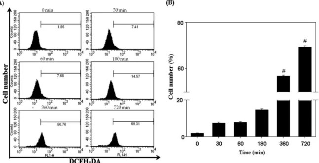
PC-3 세포에서 cordycepin의 reactive oxygen species (ROS) 생성 유도
ROS는 apoptosis를 유도하는 주요 인자 중 하나로 잘 알려
져 있다 [16]. ROS는 DNA에 손상을 줄 뿐만 아니라 unfolding
protein response (UPR)을 일으켜 ER-stress의 원인이 되기도
하고 , 미토콘드리아의 기능을 저해하기도 한다. 또한 세포내
의 ROS 증가는 세포의 산화적 stress를 증가시켜 세포의 손상
을 유발시킨다 . 따라서 cordycepin의 PC-3에 대한 apoptosis
유도에서 ROS 생성의 관여하는지 알아 보기 위해서
DCFH-DA 형광 염색법을 이용하여 측정하였다. 200 μM의
cordycepin을 15-720 분 동안 처리한 PC-3 세포를 수거하여
10 μM의 DCFH-DA를 30 분간 처리하여 생성된 ROS를 flow
cytometry를 이용하여 측정하였다. 시간에 따른 ROS의 생성
을 확인한 결과 , 1.86%에서 69.31%까지 시간 의존적으로 증가
Fig. 5. Effect of cordycepin on ROS generation in PC-3 cells. A. The ROS generation was determinded by the fluorescence of DCFH-DA by flow cytometry. PC-3 cells were treated with 200 μM cordycepin for 30-720 min. B. The percentage of ROS generation.
Data are presented as mean±SD (n=3 in each group). #
p
<0.05 vs. the control group.Fig. 6. Effect of cordycepin on MMP in PC-3 cells. A. The MMP was determined by the fluorescence of DiOC6by flow cytometry.
PC-3 cells were treated with 200 μM cordycepin for 6-24 hr. B. The percentage of MMP dysfunction. Data are presented as mean±SD (n=3 in each group). #
p
<0.05 vs. the control group.하는 것을 확인하였고 , 특히 6시간 이후부터 유의적으로 증가 함을 확인하였다 (Fig. 5). 하지만, 항산화제인 NAC이나 Ca
2+chelator 인 BAPTA/AM을 처리한 세포에서는 ROS의 변화가 유의성을 보이지 않았다 (data not shown).
PC-3 세포에서 cordycepin의 MMP 불균형의 유발 MMP의 변화는 apoptosis의 시작과 활성화에 관련이 있다.
이러한 MMP의 변화로 인해 미토콘드리아의 세포막이 파괴
되고 세포질에 있던 Bax나 Bak 등의 pro-apoptotosis 관련 단
백질들이 미토콘드리아 내부로 유입되게 되며 , cytocrome c와
같은 caspase 활성화 물질이 세포질로 분비되어 apoptosis가
일어나게 된다 [5]. 따라서 cordycepin에 의한 PC-3의 MMP 변
화를 측정하기 위해 DiOC
6염색법을 이용하여 확인하였다 .
PC-3 세포에 200 μM cordycepin을 6-24 시간 동안 처리한 후,
100 nM의 DiOC
6시약으로 30 분간 반응하여 flow cytometry 로 측정하였다 . 이 결과에서 cordycepin에 노출된 PC-3의 MMP는 시간 의존적으로 1.74%에서 32.45%로 과분극 되는 것을 확인하였다 (Fig. 6). 하지만 NAC과 BAPTA에 의한 MMP 불균형 저해는 큰 변화를 보이지 않았다 (data not shown).
이상의 결과들은 cordycepin이 PC-3 세포에 apoptosis를 유도하여 암세포의 증식을 농도 의존적으로 억제함을 나타낸 다 . 특히 cordycepin은 apoptosis의 주요한 인자인 Bcl-2 fam- ily 단백질 발현 변화에 의한 미토콘드리아의 막 전위의 변화 유도와 caspase-3 활성 증가 및 PARP의 분절을 통해 apopto- sis를 유도하는 것으로 사료된다. 또한 세포 내 Ca
2+농도를 증가시키고 이를 통해 ROS의 생산을 통해서 apoptosis가 조 절되는 것으로 사료된다 . 본 연구를 통해 cordycepin이 전립선 암에 대한 apoptosis 기전을 밝혔으나 향후 암 예방 및 암 치료 제로서 cordycepin을 활용하기 위해서는 다양한 동물실험 및 임상실험을 수행해야 할 것으로 사료된다 .
감사의 글
이 논문은 부산대학교 자유과제 학술연구비 (2년)에 의해 연 구되었음 .
References
1. ACS. 2010. Prostate cancer overview: What is prostate can- cer?.
American cancer society
.2. Chang, M. C., Y. S. Ho, P. H. Lee, C. P. Chan, J. J. Lee, L. J. Hahn, Y. J. Wang, and J. H. Jeng. 2001. Areca nut extract and arecoline induced the cell cycle arrest but not apoptosis of cultured oral KB epithelial cells: association of gluta- thione, reactive oxygen species and mitochondrial mem- brane potential.
Carcinogenesis
22, 1527-1535.3. Chi, B. H. and I. H. Chang. 2010. Prostate cancer: recent trends in Korea.
Urol. Int
. 85, 88-93.4. Crompton, M. 1999. The mitochondrial permeability tran- sition pore and its role in cell death.
Biochem. J.
341, 233-249.5. Green, D. R. and Jc. Reed. Mitochondria and apoptosis. 1998.
Science
281, 1309-1312.6. Guo, P., Q. Kai, J. Gao, Z. Q. Lian, C. M. Wu, C. A. Wu, and H. B. Zhu. 2010. Cordycepin prevents hyperlipidemia in hamsters fed a high-fat diet via activation of AMP-acti- vated protein kinase.
J. Pharmacol. Sci.
113, 395-403.7. Hail, Jr. N., E. M. Youssef, and R. Lotan. 2001. Evidence supporting a role for mitochondrial respiration in apoptosis induction by the syntheric retinoid CD437.
Cancer Res.
61, 6698-6702.8. Hsu, C. H., H. L. Sun, J. N. Sheu, M. S. Ku, C. M. Hu, Y. Chan, and K. H. Lue. 2008. Effects of the im- munomodulatory agent Cordyceps militaris on airway in- flammation in a mouse asthma model.
Pediatr. Neonatol.
49, 171-178.9. Jang, E. S., J. Y. Kim, and B. G. Kim. 2007. Tamoxifen induces mitochondrial-dependent apoptosis via intracelluar Ca2+
modulation.
J. Life Sci.
17, 1053-1062.10. Jarrett, S. G., J. Albon, and M. Boulton. 2006. The con- tribution of DNA repair and antioxidants in determining cell type-specific resistance to oxidative stress.
Free Radic.
Res.
40, 1155-1165.11. Jemal, A., R. Siegel, J. Xu, and E. Ward. 2010. Cancer statistics.
CA. Cancer J. Clin.
60, 277-300.12. Kaur, J. and S. N. Sanyal. 2010. Intrinsic mitochondrial mem- brane potential change and assoiated events mediate apop- tosis in chemopreventive effect of diclofenac in colon cancer.
Oncol. Res.
18, 481-492.13. Kim, E. J., J. J. Lee, H. K. Shin, and J. H. Yoon. 2006. A study on the mechanisms by which the aqueous extract of Inonotus obliquus induces apoptosis and inhibits pro- liferation in HT-29 human colon cancer cells.
J. Korean Soc.
Food Sci. Nutr.
35, 516-523.14. Koh, J. H., K. M. Kim, J. M. Kim, J. C. Song, and H. J. Suh.
2003. Antifatigue and antistress effect of the hot-water frac- tion from mycelia of Cordyceps sinensis.
Biol. Pharm. Bull.
26, 691-694.
15. Lee, S. J., G. S. Moon, K. H. Jung, W. J. Kim, and S. K.
Moon. 2010. C-Jun N-terminal kinase 1 is required for cor- dycepin-mediated induction of G2/M cell-cycle arrest via p21WAF1 expression in human colon cancer.
Food Chem.
Toxicol.
48, 277-283.16. Ling, Y. H., L. Liebes, Y. Zou, and R. Perez-Soler. 2003.
Reactive oxygen species generation and mitochondrial dys- function in the apoptotic response to bortezomib, a novel proteasome inhibitor, in human h460 non-small cell lung cancer cells.
J. Biol. Chem.
278, 33714-33723.17. Ron, D. and P. Walter. 2007. Signal integration in the endo- plasmic reticulum unfolded protein response.
Nat. Rev. Mol.
Cell Biol.
8, 519-529.18. Moon, D. O., M. O. Kim, Y. H. Choi, J. W. Hyun, W. Y.
Chang, and G. Y. Kim. 2010. Butein induces G(2)/M phase arrest and apoptosis in human hepatoma cancer cells through ROS generation.
Cancer Lett
. 288, 204-213.19. Paterson, R. R. 2008. Cordyceps: a traditional Chinese medi- cine and another fungal therapeutic biofactory?.
Phytochemistry
69, 1469-1495.20. Shin, S., S. Lee, J. Kwon, S. Moon, S. Lee, C.K. Lee, K. Cho, N. J. Ha, and K. Kim. 2009. Cordycepin suppresses ex- pression of diabetes regulating genes by inhibition of lip- opolysaccharide-induced inflammation in macrophages.
Immune Netw.
9, 98-105.21. Surh, Y. J. 2003. Cancer chemoprevention with dietary phytochemicals.
Nat. Rev. Cancer
3, 768-780.22. Vermes, I., C. Haanen, H. Steffens-Nakken, and C.
Reutelingsperger. 1995. A novel assay for apoptosis: Flow cytometric detection of phosphatidylserine expression on early apoptotic cells using fluorescein labelled Annexin V.
J. Immunol. Methods
184, 39-51.23. Wong, W. W. and H. Puthalakath. 2008. Bcl-2 family proteins : the sentinels of mitochondrial apoptosis pathway.
IUBMB
초록 :전립선암 세포주인 PC-3에서 cordycepin에 의해 유도된 세포 내 칼슘농도 변화와 미토콘드리 아 기능 상실을 통한 세포사멸 유도
강동민
1․김광연
1․유선녕
1․진영랑
1․전현주
1․김상헌
1․전성식
2․고학룡
3․안순철
1,4*
(
1부산대학교 의학전문대학원 미생물학 및 면역학 교실 ,
2한국국제대학교 보건과학대학 식품과학과 ,
3바이오 21센터,
4부산대학교 의학연구소 )
Cordycepin은 동충하초로부터 분리한 생리활성 물질로써 항암활성을 가진다고 보고되어 있다. 하지만 그 정 확한 항암 기전은 아직 확실하게 밝혀져 있지 않다 . 이에 인간 전립선 암 세포주인 PC-3 세포를 이용하여 apopto- sis와 그에 관련한 경로를 조사함으로써 cordycepin의 항암효과를 연구하였다. MTT assay를 통해 세포독성을 알 아보았고 Annexin-V/PI 염색과 Ca
2+농도 , ROS의 생성, MMP의 변화를 관찰하여 apoptosis 경로를 확인하였다.
뿐만 아니라 Western blot analysis를 이용하여 apoptosis와 관련된 단백질의 발현 정도를 확인하였다. 본 연구의 결과에서 cordycepin은 apoptosis 관련 단백질의 발현을 조절함으로써 apoptosis와 관련이 있음을 확인할 수 있 었고 , 미토콘드리아 관련 apoptosis 경로를 확인한 결과, ROS의 생성, Ca
2+의 증가 그리고 미토콘드리아 막 전위 의 붕괴를 통해 apoptosis 기전이 유도됨을 알 수 있었다. 이상의 결과로부터 cordycepin은 PC-3 세포에 대하여 ROS와 Ca
2+의 농도 증가를 통해 MMP를 변화시켜 미토콘드리아 관련 apoptosis 기전을 거쳐 caspase의 활성을 증가시킴으로써 apoptosis를 유도함을 알 수 있었다.
Life
60, 390-397.24. Yu, H. M., B. S. Wang, S. C. Huang, and P. D. Duh. 2006.
Comparison of protective effect between cultured Cordyceps militaris and natural Cordyceps sinensis against oxidative damage.
J. Agric. Food Chem.
54, 3132-3138.25. Zhang, H. N., C. X. Yu, W. W. Chen, A. L. Jiang, F. Kong, J. T. Deng, J. Y. Zhang, and C. Y. Young. 2007. Curcumin
downregulates homeobox gene NKX3.1 in prostate cancer cell LNCaP.
Acta. Pharmacol. Sin.
28, 423-430.26. Zhou, X., Z. Gong, Y. Su, J. Lin, and K. Tang. 2009.
Cordyceps fungi: natural products, pharmacological dunc- tions and developmental products.