Intersubunit Communication of Escherichia coli Tryptophan Synthase
Won Jin Cho and Woon Ki Lim*
Department of Molecular Biology, College of Natural Sciences, Pusan National University, Busan 46241, Korea Received October 20, 2017 /Revised November 15, 2017 /Accepted November 16, 2017
Escherichia coli tryptophan synthase (TS) contains α2
β
2, which catalyzes the final two steps in Trp biosynthesis. A molecular tunnel exists between the two active sites of α and β subunits in TS. Via intersubunit communication, TS increases catalytic efficiency, including substrate channeling. The β subunit of TS is composed of two domains, one of which, the COMM (communication) domain, plays an important role in intersubunit communication. The α subunit has a TIM barrel structure. This pro- tein has functional regions at the C terminal of β pleated sheets and in its loop regions. Three regions of the α subunit (αL6 [α-loop L6], αL2, and αL3) are implicated in intersubunit communication. In the present study, conformational changes in αL6 were monitored by measuring the sensitivity of mutant proteins in these regions to trypsin. The addition of a α subunit-specific ligand, D,L-α-glycerophos- phate (GP), partially restored the sensitivity of mutant proteins to trypsin. In contrast, the addition of the β subunit-specific ligand L-serine (Ser) resulted in varied sensitivity to trypsin, with an increase in PT53 (substitution of Pro with Thr at residue 53) and DG56, decrease in NS104 and wild type, and no change in GD51 and PH53. This finding may be related to several reaction intermediates formed under this condition. The addition of both GP and Ser led to a highly stable state of the complex.
The present results are consistent with the current model. The method used herein may be useful for screening residues involved in intersubunit communication.
Key words : Intersubunit communication, loop, substrate tunneling, trypsin digestion, tryptophan
synthase
*Corresponding author
*Tel : +82-51-510-2289, Fax : +82-51-513-9258
*E-mail : [email protected]
This is an Open-Access article distributed under the terms of the Creative Commons Attribution Non-Commercial License (http://creativecommons.org/licenses/by-nc/3.0) which permits unrestricted non-commercial use, distribution, and reproduction in any medium, provided the original work is properly cited.
Journal of Life Science 2017 Vol. 27. No. 12. 1410~1414 DOI : https://doi.org/10.5352/JLS.2017.27.12.1410
서 론
효소의 구조 변화는 촉매반응뿐 만 아니라 리간드의 결합 및 조절 등 다양한 기능에서 중요하다. 대장균 트립토판 생성 효소(tryptophan synthase, TS, EC 4.2.1.20)는 이러한 구조 변 화의 연구에 활용되어 왔다[2]. 이 효소는 α
2β
2복합체로 구성 되며, 트립토판 생합성에서 최종 2 단계의 반응에 관여한다.
α 소단위체(αTS, 286잔기, 28,600달톤)는 indole 3-glycerol phosphate를 indole과
D-glyceraldehyde 3-phosphate로 분해 한다. β소단위체(βTS, 397잔기, 43,500달톤)는 indole과 L-ser- ine를 L-tryptophan으로 합성한다. 두 개의 소단위체는 사실 상 별개의 효소로서 두 효소의 활성부위는 약 25Å 떨어져 있으며 분자 터널로 연결되어 있어, αTS에서 생성된 indole이 βTS의 활성부위로 이동하는 기질 채널링(substrate channel- ing)이 일어난다[2, 6]. 활성 부위간 상호 조절하는 정교한 조절 기작은 복합체의 닫힌 구조와 열린 구조간의 전환과 연관되어
있다. αTS는 (βα)
8의 TIM barrel 구조를 가지고 있으며, β 병풍 구조의 C 말단부위와 이를 연결하는 루프 부위가 활성과 기질 채널링에 관여한다. α-루프 L6 (αL6, 잔기 α176-196)가 무질서 구조(disordered 또는 open)에서 질서구조(ordered, closed)로 전환되는 것이 α 소단위체의 닫힌 구조의 형성에 관계한다.
이 전환에는 αL2와 αL3도 관여하는 것으로 알려져 있다[2, 5].
여기서는 αL6의 구조변화에 미치는 αL2와 αL3의 영향을 보았다. αL6의 구조는 트립신에 잘리는 Arg188자리를 포함하 고 있어 그 구조 변화를 트립신으로 확인할 수 있다[4, 11].
이를 활용하여 αL2 (GD51(Gly to Asp at residue 51), PH53, PT53, DG56)와 αL3 (NS104)의 잔기치환체를 대상으로 소단 위체에 특이적으로 결합하는 리간드의 영향을 조사하여 소단 위체간 상호 조절기작에 따른 구조 변화를 살펴보았다.
재료 및 방법
박테리아 균주와 플라스미드
대장균 균주 RB797 (F' lacI
q proL8/arg Nalr Rifr recA sup lac proXIII)는 돌연변이 단백질들을 발현하는데 사용하였다. trpA유전자를 포함하고 있는 플라스미드와 돌연변이는 단백질의 과생산 과정에 사용하였다[15].
효소와 시약
모든 시약은 순도가 높은 것을 Sigma (MO, USA)로부터
Fig. 1. Trypsin Treatment of Wild Type Tryptophan Synthase (TS). Various forms of TS (α, β, and α2β2) were treated with 1 ug/ml trypsin (A) or 5 ug/ml trypsin (B). The digestion had been stopped by adding soybean trypsin inhibitor at various times (min) and analyzed by SDS PAGE. The last lane (*) of B panel shows α subunits treated with trypsin in the presence of soybean trypsin inhibitor to confirm the activity of soybean trypsin inhibitor. (C) The trypsin cleavage sites of α and β subunits are shown. Two fragments, α-1 and α-2 are produced by cleavage of Arg-188 from α subunit. There are three cleavage sites (Lys-272, Arg-275, and Lys-283) in β subunit. F1B and F2B are produced by the cleavage at Arg-275. F1A and F2A are the N terminal fragment produced by the cleavage at Lys-272 and C-terminal fragments by the cut at Lys-283, respectively.
M, molecular markers; bottom numbers, lane numbers.
구입하여 사용하였다.
단백질의 과생산 및 분리방법
야생형 및 잔기치환 단백질들은 대장균 RB797에서 과생산 하였다[9, 14]. 단백질의 과생산은 플라스미드를 가진 대장균 을 20 ml TYS (1% bactotrypton, 0.5% yeast extract, 0.5%
NaCl)배지에서 약 16시간 배양한 후, 이를 1 l 새로운 TYS배지 로 옮겨 600 nm에서 흡광도가 약 0.6에서 1% 젖당을 첨가하여 24시간을 더 배양하였다. 대장균은 10 mM KPO
4(pH 7.8), 5 mM EDTA, 0.2 mM PMSF, 0.2 mM DTT에서 초음파 파쇄기 로 깬 후 단백질 추출액을 얻었다. 단백질 추출액은 황산암모 늄((NH4)
2SO
4) 35-50% 포화(100% 포화; 70 g/100ml) 농도에 서 침전된 분획을 얻어 이를 이온교환 HPLC (WATERS)로 순수 분리하였다. Protein-Pak
TMDEAE 5PW (WATERS사) 컬 럼을 사용하여 5 mM EDTA, 0.2 mM PMSF, 0.2 mM DTT가 포함된 KPO
4(pH 7.8)용액의 KPO
4(pH 7.8) 농도를 10 mM에 서 500 mM로 증가시켜 용출시켰다. 정제된 단백질들은 SDS-PAGE에서 95% 이상의 순도를 보였다.
단백질의 농도결정
순수 정제된 야생형의 농도는 흡광계수를 사용하였다. αTS 는 E
1%278nm= 4.4를, βTS는 E
1%278nm= 6.5를 사용하였다[1]. 잔 기치환체 단백질은 야생형과 비교하여 microbiuret 방법을 사 용하였다[12].
트립신 처리
트립신(소이자, N-tosyl-L-phenylalanine chloromethyl ke- tone처리)는 5 mg/ml 농도로 1 mM HCl에 녹인 후 -70℃에 소량 분산 저장한 후 필요할 때 한번씩만 사용하였다. 단백질
은 50 mM Bicine (pH 7.8), 1 mM EDTA, 1 mM DTT, 0.1 mM PLP 용액에서 20℃ 20분간 온도가 평형상태에 도달하도 록 한 후, 트립신(최종 농도 0.6, 1, 5 또는 6 ug/ml)을 첨가하였 다. 트립신 효소 활성은 soybean trypsin inhibitor를 첨가하여 반응을 종료하였다. 기질 또는 기질 유사체인 40 mM D,L-α- glycerophosphate (GP)와 40 mM L-serine (Ser)을 사용하였다.
SDS PAGE
전기영동은 Laemmli [13]의 방법으로 한 후, Coomassie Brilliant Blue R로 염색하였다.
결과 및 고찰
야생형 트립토판 생성효소(TS)
트립신 분해 속도는 트립신의 양, 단백질 안정도, 잘리는 자리의 유연성 등에 의해 달라짐으로 여러 조건에서 실험을 통해 적정 실험조건을 설정하였다(데이터 생략).
Fig. 1은 트립토판 생성효소에 트립신을 처리했을 때 만들 어지는 절편을 보여준다. α소단위체(αTS)와 β소단위체(βTS) 유리상태 또는 α
2β
2복합체 상태로 트립신을 처리하였다. αTS 는 유리상태에서 α-1과 α-2 두 절편으로 잘렸다(Fig. 1A와 Fig.
1B). Fig. 1C에 잘리는 위치와 두 절편을 볼 수 있다[4](α-2 절편 은 매우 빠르게 분해되어 대개 보이지 않음). 유리 βTS는 급속 히 한 개의 밴드가 생성되는 데, 이는 실제 두 가지(βF1A와 B)임이 입증되었다[4]. 이는 272와 275자리의 절단체이다(Fig.
1C). 반면 α
2β
2복합체 상태에서 αTS의 α-1 절단체는 유리 상태
에 있을 때에 비해 훨씬 빠르게 나타나나 그 분해는 느리다
(Fig. 1B). βTS는 훨씬 안정하여 거의 절단되지 않은 상태로
나타났다. 이는 복합체 상태에서 다소 변화된 구조 및 환경을
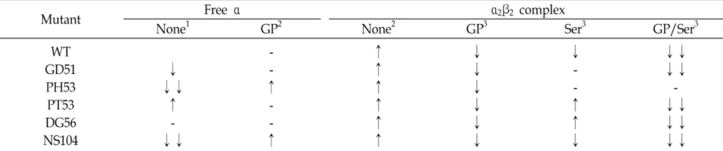
Table 1. Sensitivities of wild type and various mutant α subunits to trypsin digestion
Mutant Free α α2β2 complex
None1 GP2 None2 GP3 Ser3 GP/Ser3
WT GD51 PH53 PT53 DG56 NS104
↓
↓↓
↑ -
↓↓
- -
↑ - -
↑
↑
↑
↑
↑
↑
↑
↓
↓
↓
↓
↓
↓
↓ - -
↑
↑
↓
↓↓
↓↓
-
↓↓
↓↓
↓↓
This is summary of Fig. 1 and 2. 1The effects of altered residues on trypsin digestion of αL6 of α subunit. 2The effects of GP and β subunit on trypsin digestion of αL6 of α subunits were compared with that of the corresponding α subunits in the absence of GP, Ser and both. 3The effects of GP, Ser and both on trypsin digestion of αL6 of α subunits were compared with the corresponding α2β2 complex.
↑, increased sensitivity to trypsin: ↓, decreased sensitivity to trypsin; -, similar sensitivity; ↓↓, pronounced decreased sensitivity.
나타낸다. 이러한 관찰은 다른 연구 결과와 일치한다[1, 3].
잔기 치환체
αL6의 구조변화에 미치는 αL2와 αL3의 영향을 보고자 αL2 에 있는 GD51 (Gly to Asp at residue 51), PH53, PT53, DG56 와 αL3에 있는 NS104 잔기치환체를 활용하였다(Fig. 2). 그림 2에서 처리한 트립신의 농도보다 10배 높은 6 ug/ml를 처리하 여(데이터 생략) 이들 결과를 표1에 정리하였다. 유리 α 소단위 체는 3가지 잔기 치환체(GD51, PT53, DG56)는 야생형과 비슷 한 반면, PH53와 NS104는 그 분해 속도가 현저히 감소하였다 (Fig. 2, lane 1~3; Table 1). PH53와 NS104는 20분 후에도 많은 양의 온전한 αTS가 남아 있음을 보여준다(Fig. 2, lane3). 이는 두 치환자리인 53과 104가 있는 αL2와 αL3이 αL6 구조에 영향 을 미치고 있음을 나타낸다. 잔기치환체 αTS는 트립신에 의한 분해 속도를 복합체와 유리 α 소단위체 두 상태에서 비교해 보면, 복합체에서 그 속도가 증가하였다(Fig. 2, Table 1). 이 결과는 야생형과 비슷하였다.
αTS 리간드 GP의 영향
αTS의 활성부위에 결합하는 D,L-α-glycerophosphate (GP, 기질 D-glyceraldehyde 3-phosphate의 유사체)는 이 단백질의 단위체간의 상호작용을 조사하는 데 많이 유용하게 활용되어 왔다[11]. GP는 βTS에 진행되는 여러 단계의 반응속도를 변화 시키는 영향을 주는 것으로 보고되었다[2, 3]. GP가 첨가된 상태는 모든 잔기치환체가 복합체구조에서 분해속도가 다소 지연되었다(Fig. 2, lane 7~9 vs. 10~12; Table 1). 앞서 언급한 바 PH53는 유리 상태에서 큰 차이를 보였으나 복합체 구조에 서 야생형과 같은 양상을 보여주고 있다. 이는 GP 결합에 의해 구조가 다소 회복되고 있음을 나타낸다.
βTS 리간드 Ser의 영향
βTS의 기질인 L-Ser를 첨가하여 살펴보면, 다양한 효과를 나타낸다. 야생형과 NS104에서는 속도가 감소한 반면, GD51
Fig. 2. Trypsin Treatment of Wild Type and Various Mutant TS in the Presence of Various Ligands. The α subunit (0.39 mg/ml), β subunit (0.6 mg/ml) or α2β2 complex was treated with 0.6 ug/ml trypsin in the absence of any ligands or in the presence of 80 mM GP or 40 mM Ser or both (GP/Ser). Aliquots of solution was stopped by adding soybean trypsin inhibitor at various time in- terval (2, 5, 20 min) and was analyzed by SDS-PAGE.
WT, wild type; Mutants, for example with GD51, sub- stitution of Gly with Asp at residue 51 of α subunit.
과 PH53에서는 거의 영향이 없었고, PT53와 DG56은 증가하 였다(Fig. 2, lane 13~15; Table 1). βTS에 Ser이 결합하면 αTS 에 결합한 기질의 결합력을 증가시키는 것으로 알려져 있다[8, 10]. 또한 복합체에서는 조효소인 PLP (pyridoxal phosphate) 와 Ser이 결합하여 다섯 가지의 화학적으로 다른 반응 중간 화학종이 형성된다[2]. 마지막 화학종은 aminoacrylate인데 이 것은 분자터널을 통해 이동한 인돌과 결합하여 반응이 진행하 게 된다. 따라서 L-Ser의 결합한 상태에서 다양한 결과가 나오 는 것은 반응 중간 화학종의 분포의 변화와 연관될 가능성도 있다.
GP와 L-Ser를 동시에 처리했을 때는, 모든 단백질에서 αTS 의 절단이 현저히 감소하였다(Fig. 2, lane 16~18; Table 1). 일 반적으로 단백질은 리간드가 결합하여 구조가 안정화된다. 두 소단위체가 안정화되어 상승작용을 나타났다. 특별히 주목할 것은 이 중에서도 PH53는 가장 안정한 잔기치환체였다. 이는 Pro53가 소단위체간의 조절기작에서 중요한 역할을 하는 것 을 시사한다.
감사의 글
이 논문은 부산대학교 기본연구지원사업(2년, 2013~2015) 에 의하여 연구되었음.
References
1. Adachi, O., Kohn, L. D. and Miles, E. W. 1974. A rapid method for preparing crystalline β2 subunit of tryptophan synthase of Escherichia coli in high yield. J. Biol. Chem. 249, 7756-7763.
2. Dunn, M. F. 2012. Allosteric regulation of substrate channel- ing and catalysis in the tryptophan synthase bienzyme complex. Arch. Biochem. Biophys. 519, 154-166.
3. Faeder, E. J. and Hammes, G. G. 1970. Kinetic studies of tryptophan synthase. Interaction of substrates with β subunit. Biochemistry 9, 4043-4049.
4. Higgins, W., Fairwell, T. and Miles, E. W. 1979. An active proteolytic derivative of the alpha subunit of tryptophan synthase: Identification of the site of cleavage and character- ization of the fragment. Biochemistry 22, 4827-4835.
5. Hilario, E., Caulkins, B. G., Huang, Y. M., You, W., Chang, C. A., Mueller, L. J., Dunn, M. F. and Fan, L. 2016.
Visualizing the tunnel in tryptophan synthase with crys- tallography: Insights into a selective filter for accommodat- ing indole and rejecting water. Biochim. Biophys. Acta 1864, 268-279.
6. Hyde, C. C., Ahmed, S. A., Padlan, E. A., Miles, E. W and Davies, D. R. 1988. Three-dimensional structure of the tryp- tophan synthase α2β2 multienzyme complex from Salmonella typhimurium. J. Biol. Chem. 267, 17857-17871.
7. Jeong, M. S., Jeong, J. K., Park, K. S., Kim, H. T., Lee, K.
M., Lim, W. K. and Jang, S. B. 2004. Crystallization and pre- liminary X-ray analysis of tryptophan synthase α-subunits from Escherichia coli. Acta Crystallogr. D Biol. Crystallogr. 60, 132-134.
8. Kayastha, A. M., Sawa, U., Nagata S. and Miles, E. W. 1990.
Site-directed mutagenesis of the β subunit of tryptophan synthase from Salmonella typhimurium. J. Biol. Chem. 266, 7618-7825.
9. Kim, J. W., Kim, E. Y., Park, H. H., Jung, J. E., Kim, H.
D., Shin, H. J. and Lim, W. K. 2001. Homodimers of mutant tryptophan synthase α-subunits in Escherichia coli. Biochem.
Biophys. Res. Commun. 289, 568-572.
10. Kirschner, K., Wiskocil, R. L., Foehn, M. and Rezeau, L.
1975. The tryptophan synthase from Escherichia coli. An im- proved purification procedure for the alpha-subunit and binding studies with substrate analogues. Eur. J. Biochem.
60, 513-523.
11. Miles, E. W., Yutani, K. and Ogasahara, K. 1982. Guanidine hydrochloride induced unfolding of the alpha subunit of tryptophan synthase and of the two alpha proteolytic frag- ments: evidence for stepwise unfolding of the two alpha domains. Biochemistry 21, 2586-2592.
12. Leggett-Bailey, J. 1962. Techniques in Protein Chemistry.
Elsevier Scientific Publishing Co. New York.
13. Laemmli, U. K. 1970. Cleavage of structural proteins during the assembly of the head of bacteriophage T4. Nature 227, 680-685.
14. Lim, W. K., Shin, H. J., Milton, D. L. and Hardman, J. K.
1991. Relative activities and stabilities of mutant Escherichia coli tryptophan synthase α- subunits. J. Bacteriol. 173, 1886- 1893.
15. Milton, D. L., Napier M. L., Muers, R. W. and Hardman, J. K. 1986. In vitro mutagenesis and overexpression of the Escherichia coli trpA gene and the partial characterization of the resultant tryptophan synthase mutant alpha subunits.
J. Biol. Chem. 261, 16604-16615.
초록:대장균 트립토판 생성효소의 소단위체간 상호조절
조원진․임운기*
(