Various Aggregate Forms of Tryptophan Synthase α-Subunit
Myung Won Park and Woon Ki Lim*
Department of Molecular Biology, College of Natural Sciences, Pusan National University, Busan 609-735, Korea Received January 15, 2013 /Revised January 26, 2013 /Accepted January 26, 2013
Protein aggregation can cause diseases and hinder the production of useful recombinant proteins. The present study showed that at least three types of aggregates can be formed from tryptophan synthase α -subunit (αTS) by varying conditions: (1) an opaque white precipitous aggregate, (2) a transparent gel-like precipitous aggregate, and (3) an unprecipitous aggregate. Macroscopically different aggregate types might suggest different mechanisms underlying aggregation processes.
Key words : Opaque white aggregate, protein aggregate, transparent gel-like aggregate, tryptophan synthase α-subunit, unprecipitous aggregate
*Corresponding author
*Tel:+82-51-510-2289, Fax:+82-51-513-9258.
*E-mail : [email protected]
This is an Open-Access article distributed under the terms of the Creative Commons Attribution Non-Commercial License (http://creativecommons.org/licenses/by-nc/3.0) which permits unrestricted non-commercial use, distribution, and reproduction in any medium, provided the original work is properly cited.
Journal of Life Science 2013 Vol. 23. No. 2. 319~323 DOI : http://dx.doi.org/10.5352/JLS.2013.23.2.319
Introduction
Proteins can fail to form native structures and such fail- ures result in a wide range of symptoms [13]. Pathological states such as Parkinson’s diseases and Filaminopathy be- long to this category. In addition, proteins are often found in large intracellular aggregates during biotechnical pro- duction in bacteria, hindering their utilization. Protein ag- gregation occurs in competition with the normal folding pathway [2], and it takes place from misfolded and partially unfolded states. Although this area expanded rapidly in re- cent years, many things, especially when it comes to non- fibrillar protein aggregation, remain largely unknown.
Tryptophan synthase α subunit (αTS) from E. coli consists of 268 residues, and has no disulfide bond or prosthetic group. It was shown by X-ray structure to have (βα)
8barrel motif, a popular protein fold [5, 10]. The αTS has been ex- tensively studied for its folding property [14], but its ag- gregation properties were not. The mutational studies on α TS had produced a variety of mutant proteins with different tendency of aggregation [6, 9]. Investigation of aggregation property of these proteins could help to understand under- lying mechanisms of protein aggregation. In the course of characterizing such abnormalities, we found different types of aggregates.
Materials and Methods Chemicals
All chemicals were of reagent grade or ultrapure quality and purchased from Sigma (St. Louis, MO).
Overexpression and purification of wild-type and mutant proteins
Construction of plasmids for wild type, T24A/E49G/
F139W and T24A/D60N/F139W αTSs were described [11].
The wild-type and the mutant proteins were overproduced in E. coli RB797 and then purified as described elsewhere [6]. Proteins were kept by addition of 85% saturated ammo- nium sulfate. The pellet was dissolved in 10 mM potassium phosphate (pH 7.8), 0.2 mM EDTA, and 1 mM β -mercaptoethanol and was dialyzed against this buffer. Each purified protein appeared as a major band by SDS-PAGE.
The concentration of purified wild-type α-subunit was meas- ured by extinction values using E
1%278nm=4.4 [11]. The mutant α TSs were estimated by microbiuret assay [8] using wild-type αTS as a standard.
Nondenaturing- and SDS-PAGE
The purity and homogeneity of the protein were analyzed by nondenaturing and SDS-PAGE [7]. Nondenaturing PAGE was performed using a discontinuous buffer system [3].
Bands were visualized by Coomassie Brilliant Blue R250.
In vitro aggregation reactions
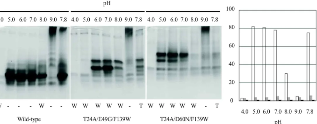
Native forms of wild-type and mutant αTSs in 10 mM
potassium phosphate (pH 7.8), 0.2 mM EDTA, and 1 mM
β -mercaptoethanol were incubated at varying conditions of
- Note -
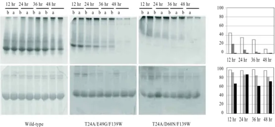
12 hr 24 hr 36 hr 48 hr b a b a b a b a
12 hr 24 hr 36 hr 48 hr b a b a b a b a
12 hr 24 hr 36 hr 48 hr b a b a b a b a
Wild-type T24A/E49G/F139W T24A/D60N/F139W
100 80 60 40 20 0 100 80 60 40 20 0
12 hr 24 hr 36 hr 48 hr
100 80 60 40 20 0