Suppressive Effects of Lees from Sweet Potato Soju on LPS-induced Inflammatory Responses in RAW 264.7 Cells
Seung-Hoon Lee1, Min-Jeong Kwon1, Soon Young Kim1, Ho-Yong Sohn2, Woo-Chang Shin3 and Jong-Sik Kim1*
1Department of Biological Sciences, Andong National University, Andong 36729, Korea
2Department of Food and Nutrition, Andong National University, Andong 36729, Korea
3Research Institute, Kooksoondang Brewery Co., Ltd., Seongnam 13202, Korea
Received September 11, 2015 /Revised November 7, 2015 /Accepted November 7, 2015
In the current study, the ethanol extracts and their subsequent organic solvent fractions from lees of sweet potato soju were prepared and the prepared samples were designated as from KSD-E8-1 to KSD-E8-5. Their effects on cell viability and nitric oxide (NO) production in mouse macrophage RAW 264.7 cells were investigated. The results showed that the ethyl acetate fraction (KSD-E8-3) of lees ex- tracts from sweet potato soju significantly decreased nitric oxide (NO) production in LPS-activated RAW 264.7 cells, whereas they did not affect cell viabilities. The fraction KSD-E8-3 reduced the ex- pression of pro-inflammatory genes such as COX-2, iNOS and TNF-alpha and also decreased protein expression of iNOS in a dose dependent manner, which were detected with RT-PCR and Western blot analysis, respectively. In addition, we detected the expression of mitogen-activated protein kinases (MAPKs) such as p38, JNK, and ERK1/2 and their phosphorylated forms. The results indicated that the treatment of the fraction KSD-E8-3 did inhibit phosphorylation of p38, JNK, and ERK1/2 MAPKs, indicating that the fraction KSD-E8-3 regulates LPS-induced inflammatory response via suppressing MAPK signaling pathway. Overall, these results may contribute to understand the molecular mecha- nism of anti-inflammatory effects by the ethyl acetate fraction of lees extracts from sweet potato soju.
Key words : Anti-inflammation, lees extracts, organic solvent fractions, RAW 264.7 cell, sweet potato soju
*Corresponding author
*Tel : +82-54-820-5798, Fax : +82-54-820-7705
*E-mail : [email protected]
This is an Open-Access article distributed under the terms of the Creative Commons Attribution Non-Commercial License (http://creativecommons.org/licenses/by-nc/3.0) which permits unrestricted non-commercial use, distribution, and reproduction in any medium, provided the original work is properly cited.
Journal of Life Science 2016 Vol. 26. No. 1. 117~122 DOI : http://dx.doi.org/10.5352/JLS.2016.26.1.117
서 론
염증(inflammation)은 다양한 외부 자극으로 인해 발생하 는 신체 방어 기작으로서[1] LPS를 비롯한 미생물 유래 내독소 (endotoxin)는 대표적인 염증 유도 물질로 알려져 있다. 이러 한 미생물에 의한 염증 반응은 국부 괴사 및 궤양 형성, 그리고 2차감염의 원인이 되기도 한다[11]. 이러한 염증 반응을 적절 하게 억제하지 못할 경우 만성염증으로 심화되어 암화과정 (carcinogenesis)을 통해 암으로 발전할 가능성도 있어 이러한 염증 반응을 조절하기 위한 다양한 연구가 진행되고 있다[4, 17].
Lipopolysaccharide (LPS)는 그람 음성균의 세포벽의 구성 성분으로서, 대식세포에 의한 염증반응을 촉발하는 인자로 알 려져 있다[3]. LPS는 대표적인 pattern recognition receptor (패턴 인식 수용체)인 toll-like receptor 4와 MD-2 복합체에
의해 인식되며 그 결과, 세포 내 주요 신호전달 경로 중 하나인 mitogen-activated protein kinase (MAPK) family 를 활성화 시키게 된다[2]. 이를 구성하는 extracellular signal-regulated kinase 1/2 (ERK1/2), jun N-terminal kinase (JNK) 그리고 p38은 전사 조절 인자인 ATF2 [5], CREB [9], STAT3 [3] 그리 고 NF-κB [19]와 같은 다양한 전사조절인자들을 통해 in- ducible nitric oxide synthase (iNOS) 및 TNF-alpha [7], cyclo- oxygenase-2 (COX-2) 그리고 cytokine [20]을 생산하는 것으 로 알려져 있다. 최종적으로 iNOS와 COX-2로부터 생성 유도 된 nitric oxide (NO)와 prostaglandin E2 (PGE2)는 cytokine과 함께 통증, 부종, 발열, 염증 부위로의 면역세포 이동을 시키는 등 염증 반응을 매개하는 것으로 알려져 있어 염증반응 정도 를 판단하는 중요한 척도로 이용되고 있다[14]. 또한, 2차 면역 반응을 유도하여 방어기작에 관여하는 것으로 알려져 있으나 [21], 감염원과 반응기전에 따라 다양한 염증성 질환의 원인이 되기도 한다[15].
주박(lees)이란 한국 전통주 제조과정에서 생성되는 발효 부산물로서 무기질, 섬유소, 비타민 등 다양한 유효성분을 포 함하고 있으나, 현재 식품위생법 상 식품 및 식품 첨가 제한 원료로 지정되어있다. 최근에는 주박이 가지는 다양한 유효성 분에 의한 항당뇨 활성[12], 항균 및 항산화 활성[6], 암세포의
- Note -
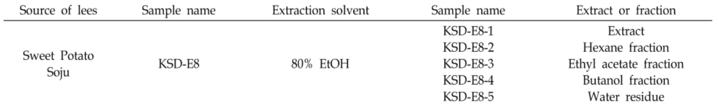
Table 1. List of lees extract and its subsequent solvent fractions of sweet potato soju
Source of lees Sample name Extraction solvent Sample name Extract or fraction
Sweet Potato
Soju KSD-E8 80% EtOH
KSD-E8-1 KSD-E8-2 KSD-E8-3 KSD-E8-4 KSD-E8-5
Extract Hexane fraction Ethyl acetate fraction
Butanol fraction Water residue 항성장 활성[8] 그리고 항혈전 활성[10]에 대한 연구 결과가
보고되었으며, 주박 발효 시 이용되는 누룩으로부터 추출한 지질성분이 NO 생성을 억제함으로써 염증반응을 억제한다는 사실이 보고되었다[13].
본 연구에서는 고구마소주 제조 시 발생하는 주박으로부터 에탄올 추출물 및 이들의 순차적 유기용매 분획물을 제조하고 이들 시료에 의한 NO 생성 억제와 pro-inflammatory 유전자 및 단백질의 발현에 미치는 영향, 그리고 MAPK 신호전달 과 정과의 관련성에 대해 연구하였다. 이러한 연구결과는 고구마 소주 제조 시 파생하는 주박에 포함된 생리활성 물질의 항염 증 활성과 작용기전을 이해하는데 도움을 줄 것으로 기대된다.
재료 및 방법
고구마 소주 주박 추출물 및 분획물의 제조
본 연구에서 사용된 고구마 소주 주박은 (주)국순당(Seong- nam, Korea)에서 제공받아 사용하였으며, 다음의 절차에 따라 에탄올 추출물 및 순차적인 유기용매 분획물을 제조하였다.
고구마 소주 주박 시료 1.5 kg에 95% 에탄올 6 L를 가하여 상온에서 3일간 2회 추출 하였고, 여과한 후 60℃에서 감압 농축하여 분말로 제조하였다. 추출된 추출물 4 g을 물에 현탁 한 후, n-hexane, ethyl acetate 및 butanol을 이용하여 순차적 으로 분획하고 최종적으로 물 잔류물을 회수하였다. 이렇게 준비된 시료를 각각 KSD-E8-1 ~ KSD-E8-5로 명명하였다 (Table 1). 각 시료는 dimethyl sulfoxide (DMSO, Sigma, St.
Louis, MO, USA)에 녹여 사용하였다.
마우스 대식세포 RAW 264.7 세포의 배양
본 연구에 사용된 세포주는 마우스 대식세포 RAW 264.7 세포주를 사용하였고 American Type Culture Collection (ATCC, Rockville, MD, USA)에서 구입하였다. 세포주의 배양 은 Dulbecco’s Modified Eagle Medium (DMEM, Gibco, Grand Island, NY, USA)에 10% Fetal Bovine Serum (FBS, Gibco), 1% penicillin 및 streptomycin (WelGene, Gyeongsan, Korea)을 첨가하여 사용하였다. 배양은 37℃, 5% CO2 조건의 배양기에서 실시하였다.
Nitric oxide assay
고구마소주 주박 분획물이 LPS로 활성화된 대식세포 RAW
264.7 세포주의 nitric oxide (NO) 생성에 미치는 영향을 측정 하기 위하여 nitric oxide assay를 수행하였다. 즉, RAW 264.7 세포를 96 well plate의 각 well에 1×10⁵개의 세포를 접종한 후, 18시간 동안 배양하였다. 그 후 lipopolysaccharide (LPS, Sigma)를 0.2 μg/ml 농도로 1시간 처리하고, 주박 분획물을 처리하여 16시간 동안 배양하였다. 생성된 NO의 양은 Griess 시약[1% sulfanilamide, 0.1% naphylethylenendiamine in 2.5% phosphoric acid]을 이용해 측정하였다. 세포 배양 상등 액 100 μl와 Griess 시약 100 μl를 1:1로 혼합하여 96 well plate 에서 15분간 반응시키고 NanoQuant Plate™ (Tecan Trading AG, Switzerland)를 사용하여 540 nm에서 흡광도를 측정하였 다. 실험결과 수치는 4개의 독립적인 well에서 수행한 값을 Sigma plot 10.0 program을 이용하여 분석한 후 mean±SD 값 과 그래프로 나타내었다.
세포생존율 측정
주박 분획물이 마우스 대식세포 RAW 264.7 세포주의 성장 에 미치는 영향을 연구하기 위하여 cell viability assay를 수행 하였다. 먼저 96 well plate에 1×10⁵개의 RAW 264.7 세포를 접종한 후, 고구마 소주 주박 시료를 16시간 처리한 후 MTS (3-(4,5-dimethylthi-azol-2-yl)5-(3-carboxy-methox- yphenyl)-2-(4-sulfophenyl)-2H-tetrazolium)용액(Promega, Madison, WI, USA)을 각 well 당 20 μl 씩 첨가하여 37℃, 5% CO2 배양기에서 4시간 동안 반응시켰다. 반응이 끝난 후 NanoQueant Plate™ (Tecan Trading AG, Switzerland)를 이용 하여 580 nm에서 흡광도를 측정하였다. 실험결과 수치는 4개 의 독립적인 well에서 수행한 값을 Sigma plot 10.0 program 을 이용하여 분석한 후 mean±SD 값과 그래프로 나타내었다.
Reverse Transcription-Polymerase chain reaction (RT-PCR)
RT-PCR을 수행하기 위해 RAW 264.7 세포로부터 RNeasy mini kit (Qiagen, Valencia, CA, USA)을 이용하여 제조사의 매뉴얼에 따라 수행하여 Total RNA를 분리하였다. 분리한 to- tal RNA 2 μg을 주형으로 PrimeScript™ RT-PCR Kit (Ta- KaRa, Japan)을 이용하여 제조사의 매뉴얼에 따라 cDNA를 합성하였다. 합성된 cDNA를 주형으로 하여 유전자 특이적인 oligo primer를 이용하여 PCR (polymerase chain reaction) 과정을 수행하였다. PCR에 사용된 primer는 Table 1과 같고,
Table 2. Sequences of the oligonucleotide primers of the mouse COX-2, iNOS, and TNF-alpha genes
Gene name Gene bank acc. no. Primer sequences
COX-2 NM_011198.3 F : 5'-CCGTGGTGAATGTATGAGCA-3'
R : 5'-CCTCGCTTCTGATCTGTCTT-3'
iNOS NM_010927.3 F : 5'-CTGCAGCACTTGGATCAGGAACCTG-3'
R : 5'-GGGAGTAGCCTGTGTGCACCTGGAA-3'
TNF-α NM_013693.2 F : 5'-CGTCAGCCGATTTGCTATCT-3'
R : 5'-CGGACTCCGCAAAGTCTAAG-3'
GAPDH NM_008084.2 F : 5'-TGCACCACCAACTGCTTA-3'
R : 5'-GGATGCAGGGATGATGTT-3'
A B
C D
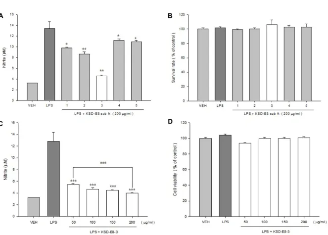
Fig. 1. Effects of ethanol extract and its subsequent solvent fractions from lees of sweet potato soju on nitric oxide production and cell viabilities. Mouse macrophage RAW 264.7 cells were plated at 1×10⁵ cells/well in a 96-well plate and (A, B) incubated with 200 μg/ml of each lees sample or (C, D) four different concentrations of KSD-E8-3. After 24 hr treatment, (A, C) nitric oxide production was measured by nitric oxide assay and (B, D) cell viability was measured using MTS proliferation assay kit. *p<0.05, **p<0.01, ***p<0.001.
primer는 Bioneer사(Daejeon, Korea)에 주문 제작하였다.
PCR은 TaKaRa Ex Taq (TaKaRa)을 이용하여 제조사의 프로 토콜에 따라 수행하였다.
Western blot analysis
마우스 대식 세포주인 RAW 264.7 세포주를 60 mm dish에 접종 후, 18시간 뒤 0.2 μg/ml의 LPS 를 처리하여 염증반응을 유도한 후, 다시 1 시간 후 고구마소주 주박 시료를 처리하여 16시간 배양 한 뒤 세포를 수확하였다. 수확한 세포는 10X
RIPA buffer (Cell Signaling, Beverly, MA, USA)를 4X 로 희석 하여 첨가한 후 sonication 하였다. MAPKs와 phospho-MAPKs 단백질의 경우, 주박 시료를 serum free media에 혼합하여 4시 간 동안 처리한 후, LPS 를 1.0 μg/ml의 농도로 30분 동안 처리하고 얼음을 이용하여 세포활동을 정지시킨 후 동일한 방법으로 단백질을 추출하였다. 본 실험에서 사용된 1차 항체 로는 iNOS, COX-2, p38, p-p38, ERK1/2, p-ERK1/2, JNK, p-JNK 그리고 Actin을 사용하였으며, 2차 항체는 HRP-con- jugated rabbit antibody 그리고 HRP-conjugated mouse anti-
A B
Fig. 2. Down-regulation of COX-2, iNOS and TNF-α genes by KSD-E8-3 treatment. Mouse macrophage RAW 264.7 cells were treated with KSD-E8-3, and then total RNA and total protein were prepared from KSD-E8-3 treated cells. (A) Total RNA was used for RT-PCR with COX-2, iNOS, and TNF-α gene specific primers. (B) Western blot was carried out by using COX-2, iNOS, and Actin antibodies.
body를 사용하였다. Actin과 2차 항체는 Santa Cruz 사(Santa Cruz, CA, USA)로부터 구입하였고, 그 외 모든 1차 항체는 Cell Signaling 사(Beverly, MA, USA)로 부터 구입하여 사용 하였다.
통계처리
NO assay와 세포 생존율 실험은 3회 반복실험을 실시하였 으며, 실험결과는 평균±표준편차로 나타내었고, 각 실험결과 의 유의성 검토는 시료가 포함되지 않은 대조구와 비교하여 Student’s t-test에 의해 판정하였으며 p값이 0.05 미만일 때 유의성이 있다고 판단하였다.
결과 및 고찰
고구마 소주 주박 추출물의 유기용매 분획물이 nitric oxide 생산과 세포생존율에 미치는 영향
본 연구팀은 선행 연구로서 주박과 누룩 추출물 및 유기용 매 분획물 85종에 의한 nitric oxide (NO) 생산 저해 활성을 보고 한 바 있다[18]. 본 연구에서는 이 중 nitric oxide 생산 저해율은 매우 높으나, 현재까지 전혀 보고되지 않은 고구마 소주 주박 에탄올 추출물 및 유기용매 분획물(Table 1)이 NO 생산에 미치는 영향과 세포 생존에 미치는 영향을 연구하였 다. 즉, 고구마 소주 주박 추출물 및 유기용매 분획물(KSD- E8-1 ~ KSD-E8-5)이 NO 생산 및 세포 생존율에 미치는 영향 을 연구하기 위해 LPS로 염증이 유도된 RAW 264.7 세포주에 각 시료를 200 μg/ml의 농도로 처리한 결과 Fig. 1A 에서 보는 바와 같이 KSD-E8-3 (ethyl acetate 분획물)의 처리에 의해 가 장 높게 NO 생산이 저해되었다. 5종의 시료의 처리에 의한 세포 생존율에 미치는 영향은 없는 것으로 확인되었다(Fig.
1B). KSD-E8-3의 농도 의존적인 처리에 따른 NO 생산에 미치 는 영향을 연구하기 위하여 시료를 LPS로 활성화된 RAW 264.7 세포주에 50, 100, 150, 200 μg/ml의 농도로 처리한 후 NO 생산을 측정하였다. 그 결과, 시료를 50 μg/ml의 농도로
처리한 군부터 현저하게 NO 생산이 저해됨을 확인하였고, 시 료의 처리 농도가 증가됨에 따라 NO 생산이 저해되는 농도 의존적인 양상을 보여 주었다(Fig. 1C). 한편, 처리한 시료의 농도에서 세포 생존율에는 영향이 없는 것으로 확인되었다 (Fig. 1D). 이후 연구는 5종의 시료중 NO 생산의 저해율이 가 장 높은 KSD-E8-3 시료를 이용하여 수행하였다.
Ethyl-acetate 분획물에 의한 pro-inflammatory 유전자 및 단백질 발현 감소
KSD-E8-3에 의한 NO 생산 억제 기전을 연구하기 위하여 시료를 100, 200 μg/ml 의 농도로 LPS로 염증 유도된 RAW 264.7 세포주에 처리 한 후, pro-inflammatory 유전자인 COX- 2, iNOS 그리고 TNF-alpha 의 유전자 및 단백질 발현 변화를 연구하였다. 그 결과, LPS에 의해 염증이 유도된 대조구에 비 해 COX-2, iNOS, 그리고 TNF-alpha 유전자의 발현이 현저하 게 감소됨을 확인하였다(Fig. 2A). 단백질의 발현을 확인한 결 과 iNOS 단백질은 시료의 처리에 의해 현저하게 발현이 감소 될 뿐만 아니라, 농도 의존적으로 발현이 감소됨을 확인한 반 면, COX-2의 경우 KSD-E8-3의 처리에 의해 미미한 발현 감소 만을 확인할 수 있었다(Fig. 2B). 이는 KSD-E8-3 처리에 의해 NO 생성이 크게 억제되는 Fig. 1C의 결과와 부합하는 것으로 생각된다. LPS에 의한 염증 유도 시 MAPK pathway 및 NF-κ B 경로를 통해 COX-2, iNOS 그리고 TNF-alpha 등이 생산되 며 PGE2 및 NO 생성 등을 통해 염증반응을 매개하는 것으로 알려져 있다[14]. 이러한 결과는 KSD-E8-3 분획물에 함유된 생리활성 물질이 NO 생성을 억제하고 pro-inflammatory 반 응을 매개하는 유전자들의 발현을 조절 할 수 있음을 보여준 다.
KSD-E8-3 분획물에 의한 MAPK 신호 억제
MAPK는 세포 내 주요 신호전달 경로 중 하나로서 인산화 를 통해 활성화되며 수 많은 전사인자를 통해 다양한 단백질 의 발현을 조절하는 것으로 알려져 있다. KSD-E8-3 에 의한
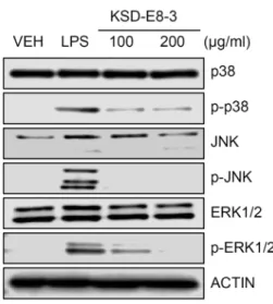
Fig. 3. Inhibition of phosphorylation of MAPK by KSD-E8-3.
Mouse RAW 264.7 cells were treated with 100 μg/ml or 200 μg/ml of KSD-E8-3 and total proteins were pre- pared from treated cells and Western blotting was car- ried out by using p38, phospho-p38, JNK, phospho-JNK, ERK1/2, phospho-ERK1/2, and Actin antibodies.
NO 생산의 저해와 MAPK 경로와의 관련성을 검증 하기 위하 여 LPS로 활성화된 RAW 264.7 세포에 KSD-E8-3를 100, 200 μg/ml의 농도로 처리하고, MAPK family 에 속하는 p38, JNK 그리고 ERK1/2의 발현과 이들 MAPK의 인산화 수준을 확인 하였다. 그 결과, Fig. 3에서 보는 바와 같이 모든 MAPK 단백 질의 인산화가 LPS 만 처리된 대조구에 비해 현저하게 감소하 는 것을 확인하였다. MAPK 단백질이 다양한 자극에서 pro-inflammatory 인자들의 전사와 발현을 조절하는 것으로 알려져 있어[16, 22] KSD-E8-3 분획물에 포함된 생리활성 물질 이 LPS 에 의해 유도된 염증 반응을 신호전달 단계에서 조절 할 수 있는 것으로 생각된다. 따라서, 향후 추출물에 함유된 핵심 생리활성 물질을 찾는 연구가 지속되어야 하며, 이러한 핵심물질에 의한 신호전달체계가 연구되어야 할 것으로 판단 된다.
감사의 글
본 연구는 2012년도 농림수산식품부 고부가가치식품기술 개발사업(과제번호 112073-3)에 의해 수행되었으며, 이에 감 사드립니다.
References
1. Akira, S. and Takeda, K. 2004. Toll-like receptor signaling.
Nat. Rev. Immunol. 4, 499-511.
2. Anderson, N. and Borlak, J. 2008. Molecular mechanisms and therapeutic targets in steatosis and steatoheptitis.
Pharmacol. Rev. 60, 311-357.
3. Chelsky, Z. L., Yue, P., Kondratyuk, T. P., Paladino, D., Pezzuto, J. M., Cushman, M. and Turkson, J. 2015. A resver- atrol analogue promotes ERK MAPK-dependent STAT3 ser- ine and tyrosine phosphorylation alterations and antitumor effects in vitro against human tumor cells. Mol. Pharmacol.
88, 524-533.
4. Eisinger, A. L., Prescott, S. M., Jones, D. A. and Stafforini, D. M. 2007. The role of cyclooxygenase-2 and prostaglandins in colon cancer. Prostaglandins Other Lipid Mediat. 82, 147- 154.
5. Jacobsen, M. C., Dusart, P. J., Korowicz, K., Bajaj-eliott, M., Hart, S. L., Klein, N. J. and Dixon, G. L. 2015. A critical role for ATF2 transcription factor in the regulation of E-se- lectin expression in response to non-endotoxin components of Neisseria meningitides. Cell. Microbiol. DOI: 10.1111/cmi.
12483.
6. Jin, J. H., Kim, J. S., Kang, S. S., Son, K. H., Chang, H. W.
and Kim, H. P. 2010. Anti-inflammatory and anti-arthritic activity of the roots of sophora flavescens. J. Ethnopharmacol.
127, 589-595.
7. Ju, A., Cho, Y. C. and Cho, S. 2015. Methanol extracts of Xanthium sibiricum roots inhibit inflammatory response via the inhibition of nuclear factor-κB (NF-κB) and signal trans- ducer and activator of transctiption 3 (STAT3) in murine macrophage. J. Ethnopharmacol. DOI: 10.1016/j.jep.2015.
07.038.
8. Kang, H. T., Lee, S. H., Kim, S. Y., Kim, M. S., Shin, W.
C., Sohn, H. Y. and Kim, J. S. 2014. Anti-proliferative activ- ities of solvent fractions of lees extracts in human colorectal HCT 116 ells. J. Life Sci. 24, 967-972.
9. Kim, H. G., Shi, C., Bode, A. M. and Dong, Z. 2015. p38α MAPK is required for arsenic-induced cell transformation.
Mol. Carcinog. DOI: 10.1002/mc.22331.
10. Kim, M. S., Lee, Y, S., Kim, J. S., Shin, W. C. and Sohn, H. Y. 2014. Evaluation of in-vitro antithrombosis activity of lees of Korean traditional wine. J. Life Sci. 24, 865-872.
11. Kindt, T. J., Goldsby, R. A. and Osborne, B. A. 2007. Innate immunity : Tenneys. Kuby Immunology. New York. Freeman Press 6, 52-73.
12. Kim, T. Y., Jeon, T. W., Yeo, S. H., Kim, S. B., Kim, J. S.
and Kwak, J. S. 2010. Antimicrobial, antioxidant and SOD-like activity effect of Jubak extracts. Kor. J. Food Nutr.
23, 299-305.
13. Kojda, G. and Harrison, D. 1999. Interactions between NO and reactive oxygen speices : pathophysiological importance in atherosclerosis, hypertension, diabetes and heart failure.
Cardiovasc. Res. 43, 562-571.
14. Lee, E. S., Ju, H. K., Moon, T. C., Lee, E., Jahng, Y., Lee, S. H. and Chang, H. W. 2004. Inhibition of nitric oxide and tumor necrosis factor-alpha (TNF-alpha) production by pre- penone compound through blockade of nuclear factor (NF)- κB activation in cultured murine macrophages. Biol. Pharm.
Bull. 27, 617-620.
15. Li, G., Wulan, H., Song, Z., Paik, P. A., Tsao, M. L., Goodman, G. M., MacEachern, P. T., Downey, R. S.,
초록:고구마 소주 주박에 의한 RAW 264.7 세포주에서 lipopolysaccharide로 유도된 염증 반응의 억제 효과
이승훈1․권민정1․김순영1․손호용2․신우창3․김종식1*
(1국립안동대학교 생명과학과, 2국립안동대학교 식품영양학과, 3㈜국순당 연구소)
본 연구에서는 고구마 소주로부터 획득한 에탄올 추출물 및 순차적 유기용매 분획물을 제조하고, 이들 시료가 LPS로 활성화된 마우스 대식세포 RAW 264.7 세포주에서 nitric oxide (NO) 생산과 세포생존율에 미치는 영향을 연구하였다. 처리한 시료 중 ethyl acetate 분획물(KSD-E8-3) 처리군에서 LPS로 염증 유도된 RAW 264.7 세포주의 생존율에는 영향 없이 NO 생성을 현저하게 감소 시킴을 확인하였다. 또한 분획물 KSD-E8-3 처리에 의해서 염증 매개 인자인 COX-2, iNOS 그리고 TNF-alpha의 유전자의 발현이 감소되었으며, iNOS 단백질의 발현이 현저히 감 소됨을 Western blot으로 확인하였다. 또한, 분획물 KSD-E8-3 처리에 의해 세포내 대표적인 신호 전달 단백질인 3가지의 MAPK 단백질(p38, JNK, ERK1/2)의 인산화가 모두 억제됨을 확인하였다. 종합적으로, 이러한 연구결과 는 고구마 소주 주박에 함유된 생리활성 물질에 의한 항염증 활성 및 작용 기전을 이해하는데 도움을 줄 것으로 기대된다.
Jankowska, A. J., Rabinowitz, Y. M., Learch, T. B., Song, D. Z., Yuan, J. J., Zheng, S. and Zheng, Z. 2015. Regulatory B cell function is suprresed by smoking and obesity in H.
pylori-infected subjects and is correlated with elevated risk of gastric cancer. Plos One 10, e0134591.
16. Lo, J. Y., Kamarudin, M. N., Hamdi, O. A., Awang, K. and Kadir, H. A. 2015. Curcumenol isolated from Curuma ze- doaria suppresses Akt-mediated NF-κB activation and p38 MAPK signaling pathway in LPS-stimulated BV-2 microlial cells. Food Funct. 6, 3550-3559.
17. Monick, M. M., Arter, A. B., Robeff, P. K., Flaherty, D. M., Peterson, M. W. and Hunninghake, G. W. 2001. Lipopoly- saccharide activates Akt in human alveolar macrophages re- sulting in nuclear accumulation and transcriptional activity of beta-catenin. J. Immunol. 166, 4713-4720.
18. Park, M. J., Kang, H. T., Kim, M. S., Shin, W. C., Sohn, H.
Y. and Kim, J. S. 2014. Anti-inflammatory effects of extracts and their solvent fractions of rice wine lees. J. Life Sci. 24, 843-850.
19. Sahu, B. D., Kumar, J. M. and Sistla, R. 2015. Baicalein, a bioflavonoid, prevents cisplatin-induced acute kidney injury by up-regulating antioxidant defense and down-regulating the MAPKs and NF-κB pathways. Plos One 10, e0134139.
20. Syed Hussein, S. S., Kamarudin, M. N. and Kadir, H. A.
2015. (+)-Catechin attenuates NF-κB activation through reg- ulation of Akt, MAPK, and AMPK signaling pathways in LPS-induced BV-2 microglial cells. Am. J. Chin. Med. 43, 927-952.
21. Vlkova, M., Ticha, O., Nechvatalova, J., Kalina, T., Litzman, J., Mauri, C. and Blair, P. A. 2015. Regulatory B cells in CVID patients fail to suppress multifunctional IFN-gam- ma+TNF-alpha+CD4+ T cells differentiation. Clin. Immunol.
29, 292-300.
22. Xu, C., Chen, G., Yang, W., Xu, Y., Xu, Y., Huang, X., Liu, J., Feng, Y., Xu, Y. and Liu, B. 2015. Hyaluronan ameliorates LPS-induced acute lung injury in mice via Toll-like receptor (TLR) 4-dependent signaling pathway. Int. Immunopharmacol.
28, 1050-1058.