豆豉 추출물을 이용한 下胎毒法이 NC/Nga 생쥐에서 유발된 아토피 유도 피부염에 미치는 항염증 효과
엄선호1․안상현2․박선영3․천진홍1,4․김기봉1,4
1부산대학교 한의학전문대학원 소아과학교실, 2부산대학교 한의과학연구소
3세명대학교 한의과대학 생리학교실. 4부산대학교한방병원 한방소아과
Abstract
The Anti-inflammatory Effects of Hataedock Taken Douchi Extracts on Atopic Dermatitis-like Skin Lesion of NC/Nga Mouse
Aum Sun Ho1․Ahn Sang Hyun2․Park Sun Young3․Cheon Jin Hong1,4․Kim Ki Bong1,4
1Department of Pediatrics, School of Korean Medicine, Pusan National University
2Research Institute for Korean Medicine, Pusan National University
3Department of Physiology, College of Korean Medicine, Semyung University
4Department of Pediatrics, Korean Medicine Hospital, Pusan National University
Objectives
Hataedock is a Korean herbal medical oral treatment that removes fetal toxic heat and meconium from new born babies. The purpose of this study is to evaluate whether Hataedock treatment of Duchi extracts has anti-inflammation effects on AD (Atopic Dermatitis)-induced NC/Nga mice.
Methods
After Hataedock treatment of Duchi extracts on days 0, 3-week-old NC/Nga mice were sensitized on days 28, 35, 42 by exposure of DNFB (dinitrochlorobenzene) and were induced to have AD. Immunohistochemistry of NF- κB p65, iNOS, COX-2 and TUNEL assay of apoptotic body was used to identify changes of skin damages and anti-inflammation effects.
Results
The alleviate effect of the skin damage and angiogenesis was observed in DT group. The damage of stratum corneum, hyperplasia, edema, infiltration of lymphocytes and distribution of capillary were decreased in DT group.
Also, the study results suggested that Hataedock treatment of Duchi extracts in DT group remarkably down- regulated levels of NF- κB p65 by 70% (p < 0.001), as well as COX-2 by 51%, iNOS by 62% (p < 0.001).
Additionally, Hataedock treatment of Duchi extracts in DT group up-regulated apoptosis of inflammatory cells by 68% in atopic dermatitis-like skin lesion.
Conclusions
From the study results, we observed that Hataedock treatment of Duchi extracts alleviates AD through diminishing various inflammatory cytokines in the skin lesions, which are involved in the initial steps of AD development. It is anticipated to have potential applications for prevention and treatment of atopic dermatitis.
Key words : Hataedock, Duchi, Atopic dermatitis, NF- κB, iNOS, Apoptosis
Received: April 22, 2016 ∙ Revised: May 13, 2016 ∙ Accepted: May 16, 2016 Corresponding Author: Kibong Kim, KMD, Ph.D.
Department of Pediatrics, Korean Medicine Hospital, Pusan National University 20, Geumo-ro, Mulgeum-eup, Yangsan-si, Gyeongsangnam-do, 50612, Republic of Korea
Tel: +82-55-360-5952, Fax: +82-55-360-5952 E-mail: [email protected]
ⓒ The Association of Pediatrics of Korean Medicine. All rights reserved. This is an open-access article distributed under the tenus of the Creative Commons Attribution Non-Commercial License (http://creativecommons.org/licenses/by-nc/3.0/), which permits unrestricted non-commercial use, distribution, and reproduction in any medium, provided the original work is properly cited.
ISSN 1226-8038(Print), 2287-9463(Online), http://dx.doi.org/10.7778/jpkm.2016.30.2.001
필요하다3).
아토피 피부염은 유전이나 환경, 약물성, 심인성, 면역 등의 여러 가지 요인들이 복합적으로 작용하여 발생하는 것으로 알려져 있다4). 최근에는 여러 가지 면 역학적 이상을 동반한 질환으로 보고되고 있는데5-6), 이는 Th1과 Th2 세포의 불균형으로 인하여 과도한 Th2 반응에 의한 면역반응으로 보고 있다. 2012년 연 령별 진료인원에서 9세 이하 소아가 전체 아토피 피부 염 진료환자 중 48%로 높게 나타난 점도 출산시 Th2 치우침의 불균형 상태에서 아토피 피부염 발병 요인에 노출된 결과로 보여진다7-8).
한의학에서는 아토피 피부염을 태열 (胎熱), 태선 (胎癬), 내선 (內癬), 습진 (濕疹) 등의 범주로 보았으며, 풍열 (風熱), 습열 (濕熱), 혈허 (血虛), 혈열 (血熱) 등의 원인으로 발생하다고 보았다9). 출산 후 이러한 태독 (胎毒)을 제거하는 방법으로 두시법 (豆豉法), 감초법 (甘草法), 황련법 (黃連法), 주밀법 (朱蜜法) 등의 하태 독법 (下胎毒法)을 사용하였다. 하태독법은 신생아에 게 한약재를 달인 약물을 소량 먹이는 방법이다10). 하 태독법의 아토피 피부염에 대한 항염증 효과가 기대되 나, 아직까지 이에 대한 체계적인 검증이나 연구가 진 행되어진 적은 없다.
본 연구는 피부염을 인위적으로 유발하는 DNFB를 Ng/Nga 생쥐의 피부 국소부위에 반복적으로 처리한 후 아토피 유사 피부염을 유발하여 아토피 피부염 유 사 동물모델을 만들었다11). DNFB는 histamine, tumor necrosis factor (TNF)-α, vascular endothelial growth fac- tor (VEGF) 같은 다양한 pro-inflammatory cytokines이나 mediators를 분비하는 것으로 알려져 있다. 두시 추출 물을 이용한 하태독법 실시 후 아토피 유사 피부염을 유발하여 일어나는 전염증효소, 염증전사인자, 염증효 소, 염증세포의 apoptosis 변화를 면역조직학적 관찰하
누었으며, 각 군에 각 10마리씩 배정하였다. 하태독법 실시 4주 후에 아토피 유사 피부염을 유발하였다. 우선 생쥐 등쪽 부위 피부를 면도한 다음 5% sodium do- deecyl sulfate (SDS: Sigma, USA) 1 ㎖을 면봉으로 20회 문질러서 각질층의 lipid lamella를 제거하고 ace- tone/olive (4 : 1)에 희석된 1% 2,4-dinitrofluorobenzene (DNFB) 100 ㎕로 감작시켰다. 감작 후 7일과 14일째 2% DNFB 100 ㎕을 도포하여 아토피 유사 피부염을 유발하였다 (Fig. 1). 피부염 유발 72시간 후 sodium pentobarbital 용액으로 마취 후 처치하였다. 얻어진 등 쪽 피부를 10% NBF에 실온에서 24시간동안 고정한 후 통상적인 방법으로 paraffin에 포매하고 5 ㎛ 두께로 연속절편을 만들었다. 만들어진 연속절편은 Phloxine- tartrazine 염색하여 표본을 제작하였다. 본 연구과정은 부산대학교 동물실험윤리위원회 승인을 받아 시행되 었으며 (IACUC number: PNU-2014-0732), 실험실 동 물의 관리와 사용에 대해서는 NIH 가이드라인에 따라 시행되었다.
2. 하태독법 약물의 제조와 처리
두시는 청호의 전탕액에 흑대두를 담그어 흡수시킨 후 남은 전탕액으로 흑대두를 삶고, 이후 흑대두를 37~38 ℃에서 3~5일간 발효시키는 방법으로 제조되었 다. 두시 100 g을 증류수 1000 ㎖에 넣고 3시간동안 전탕한 후 여과하였다. 그 여액을 rotary evaporator를 이용하여 50 ㎖으로 감압, 농축한 후 동결 건조하여 추 출물 20.2 g (수득률 20.2%) 얻어 사용하였다. 3주령 Nc/Nga 생쥐 DT군에 3일동안 두시 추출물 20 ㎎/㎏을 경구투여하는 하태독법을 실시하였다.
3. Fingerprinting analysis
지표성분의 정량분석을 위해서 binary solvent deliv- ery pump (G1312A), auto sampler (G1329A), column oven (G1316A), diode array detector (DAD; G1315D), vacuum degasser (G1322A) 등으로 구성된 HPLC system (Agilent technologies 1200 series; Agilent Technologies, CA, USA)을 사용하였다. 컬럼은 Capcell PAKMGII C18 (3.0 × 150 mm, 3.0 μm; Shiseido, Tokyo, Japan)을 사용하였고, 컬럼 온도는 35 ℃, 유속은 0.6 mL/min, 시료 주입량은 15 μL로 설정하였다. 이동상은 0.5%
formic acid가 포함된 water (v/v; A)와 acetonitrile (B)로 구성하였고, 기울기 용리법을 이용하여 분석을 진행하
였다 (기울기 용리 조건: initiation-5 min-2% B, 12 min-10% B, 20 min-25% B, 27 min-25% B, 25 min-80% B, 37 min-80% B, 40 min-30% B, 45 min-2%
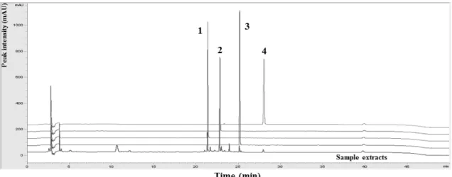
B). Palmitine, Berberine (ChemFaces, Wuhan, China) 및 Liquritin, Liquritigenin (Sigma-Aldrich, USA) 등의 지표 물질 각 10 mg을 증류수 100 mL에 녹여 지표물질 혼 합액을 제조하였다. 시료 용액은 0.45 μm syringe filter 로 여과하여 HPLC 분석에 사용하였다. 시료의 분석은 HPLC를 이용하였으며 각 시료의 주요성분에 대한 sample choromatogram 결과는 Fig. 2에서 제시하였다.
분석결과 daidzin, daidzein, genistin, genistein이 검출되 었다 (Fig. 2).
Fig. 1. Protocol of Hataedock for DNFB induced dermatitis
Mice were sensitized on days 28 by exposure of 1% DNFB 100 ㎕. On days 7 and 14 after initial sensitization, the mice were challenged with 2% DNFB 100 ㎕. Hataedock treatment was orally administered on days 1, 2 and 3. Abbreviation: PBS, phosphate-buffered saline; DNFB, 2,4-dinitrofluorobenzene
Fig. 2. Comparative chromatogram of isoflavones in standard solution and Douchi extract
Peak number 1: daidzin; 2: genistin; 3: daidzein; and 4: genistein in standard solution. Separation was performed by HPLC-DAD.
서 24시간동안 반응한 후 avidin biotin complex kit (Vector Lab, USA)를 이용하여 1시간동안 실온에서 반 응시켰다. 0.05% 3,3'-diaminobenzidine과 0.01% HCl이 포함된 0.05 M tris-HCl 완충용액 (pH 7.4)에서 발색시 킨 후, hematoxylin으로 대조염색하였다.
5. TUNEL assay
Apoptosis 변화를 조사하기 위해 in situ apoptosis de- tection kit (Apoptag, Intergen, USA)를 이용한 TUNEL (terminal deoxynucleotid transferase-mediated dUTP-bio- tin nick-end labelling) 방법을 실시하였다. 먼저 조직 절편을 proteinase K에 5분간 proteolysis 시킨 다음 equi- libration buffer에서 20초간 처리하였다. 그런 다음 strength TdT enzyme (36 ㎕ TdT enzyme : 72 ㎕ re- action buffer)을 처리하여 37 ℃의 humidified chamber 에서 1시간 동안 반응시킨 후 strength stop/wash buffer 에서 10분 동안 처리하였다. Anti-digoxigenin-perox- idase에 1시간 동안 반응시킨 후 DAB를 처리하였다.
Eosin으로 대조염색한 후 광학현미경으로 관찰하였다.
6. 영상처리, 분석과 통계처리
혈관분포는 sharpen low-filter를 사용하여 x4 배율로 촬영 후, 반전 (invert) 처리하였다. 면역조직화학 결과 와 반전 처리된 TUNEL assay의 결과는 image Pro Plus (Media cybernetics, USA)를 이용한 영상분석을 통해 수 치화 (means ± standard error) 하였다. 영상분석은 각 군의 표본에서 임의로 선정된 상피와 진피유두를 x400배율 에서 촬영한 다음 positive pixels/전체 pixel (10,000,000 pixels)로 이루어졌다. 통계는 SPSS software (SPSS 23, SPSS Inc., USA)로 처리되었으며, one-way ANOVA 시 행을 통해 유의성 (P<0.001)을 검증하고 Duncan’s mul- tiple range test로 사후 검증하였다.
피부를 절개하여 혈관의 직경 차이와 모세혈관 분 포 차이를 확인하였다. 진피 쪽 혈관을 관찰한 결과, AE군에서 혈관분포가 증가한 반면, DT군에서는 감소 하였다 (Fig. 3).
AE군의 피부에서는 각질층의 탈락 및 세포 과형성, 그리고 피부 기저층으로 과립백혈구와 림프구와 같은 면역세포의 침윤이 증가되었다. 이에 반해 DT군은 일 부 지역을 제외하고는 각질층 탈락, 상피층 증가, 과립 백혈구와 림프구의 침윤 및 출현이 감소하였다 (Fig. 3).
2. 항염증효과
AE군의 상피각질층, 상피 기저부, 진피 유두에서 NF-κB p65 양성반응은 세포 핵 주변에서 강하게 나타 났으며, DT군이 AE군에 비해 낮게 관찰되었다. 영상 분석결과 DT군에서는 AE군에 비해 70% 감소하였다 (Fig. 4).
상피 기저부, 진피 유두에서 관찰되는 COX-2 양성 반응은 세포질에서 강하게 나타났으며, AE군에 비해 DT군이 적은 것으로 관찰되었다. 영상분석결과 DT군 에서는 AE군에 비해 51% 감소하였다 (Fig. 4).
또한, 상피각질층, 상피 기저부, 진피 유두에서 관찰 되는 iNOS 양성반응은 세포질 가장자리에서 강하게 나타났으며, AE군에 비해 DT군이 적은 것으로 관찰되 었다. 영상분석결과 DT군에서는 AE군에 비해 62% 감 소했다 (Fig. 4).
진피 유두에서 관찰되는 apoptotic body는 AE군에 비해 DT군이 많은 것으로 관찰되었다. 영상분석결과 DT군에서는 AE군에 비해 68% 증가했다 (Fig. 5).
Fig. 3. The mitigative effect of Hataedock for atopic dermatitis like skin lesion
External morphology; The skin damages as eczema were mitigated in DT group. Angiogram; The distribution of capillary were increased in AE group, but decreased in DT group (x4). Phloxine-tartrazine method (P/T); The damage of intercellular space of stratum corneum, hyperplasia, edema, infiltration of lymphocytes, and increase of capillary (arrow) were increased in AE group, but decreased in DT group. Abbreviation: EP, epithelium;
DE, dermis; Bar size, 50 ㎛.
Fig. 4. Down-regulation of inflammation in atopic dermatitis like skin lesion by Hataedock (NF-κB p65, COX-2 and iNOS immunohistochemistry)
NF-κB p65 positive reaction (arrow) was activated by DNFB in the AE group. Arrows indicate NF-κB p65 positive reactions in the stratum corneum and dermal papillae. The NF-κB p65 positive reactions of DT group were remarkably decreased compared with those of the AE group. Data of NF-κB p65 image analysis was also shown same result in photograph. The production of Inflammation intricate cytokine (arrow; COX-2 and iNOS) in dermatitis elicited skin of DT group were decreased than AE group. Both image analysis were also shown same result in photograph. Bar size, 50 ㎛.
Ⅳ. Discussion
본 연구는 두시 추출물을 이용한 하태독법 실시 후 DNFB를 Ng/Nga 생쥐의 피부 국소부위에 반복적으로 처리하여 아토피 유사 피부염을 유발한 후 일어나는 전 염증효소, 염증전사인자, 염증효소, 염증세포의 apopto- sis 변화를 관찰하여 하태독법의 아토피 유사 피부염의 항염증효과를 확인하고자 하였다.
아토피 피부염은 각질층에 있는 세라마이드가 감소 하면 Protein Kinase C (PKC)가 활성화되고12), IκB kin- ase (IKKα, β)에 의한 IκB protein의 serine residue 인산 화가 일어나게 된다. IκB protein이 26S proteasom에 의 해서 분해되면 nuclear factor (NF)-κB가 핵으로 들어가 target 유전자의 NF-κB binding site에 결합하여 염증관 련 유전자, anti-apoptosis 유전자 등의 발현을 유도하게 된다13). 또한 NF-κB는 COX-2나 iNOS와 같은 유도효 소 (inducible enzyme) 등을 코드화하는 유전자를 조절 함으로서 염증반응에도 중요 역할을 수행하게 되는데, 이로 인한 prostaglandin E2 (PGE2)와 nitric oxide 등의
염증촉진 매개물은 아토피 피부염을 더욱 악화시키는 것으로 알려져 있다14-5). 이러한 매개물들은 혈관 투과 성 증가, 부종 등의 염증반응을 촉진하는데, 한의학에 서는 피부 내 축적된 열이 그 원인으로 볼 수 있으며, 각종 면역학적 변화는 정상적 면역체계가 열 (熱) 또는 화 (火)에 의해 교란되어 발생한다고 해석할 수 있다.
실험에서 보여진 다양한 염증세포의 교란 및 이로 인 해 야기된 염증증상은 한의학에서 칭하는 ‘열’이 혈류 의 흐름 증가 및 피부 내 염증세포들의 이주를 촉진시 킴으로서 발생된 것으로 사료된다.
한의학에서는 신생아에서 Th2 치우침 상태로 인해 발생하는 이러한 열성 증상을 태열 (胎熱)로 볼 수 있 으며, 아토피 피부염은 태열 혹은 태독이 제대로 제거 되지 못한 상황에서 발생하는 질환으로 보고 있다9). 이 러한 태독을 제거하는 방법으로 하태독법을 사용하였 는데, 이는 각각의 약재를 물에 달여 신생아에게 소량 을 먹이는 방법이다10). 동의보감에서는 하태독법에 두 시를 달인 물을 사용하고 있으며16), 본 연구에서는 이 를 참고하여 두시 추출물 사용하여 하태독법을 시행하 였다.
Fig. 5. Up-regulation of apoptosis in atopic dermatitis like skin lesion by Hataedock (TUNEL assay)
The apoptotic body (arrow) in the DT group were remarkably increased than the AE group. Data of TUNEL assay image analysis was also shown same result in photograph. Bar size, 50 ㎛.
두시는 辛凉解表藥에 속하며, 피부의 열을 발산시 키는 효능이 있고, 성질이 차가워서 청열 (淸熱), 해독 (解毒) 효능이 있다. 선행연구에 의하면 발효콩이 아토 피 피부염 증상 완화와 소양감 억제 효과, Th2 response 를 통한 Th1, Th2 조절 효과 및 eosinophil airway in- flammation 억제 효과를 보고하고 있다17-20). 이러한 선 행연구들은 발효콩 중 하나인 두시 또한 면역학적 변 화를 야기할 것이라는 가능성을 제시하고 있다.
두시는 isoflavone 중 하나인 genistein을 포함하고 있 으며, 이는 Protein Kinase C (PKC)와 같은 tyrosine kin- ase를 억제하는 효과를 가지는 것으로 알려져 있다21). PKC는 Th2 사이토카인 내의 PKC 의존성 Ca채널을 조 절함으로서 Th2 cell function의 활성화를 야기한다22). Genistein의 PKC의 억제효과는 PKC에 의해 야기되는 Th2 cytokine의 발현을 억제함으로서 비만세포의 활성 화를 제어하게 되는데, 이는 염증반응의 개시를 막는 역할을 하게 된다23). 따라서 두시는 인체 내 염증성 알 레르기 질환 발현을 예방하는 효과가 있을 것을 기대 하였다.
본 연구는 두시 추출물을 이용하여 하태독법을 시 행한 NC/Nga 생쥐에서 아토피 유사 피부염을 유발한 후 항염증 효과 확인을 통해 하태독법 효능을 평가하 였다. 기존 연구들과는 달리 본 연구는 실제 임상적 상 황과 유사하도록 하태독법을 먼저 시행한 후 아토피 유사 피부염을 유발하여 염증 경감의 정도를 확인했다 는 점이 가장 큰 차이점이다.
본 연구에서는 먼저 외부 형태의 변화를 관찰하여 피부 손상 정도를 파악하였다. 반복적으로 DNFB를 감 작한 결과 등 쪽 피부 각질층이 손상되었고 습진이 나 타났으며, 가장자리 부근에서는 혈병도 관찰되었다.
그러나 담두시 추출물로 하태독법을 실시하고 피부염 을 유발시킨 군에서는 피부 손상 정도가 현저히 낮았 다 (Fig. 3).
아토피 피부염과 같은 만성 염증성 질환에서는 조 직에서 신생 모세혈관이 많이 형성되어 있으며, VEGF 는 신생혈관 형성 조절에 주요한 cytokine으로 알려져 있다24). 본 연구에서 DNFB로 피부염을 유발한 후 피부 진피층의 모세혈관 분포를 조사하여 매우 많은 모세혈 관들이 형성되었음을 확인하였다. 반면 두시 추출물로 하태독법을 실시하고 피부염을 유발시킨 군에서는 모 세혈관이 상대적으로 적게 형성되어 있었다 (Fig. 3).
또한 DNFB로 피부염을 유발한 군에서는 피부 상피 부위에 세포가 과형성되어 상피층 두께가 매우 두껍게
형성되어 있었으며, 피부 기저층에는 과립백혈구와 림 프구와 같은 면역세포의 침윤이 증가되어 있었다. 반 면, 두시 추출물로 하태독법을 실시한 군에서는 상대 적으로 상피층 두께가 두껍지 않았으며, 면역세포의 침윤 정도도 낮았다 (Fig. 3). 따라서 두시 추출물을 이 용한 하태독법은 피부 보호 효과, 피부 신생혈관 형성 억제, 상피층의 세포 과형성 및 기저층에서의 면역세 포 침윤 억제를 통한 항염증 효과를 나타내는 것으로 생각된다.
알레르기 질환이나 염증질환에서는 비만세포와 대 식세포가 자극되면 NF-κB, nitric oxide 같은 물질들을 분비하게 된다. NF-κB는 염증 반응에 주요한 전사인자 로, pro-inflammtory mediators의 유전자 발현 조절을 통 하여 hypoxia-inducible factor (HIF)-1이나 cyclooxygenase (COX)-2를 조절한다25). 본 연구에서 DNFB에 의해 피 부염이 유도된 군에서는 NF-κB가 세포핵에서 상당히 높게 발현되었으며, COX-2의 발현도 상당히 높았다.
그러나 두시 추출물로 하태독법을 실시하고 피부염을 유발시킨 군에서는 유의적으로 NF-κB과 COX-2 발현 이 감소하였다 (Fig. 4).
Nitric oxide (NO)는 일반적으로 방어효과를 가지지 만, inducible nitric oxide synthase (iNOS)에 의해 과잉 생산되면 염증 반응을 오히려 심화시키게 된다26). DNFB에 의해 피부염이 유도된 군에서는 iNOS가 상당 히 높게 발현되었으나, 두시 추출물로 하태독법을 실 시하고 피부염을 유발시킨 군에서는 유의적으로 iNOS 발현이 감소하였다 (Fig. 4).
비만세포는 calcium signaling이 정상적으로 작동되 지 못하면 사멸하는 것으로 알려져 있다27). 본 연구결 과에서 염증 매개물질의 발현 감소가 염증성 세포 사 멸로 인한 것인지 확인하기 위해 TUNEL assay를 실시 하였다. DNFB에 의해 피부염이 유도된 군에서는 apoptotic cell이 거의 관찰되지 않았으나, 두시 추출물 로 하태독법을 실시하고 피부염을 유발시킨 군에서는 약 2배 많은 apoptotic cell이 관찰되었다 (Fig. 5). 따라 서 두시 추출물을 이용한 하태독법은 염증성 세포의 사멸을 유도하여 염증 매개물질의 분비와 전사인자의 발현을 유의적으로 감소시키고 아토피 피부염의 증상 을 경감시킬 것으로 기대된다.
하태독법의 항염증작용을 통한 아토피 유사 피부염 의 조절효과를 조사하기 위해 행해진 본 연구는, 태령 3주에 두시 추출물로 하태독을 한 Nc/Nga 생쥐에 DNFB를 이용하여 아토피 유사 피부염을 유발한 후 피
다. 이러한 결과들은 하태독법이 과도한 염증 반응을 유발하는 세포들을 조절함으로써 염증 매개 물질 분비 가 감소되고 염증 반응에 의한 피부 손상 역시 감소시 킬 수 있음을 의미한다. 향후 후속연구를 통하여 하태 독법의 염증조절 기전과 임상연구를 위한 안정성 및 유효성 검증이 필요할 것으로 생각된다.
Acknowledgement
이 연구는 2014년도 정부 (교육부)의 재원으로 한국 연구재단의 지원을 받아 수행된 기초연구사업임 (No.
NRF-2014R1A1A2055061).
References
1. Flohr C, Mann J. New insights into the epidemiology of childhood atopic dermatitis. Allergy. 2014;69(1):3-16.
2. Suh SH. Prevalence of allergic diseases in Korean children, 2010. Health Dis. 2011;4:425-31.
3. Schneider L, Tilles S, Lio P, Boguniewicz M, Beck L, LeBovidge J, Novak N. Atopic dermatitis: a practice parameter update 2012. J Allergy Clin Immunol.
2013;131(2):295-9.
4. Kabashima K. New concept of the pathogenesis of atopic dermatitis: interplay among the barrier, allergy, and pruri- tus as a trinity. J Invest Dermatol. 2013;70:3-11.
5. Manti S, Chimenz R, Salpietro A, Colavita L, Pennisi P, Pidone C, Sturiale M, Arrigo T, Miraglia Del Giudice M, Salpietro C, Cuppari C. Atopic dermatitis: expression
Th1/Th2 patterns and balance in cytokine production in the parents and infants of a large birth cohort. J Immunol. 2009;182(5):3285-93.
9. Im GM, Jeong HW, Kim HS, Jeong WY. Oriental medical approach on the allergic disease. Kor J Orient Physiol Pathol. 2002;16(5):831-9.
10. Kang MY, Jang GT, Kim JH. A study on fetal toxicosis removal therapy. J Pediatr Korean Med. 2003;17(1):
29-51.
11. Hopkins JE, Naisbitt DJ, Kitteringham NR, Dearman RJ, Kimber I, Park BK. Selective haptenation of cellular or extracellular protein by chemical allergens: association with cytokine polarization. Chem Res Toxicol. 2005;
18(2):375-81.
12. Park MC, Choi IH. The efficacy of Sihocheonggan-san (Chaihuqinggan-san) extract to improve the function of Stratum Corneum on mice model after atopic dermati- tis elicitation. J Kor Orient Med. 2004;25(3):137-48.
13. Baeuerle PA, Baltimore D. NF-kappa B: ten years after.
Cell. 1996;87(1):13-20.
14. Bruch-Gerharz D, Fehsel K, Suschek C, Michel G, Ruzicka T, Kolb-Bachofen V. A proinflammatory activity of inter- leukin 8 in human skin: expression of the inducible nitric oxide synthase in psoriatic lesions and cultured keratinocytes. J Exp Med. 1996;184(5):2007-12.
15. Bruch-Gerharz D, Ruzicka T, Kolb-Bachofen V. Nitric oxide and its implications in skin homeostasis and disease - a review. Arch Dermatol Res. 1998;290(12):643-51.
16. Heo J. DongUiBoGam. 2nd ed. Seoul: NamSanDang.
2004.
17. Yeh CY, Jung CJ, Huang CN, Huang YC, Lien HT, Wang WB, Wang LF, Chia JS. A legume product fer- mented by Saccharomyces cerevisiae modulates cutaneous
atopic dermatitis-like inflammation in mice. BMC Complement Altern Med. 2014;14:194.
18. Matsuda A, Tanaka A, Pan W, Okamoto N, Oida K, Kingyo N, Amagai Y, Xia Y, Jang H, Nishikawa N, Kajiwara N, Ahn G, Ohmori K, Matsuda H.
Supplementation of the fermented soy product im- mubalance effectively reduces itching behavior of atopic NC/Tnd mice. J Dermatol Sci. 2012;67(2):130-9.
19. Zhang T, Pan W, Takebe M, Schofield B, Sampson H, Li XM. Therapeutic effects of a fermented soy product on peanut hypersensitivity is associated with modulation of T-helper type 1 and T-helper type 2 responses. Clin Exp Allergy. 2008;38(11):1808-18.
20. Bao ZS, Hong L, Guan Y, Dong XW, Zheng HS, Tan GL, Xie QM. Inhibition of airway inflammation, hyperresponsiveness and remodeling by soy isoflavone in a murine model of allergic asthma. Int Immunopharmacol.
2011;11(8):899-906.
21. Akiyama T, Ishida J, Nakagawa S, Ogawara H, Watanabe S, Itoh N, Shibuya M, Fukami Y. Genistein, a specific inhibitor of tyrosine-specific protein kinases. J Biol Chem. 1987;262(12):5592-5.
22. Robert V, Triffaux E, Paulet PE, Guery JC, Pelletier L, Savignac M. Protein kinase C-dependent activation of CaV1.2 channels selectively controls human Th2- lymphocyte functions. J Allergy Clin Immunol. 2014;
133(4):1175.
23. Yang JQ, Leitges M, Duran A, Diaz-Meco MT, Moscat J. Loss of PKC lambda/iota impairs Th2 establishment and allergic airway inflammation in vivo. Proc Natl Acad Sci USA. 2009;106(4):1099-104.
24. Ferrara N, Gerber HP, LeCouter J. The biology of VEGF and its receptors. Nat Med. 2003;9(6):669-76.
25. Jung YJ, Isaacs JS, Lee S, Trepel J, Neckers L.
IL-1beta-mediated up-regulation of HIF-1alpha via an NFkappaB/COX-2 pathway identifies HIF-1 as a critical link between inflammation and oncogenesis. FASEB J. 2003;17(14):2115-7.
26. Kroncke KD, Fehsel K, Kolb-Bachofen V. Nitric oxide:
cytotoxicity versus cytoprotection-how, why, when, and where?. Nitric Oxide. 1997;1(2):107-20.
27. Dong Z, Saikumar P, Weinberg JM, Venkatachalam MA. Calcium in cell injury and death. Annu Rev Pathol.
2006;1:405-34.