https://doi.org/10.12750/JET.2017.32.3.95
Optimization of Post-Activation Systems to Improve the Embryonic Development in Porcine Parthenogenesis and Somatic Cell Nuclear Transfer
Pantu Kumar Roy, Ghangyong Kim, Xun Fang, Bahia MS Hassan, Mahanama De Soysa, Sang Tae Shin
†and Jong Ki Cho
†College of Veterinary Medicine, Chungnam National University, Daejeon 34134, Republic of Korea
ABSTRACT
This study was conducted to establish the optimal chemical post-activation conditions in porcine embryonic development after parthenogenesis (PA) and somatic cell nuclear transfer (SCNT) using 4 different chemical compositions (cytochalasin B (CB), cyclohexamide (CHX), demecolcine (DC), 6-dimethylaminopurine (DMAP).
Porcine embryos were produced by PA and SCNT and then, cultured for post-activation with CB (7.5 ㎍/mL), CB (7.5 ㎍/mL) + CHX (10 ㎍/mL), CB (7.5 ㎍/mL) +DC (0.4 ㎍/mL), and CB (7.5 ㎍/mL) + DMAP (2 mM). In PA embryonic development, cleavage rates have been significantly higher in CB group (94.7%) and CB+DMAP group (94.1%) than that of CB+CHX and CB+DC group (88.1 and 84.3%, respectively). There have been no significant differences in blastocyst formation rates among the four groups. In cell number of blastocyst was shown in CB group (42.3%) significantly higher than CB+CHX and CB+DC group (40.6 and 40.6%, respectively). In SCNT embryonic development, CB+DMAP group (89.7%) significant differences were found on embryo cleavage rates when compared with other three groups. Blastocyst formation rates in CB+DMAP group (26.9%) were significantly higher when compared with CB, CB+CHX, and CB+DC groups (25.5, 20.2, and 22.1%, respectively). In blastocyst cell number, CB+DMAP group (41.4%) was found higher significant difference compared with other three groups. Additionally, we have investigated survivin expression in early development stages of porcine SCNT embryos for more confirmation. Our results establish that CB group and CB+DMAP group for 4 h during post-activation improves pre-implantation improvement of PA and SCNT embryos.
(Key words: Embryo, Cloning, Parthenogenesis, Pig, Post-activation)
†Correspondence: Sang Tae Shin & Jongki Cho Phone: +82-42-821-6788, Fax.: +82-42-821-8903 E-mail: [email protected], [email protected]
INTRODUCTION
Somatic cell nuclear transfer (SCNT) is a process whereas somatic cell of nucleus transfers into perivitelline space of enucleated oocytes, which is used for transgenic animal production and scientific research for xenotransplantation.
SCNT embryos in early embryonic developmental stages have a role for nuclear reprogramming and remodeling of donor nuclei. Nowadays, cloned offspring production are very poor with low efficiency with SCNT embryos viability is very low (Betthauser et al., 2000a). To improve embryos quality, focus on post-activation on parthenogenesis (PA) and SCNT.
Physiologically and genomics properties in pigs are similar with human and also play importance role for different disease model and biological research with compared other species (Chieppa et al., 2014; Ito et al., 2014; Polejaeva et al., 2000;
Umeyama et al., 2013). Porcine somatic cell cloning is
essential for the conservation of endangered species, improved
livestock animals, animals as bioreactors and hybrid
bio-artificial organs production (Guo et al., 2015; Holm et al.,
2016; Ito et al., 2014; Whitelaw et al., 2016). In porcine
somatic cell cloning, the proficiency measured with embryos
developing rate to stages of a blastocyst or born offspring rate
in a relationship with a reconstructed number of embryos
remains drastically low. However, the production rate is very
low due to the maintenance of improper environmental
conditions, early development of fetal death, stillbirth,
abnormal birth and after birth neonatal death immediately
(Miyazaki et al., 2005) and severe combined immune
deficiency (SCID)(Ito et al., 2014) . Therefore, porcine cloning
is very important in practical field of SCNT technique for
developing efficient procedure of embryo production (Song et
al., 2009c). For developing the embryos quality and increasing the developmental rates have to focus on post-activation.
Post-activation is an important steps for developmental competence of porcine cloning (De Sousa et al., 2002; Kurome et al., 2003; Miyoshi et al., 2005). Thus, most of the researchers have been trying to find a valuable method to optimize different post-activation media with cultured media of oocytes in vitro.
To minimize the problems different post-activation media used to increase the production rates. Such as cytochalasin B (CB) (De Sousa et al., 2002), CB with cycloheximide (CHX) (Du et al., 2008), CB with 6-dimethylaminopurine (DMAP) (Yi and Park, 2005a) and CB with demecolcine (DC) (Song et al., 2009b).
As a result, a better understanding of optimum post-activation media could enhance the embryos quality with positive effects on production of transgenic pigs. Cytochalasin B (CB) mechanism is to suppress the actin filament polymerization and has been widely used for suppression of the extrusion of the polar body in SCNT and PA embryos (Lee et al., 2004a; Meena and Das, 2006; Wilmut et al., 1997). It also suppresses pseudo polar body (PPB) extrusion, inhibits maturation of embryos, break up the spindle structure, and diploid embryos formation. Another researcher also reported that inhibit the extrusion of the polar body after activation of SCNT embryos (Song et al., 2009a).
Cycloheximide (CHX) inhibits the protein synthesis and block the translation of eukaryotic and attached with ribosome (Schneider-Poetsch et al., 2010) reduced the maturation-promoting factor (MPF) with protein synthesis inhibition (Im et al., 2007).
Demecolcine (DC) has less cytotoxicity (Cooper, 1987) and ability to microtubule depolymerization and limited formation of microtubule (Rieder and Palazzo, 1992). Post-activation treatment with DC, induce the single pronucleus (PN) formation and assist development to delivery of pig SCNT embryos (Lee et al., 2010).
6’DMAP also reduced the MPF levels (Szollosi et al., 1993) of pig embryos and used to induce embryo activation with electrically in pig SCNT embryos (Cervera et al., 2010; Vichera et al., 2010). Additionally, we investigated survivin expression during developmental stages in different groups of SCNT embryos for more confirmation, which is a member of the inhibitor of apoptosis (IAP) gene family. It suppresses the replication of apoptotic protein domains containing a single baculovirus (Altieri, 2004), cell division regulated by inhibition of apoptosis, which convey the attachment of kinetochore, process of cytokinesis and formation of bipolar spindle (Ruchaud et al., 2007).
The objectives of this study were; (i) To investigate the
effects of the condition of post-activation with; CB, CB+CH, CB+DC, CB+DMAP development of parthenogenesis (PA) and somatic cell nuclear transfer (SCNT) changes nuclei of the donor in vitro pig embryos. (Miyazaki et al.) survivin gene expression during development of SCNT embryos using RT-PCR.
Materials and Methods
1. Culture Media
All chemicals and reagents purchased from Sigma-Aldrich Chemical Company (St. Louis, MO, USA) unless otherwise indicated. Oocytes cultured in maturation media consist of Tissue Culture Media-199 (TCM-199-Gibco). Culture media supplemented with 10% (v/v) of pFF (porcine follicular fluid), 10 µg/mL of eCG/hCG, 0.6mM cysteine, 0.91mM sodium pyruvate, and 75 µg/mL kanamycin, 10 ng/mL EGF (epidermal growth factor), 1 µg/mL insulin, 10 IU/mL human chorionic gonadotrophin (hCG; Intervet International BV, Holland) for 22 h (39°C, 5% CO
2). After 22 h of incubation, oocytes transferred to the 500 mL of culture media contains above formulation without hormone for another 22 h of incubation 39°C, 5% CO
2conditions. 44 h of total incubation, oocytes stripped of cumulus cells by denuding of 0.1% (w/v) of hyaluronidase and oocytes with a visible first polar body was used for assessment of nuclear maturation for parthenogenesis and SCNT embryo production.
2. Oocytes Collection and IVM
Pig ovaries were collected from the local slaughterhouse within 4 h. The ovaries were maintained in a temperature- controlled thermos (38°C) and placed in 0.9% saline containing beaker whereas osmolality was regulated at 280 osmols/L. Collected ovaries were transported to the laboratory washed with pre-warmed saline water (38°C), and trimming the ovaries for collecting oocytes. The fluid in the follicles was aspirated using 10 mL syringes with the aid of 18 gauges’
needle. The follicles, which have (3-8 mm) in diameter,
selected for aspiration. Then the aspirate fluid collected into a
15 mL conical tube and kept for 5 min to allow them to settle
down. After settling down the fluid washed with
HEPES-buffered Tyrode’s (TLH) media which containing
0.05% (w/v)polyvinyl alcohol (TLH-PVA) (Bavister et al.,
1983) then under stereomicroscope selected cumulus-oocyte complexes (COCs) which have at least thrice compact cumulus cells layers. COCs selected after three times with TLH-PVA, before IVM washed one time in IVM media. 50-80 COCs were selected for every 4-well multi dishes (Nunc, Denmark) for every well which containing 500 mL of IVM media including 10 IU/mL PMSG/hCG (Intervet International BV, Holland). Selected COCs were cultured at 5% CO
2humidified atmosphere with temperature 39°C. After 22 h of maturation, transferred into hormone free IVM media after washing IVM media without hormone and cultured in additionally 18-21 h in without hormone IVM media.
3. Production of PA embryos
COCs were transferred into IVM medium without hormone by using 0.1% (w/v) hyaluronidase with pipetting gently repeated for removing cumulus cells after 44 h cultured in the maturation medium. After denuding, matured and good quality oocytes were activated with 120 V/cm of direct current with 2 pulses for 60 μsec where was media used 280 mM mannitol solution containing a low concentration of 0.01 mM CaCl
2and 0.05 mM MgCl
2using a BTX 2001 Electro-cell Manipulator (BTX, San Diego, CA, USA) for parthenogenetic activation (PA). After electrically activated PA oocytes were cultured with CB, CB+CHX, CB+DC, and CB+6’DMAP for 4 h with 5% CO
2humidified atmosphere at 39°C.
4. Production of SCNT embryos 1) Donor cells preparation
Primary cell culture prepares from Sinclair's kidney cut into small pieces and centrifuge several times and culture in the incubator until 3/4 passages. Sinclair kidney fibroblasts were placed in 60 mm tissue culture dish that cultured with DMEM (Dulbecco’s Modified Eagle Medium) from Sigma-Aldrich (De Sousa et al.) containing 10% (v/v) fetal bovine serum (FBS) from a single group before the formation of complete monolayers cells. G0/G1 stages of donor cells cycle synchronized for 48-72 h and a similar number of passages were used for each replicate (3-8 passages). Prior to nuclear transfer by using 0.4% (w/v) BSA with TLH prepared donor cells resuspension from trypsinization of cultured cells.
2) Transfer of nucleus
COCs were transferred into IVM media without hormone
by using 0.1% (w/v) hyaluronidase with pipetting gently repeated for removing cumulus cells after 40 h cultured in maturation media. After denuding oocytes washed thrice in without hormone IVM media and put in an incubator for 15 min with 5 µg/mL Hoechst 33342 media of manipulation, then put into manipulation media which overlaid by mineral oil.
Metaphase II and first polar body (PB) were removed with 17 µm beveled glass pipette (Humagen, Charlottesville, VA, USA) from metaphase II oocytes enucleating, after that enucleation confirmed by using epifluorescence microscope.
After enucleating, inserted a fresh clean single cell into the space between zona pellucida and cell membrane.
3) Fusion and electrical activation
After enucleation, mature and good quality embryos were activated with 120 V/cm of direct current with 2 pulses for 60 μ sec where was media used 280 mM mannitol solution containing a low concentration of 0.01 mM CaCl
2and 0.05 mM MgCl
2for SCNT and parthenogenetic activation (PA).
When enucleation was completed for SCNT oocytes, electric cell fusion by 2 DC pulses of 160V/cm with 40 μsec, alternative current of 2V/cm, 2 sec (BTX, ECM 2001) 280 mM mannitol solution with low Ca concentration (0.001 mM).
After electrically activated PA and SCNT oocytes were cultured with CB, CB+CH, CB+DC, CB+6’DMAP for 4 h with 5% CO
2humidified atmosphere at 39°C.PA and SCNT embryos culture in vitro.
5. In vitro culture of embryos
PZM-5 (porcine zygote medium) was used for IVC medium that was made by 25 μL IVC droplet covered with mineral oil.
Embryos washed thrice in PZM-5 medium and put into an incubator for 6 days with 39°C, 5% CO
2humidified atmosphere, 5% O
2, and 90% N
2. Day of PA or SCNT designated at day 0, whereas cleavages and formation of blastocysts evaluated on day 2 and 6, respectively. By using Hoechst 33342 staining with the stereomicroscope total cells number in blastocysts were counted.
6. Comparison of gene expression
1) Total RNA Extraction and cDNA Synthesis
Embryos were harvested at different stages for analysis of
total RNA transcript of survivin and GAPDH genes. For
homogenization of the sample, used 10% volume of TRI
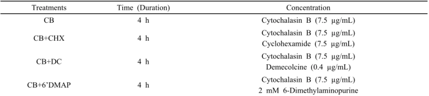
Table 1. Different post-activation treatment groups, time duration (h) and concentrations
Treatments Time (Duration) Concentration
CB 4 h Cytochalasin B (7.5 µg/mL)
CB+CHX 4 h Cytochalasin B (7.5 µg/mL)
Cyclohexamide (7.5 µg/mL)
CB+DC 4 h Cytochalasin B (7.5 µg/mL)
Demecolcine (0.4 µg/mL)
CB+6’DMAP 4 h Cytochalasin B (7.5 µg/mL)
2 mM 6-Dimethylaminopurine
Table 2. Primers with a base pair (bp) used for reverse transcriptase RT-PCR.
No. Gene Name Primer Size (bp)
1 Survivin (F) 5′-GAC GAC GAC CCC ATA GAA GA-3′ 149
2 Survivin (R) 5′-TTT GAC GTT TCT TTC AGG CG-3′ 149
4 GAPDH (F) 5′–TCG GAG TGA ACG GAT TTG-3′ 219
5 GAPDH (R) 5′–CCT GGA AGA TGG TGA TGG-3′ 219
REAGENT (MOLECULAR RESEARCH CENTER, Ohio, USA). Store the homogenate 2-3 min at RT (room temperature).
Supplement with 500 μL chloroform/1mL TRI REAGENT, vigorously shakes by hand for 15 sec spin at 12000 rpm, 4°C at 15 min. Transfer the 60% of colorless upper phase in a clean eppendorf tube. Adding 500 μL 0.5 mL isopropyl alcohol and 20 µg glycogen. Mixed well by hand and store at 4°C overnight.
Centrifuge the store sample 12000 rpm for 10 min, 4°C. Discard the upper fluid slowly and wash with 75% of EtOH of RNA pellet. Following the manufacturer’s instructions RNA were converted to cDNA 20 µL with 10 µL of 2X RT Reaction Solution, 1 μL Enzyme mix solution, 5 μL Template RNA, 4 μ L DNase/RNase free water (cDNA synthesis kit, iNtRON Bio Inc.). The cDNA synthesis was completed reverse transcription at 50°C, 30 min, and RTase inactivation at 95°C, 5 min.
2) RT-PCR
Different numbers of cycles were amplified by using aliquots (1 µg) for target mRNA different conditions of PCR amplification logarithmic phase. RNA degradation possibility was ruled out and different concentration of mRNA was used for gene PCR amplification. The Survivin gene was quantified using 35 cycles. The cDNA extended with 20 µL of PCR reaction supplemented with 2.5 U i-StarTaqTM DNA polymerase, 2.5 mM dNTPs (iNtRON Bio. Inc.) including 10 pmol/µL specific primer. Initially, denaturation at 94°C for 2 min, denatured at 94°C with 20 sec, annealing at 58°C with
10 sec, extended at 72°C with 30 sec and finally extended at 72°C with 5 min. PCR reaction for oligo primers was listed Table 2. By using 1.5 %, agarose gel PCR reactions fractionated and stained by ethidium bromide and illumination under UV light. Pictures were taken, analyzed by Gel Doc EQ system (Bio-Rad Laboratories, Inc.).
7. Experimental Design
Experiment 1: Development of in vitro PA embryos treated with CB (cytochalasin B); CB+CHX (cycloheximide); CB+DC (demecolcine); CB+6’DMAP (6-Dimethylaminopurine) on embryonic development. Experiment 2: Development of in vitro SCNT embryos treated with CB (cytochalasin B);
CB+CHX (cycloheximide); CB+DC (demecolcine); CB+6’
DMAP (6-Dimethylaminopurine) on embryonic development.
Experiment 3: Expression of survivin gene in different SCNT
embryos using RT-PCR. In this study, four different
post-activation media such as CB, CB+CHX, CB+DC, and
CB+6’DMAP compared during post-activation in porcine
embryos. Maturation rates were evaluating after denuding
oocytes with a visible first polar body was used assessment
and mature oocytes taken for the parthenogenetic and SCNT
activation. Then activated embryos post-activated and cultured
in PZM-5 medium for further development. After 2 and 6 days
of incubation, embryos were examined, counted cleavage,
blastocysts rates, and cells number, respectively. Blastocysts
were stained for counting total cell numbers.
Table 3. Effects of post-activation treatments on development of PA embryos with different media of CB, CB+CHX, CB+DC) and CB+6`DMAP on PA porcine embryos in vitro.
Treatment groups No. of embryos cultured
% of embryos developed to*
Blastocysts cell no.
≥2 cells Blastocysts
CB 295 94.7±1.5
a43.5±4.0
a42.3±0.9
aCB+CHX 295 88.1±4.0
ab32.2±5.5
a40.6±0.7
bCB+DC 295 84.3.±3.8
b31.8±3.3
a40.6±0.7
bCB+6`DMAP 295 94.1±1.6
a41.5±2.4
a41.7±0.8
aThe experiment was replicated 8 times (n=8).
*Percentage of the number of embryos cultured.
a, b
Values in the same column with different superscript letters are different (P<0.05).
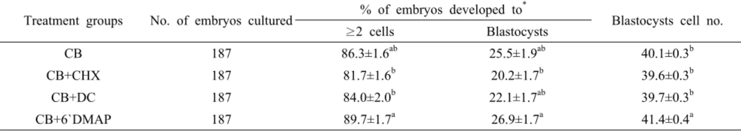
Table 4. Effects of post-activation treatments on development of SCNT embryos with different media of CB, CB+CHX, CB+DC) and CB+6`DMAP on SCNT porcine embryos in vitro.
Treatment groups No. of embryos cultured % of embryos developed to
*Blastocysts cell no.
≥2 cells Blastocysts
CB 187 86.3±1.6
ab25.5±1.9
ab40.1±0.3
bCB+CHX 187 81.7±1.6
b20.2±1.7
b39.6±0.3
bCB+DC 187 84.0±2.0
b22.1±1.7
ab39.7±0.3
bCB+6`DMAP 187 89.7±1.7
a26.9±1.7
a41.4±0.4
aThe experiment was replicated 8 times (n=8).
*Percentage of the number of embryos cultured.
a, b