Exogenous Nitric Oxide Donation During In Vitro Maturation
Improves Embryonic Development after Parthenogenesis
and Somatic Cell Nuclear Transfer in Pigs
Fazle Elahi1, Hyeji Shin1, Joohyeong Lee2, Seung Tae Lee3,
Geun-Shik Lee1,2 and Eunsong Lee1,2,
1College of Veterinary Medicine, Kangwon National University, Gangwon 24341, Korea 2Institute of Veterinary Science, Kangwon National University, Gangwon 24341, Korea 3Division of Applied Animal Science, College of Animal Life Science, Kangwon National
University, Gangwon 24341, Korea
Abstract
Nitric oxide (NO) has an important role in oocyte maturation and embryonic development in mammals. This study examined the effect of exogenous NO donor S-nitroso-N-acetylpenicillamine (SNAP) in a maturation medium on meiotic progression and embryonic development after parthenogenesis (PA) and somatic cell nuclear transfer (SCNT) in pigs. When oocytes were exposed to 0.1 µM SNAP for first 22 h of in vitro maturation (IVM) in Experiment 1, SNAP significantly improved blastocyst development in both defined and standard follicular fluid-supplemented media compared to untreated control (48.4 vs. 31.7-42.5%). SNAP treatment significantly arrested meiotic progression of oocytes at the germinal vesicle stage at 11 h of IVM (61.2 vs. 38.7%). However, there was no effect on meiotic progression at 22 h of IVM (Experiment 2). In Experiment 3, when oocytes were treated with SNAP at 0.001, 0.1 and 10 µM during the first 22 h of IVM to determine a suitable concentration, 0.1 µM SNAP (54.2%) exhibited a higher blastocyst formation than 0 and 10 µM SNAP (36.6 and 36.6%, respectively). Time-dependent effect of SNAP treatment was evaluated in Experiment 4. It was observed that SNAP treatment for the first 22 h of IVM significantly increased blastocyst formation compared to no treatment (57.1% vs. 46.2%). Antioxidant effect of SNAP was compared with that of cysteine. SNAP treatment significantly improved embryonic development to the blastocyst stage (49.1-51.5% vs. 34.4-37.5%) irrespective of the presence or absence of cysteine (Experiment 5). Moreover, SNAP significantly increased glutathione (GSH) content and inversely decreased the reactive oxygen species (ROS) level and mitochondrial oxidative activity in IVM oocytes. SNAP treatment during IVM showed a stimulating effect on in vitro development of SCNT embryos (Experiment 7). These results demonstrates that SNAP improves developmental competence of PA and SCNT embryos probably by maintaining the redox homeostasis through increasing GSH content and mitochondrial quality and decreasing ROS in IVM oocytes.
Received 23 November 2018 Revised 13 December 2018
Accepted 13 December 2018 Key Words : Nitric oxide, SNAP, Oocyte maturation, Embryonic development, Pig
INTRODUCTION
Nitric oxide (NO) plays an important role as a signaling molecule in many biological functions. Specially, NO stimulates the physiological activity of the reproductive system. According to the in vitro and in vivo research, it has been revealed that NO may regulate the endocrine function (Vargas et al. 2007). In mammals, NO is synthesized by NO synthase (NOS), which has three functional isoforms, inducible (iNOS), endothelial eNOS (eNOS) and neuronal NOS (nNOS). The mechanisms regulating meiotic progression during oocyte maturation process are not fully understood yet, but substantial indication shows a contribution of the NO/NOS system in oocyte maturation. Recently, NO is considered as a crucial constituent for the oocyte microenvironment as it influences oocyte maturation and early development to the blastocyst stage (Sengoku et al., 2001, Bergandi et al., 2014). The isoform eNOS and iNOS expression has been found in oocytes of mice (Mitchell et al., 2004), rat (Jablonka-Shariff & Olson, 1998), cattle (Pires et al., 2009), and pigs (Chmelikova et al., 2009) and their presence was confirmed throughout folliculogenesis and follicle maturation (Chmelıkova et al., 2009; Pires et al., 2009) while nNOS is only found in oocytes of mice and pigs (Abe et al., 1999; Chmelıkova et al., 2009). Another study shows that NO has an effect on oocyte maturation in mice (Jablonka-Shariff & Olson, 1998). Inhibition of NO synthesis induces anovulation, increases abnormal oocytes, and the number of blastocysts decreases (Matta et al., 2009) as well as apoptosis increases in preimplantation embryos (Schwarz et al., 2010). Beside this, Goud et al. (2014) have reported that NO increases developmental competence of mouse oocytes. In contrast, high NO levels impair meiotic progression and blastocyst development in cattle (Schwarz et al., 2008) and delay or arrest resumption in the rat (Nakamura et al., 2002; Bu et al., 2004; Sela-Abramovich et al., 2006), cattle (Schwarz et al., 2014), and pigs (Tao et al., 2005), while iNOS-specific inhibitors induce meiotic resumption (Nakamura et al., 2002).
NO has a dual function in reproduction depending on its concentration. At low concentrations, NO positively influences early embryonic development, in contrast, both abundant and insufficient level of NO show negative effects (Barroso et al., 1998; Lee et al., 2013). In mammalian oocytes, a higher dosage of NO arrests meiotic progression during in vitro maturation (IVM) and enhances apoptosis and oxidative stress (Barroso et al., 1998; Jablonka-Shariff & Olson, 2000), while low dosage protects against oxidative stress and modulates meiotic progression (Kuo et
al., 2000; Bu et al., 2003; Tao et al., 2005).
The glutathione (GSH) is an important non-enzymatic antioxidants in cells of mammal and is an essential for maintenance, formation, and protection of the meiotic spindle against oxidative stress (Luberda, 2005). GSH is synthesized during oocyte maturation in the cytosol and stored as a separate redox pools in mitochondria, nucleus and endoplasmic reticulum. Metaphase II (MII) oocytes have higher GSH content and it decreases during the preimplantation development. Generally, GSH is synthesized during oocyte maturation, reaches its lowest concentration in the blastocyst. Changes in intracellular GSH level or EGSH impair embryonic development and can increase apoptosis (Furnus et al., 2008; Li et al., 2011). GSH content or EGSH was significantly decrease in different species after IVM (Somfai et al., 2007; Curnow et al., 2010). Since cumulus cells can influence intra-oocyte GSH contents which synthesize GSH and transfer to the oocyte during IVM (Curnow et al., 2010).
The efficacy of reproductive technologies such as SCNT and in vitro fertilization (IVF) depends on many factors such as the culture conditions, oocyte activation method and quality of oocytes (Mizutani et al., 2006; Whitworth et al., 2009). The quality of oocytes substantially influence the developmental competence from oocytes to early embryonic development. It was hypothesized that increasing NO after supplementing the NO donor like S-Nitroso-N-acetyl-DL-penicillamine (SNAP) IVM media would positively influence oocyte maturation as well as subsequent embryonic development in case of PA and SCNT in pigs. To test this hypothesis, this study investigated the effect of SNAP during IVM on oocyte meiotic progression, intra-oocyte GSH and reactive oxygen species (ROS) content, mitochondrial oxidative activity, and embryonic development to the blastocyst stage after PA and SCNT.
MATERIALS AND METHODS
1. Culture media
All chemicals were purchased from Sigma-Aldrich (St. Louis, MO, USA) unless otherwise specified. The base IVM medium for oocytes was medium-199 (M199; Invitrogen, Grand Island, NY, USA). M199 was added with 0.91 mM pyruvate, 0.6 mM cysteine, 10 ng/ml epidermal growth factor, 1 μg/ml insulin and 75 μg/ml kanamycin and 10% (v/v) porcine follicular fluid (PFF). The in vitro culture (IVC) medium for PA and SCNT embryos was porcine zygote medium-3 (PZM-3) (Yoshioka et
al, 2002), which was supplemented with 0.34 mM trisodium citrate, 2.77 mM myo-inositol, and 10 μM β-mercaptoethanol (You et al, 2012).
2. Oocyte collection and IVM
Pig ovaries were obtained from a local abattoir and then transported to the laboratory in warm physiological saline. The cumulus oocytes complex (COCs) were subsequently aspirated from follicles (3 8 mm in diameter). COCs with multiple layers of compact cumulus cells and uniform ooplasm were selected and washed three times in HEPES-buffered Tyrode's medium containing 0.05% (wt/vol) polyvinyl alcohol (PVA). The COCs were then cultured in of IVM (500 μl) medium in the presence of 10 IU/ml hCG (Intervet International BV, Boxmeer, Holland) and 80 μg/ml FSH (Antrin R-10; Kyoritsu Seiyaku, Tokyo, Japan). COCs were matured at 39°C with 5% CO2 at maximum
humidity for 22 h. For an additional 22 h or 20 h oocytes were cultured in hormone-free IVM medium after washing in fresh hormone-free IVM medium for PA and SCNT, respectively. 3. Somatic cell nuclear transfer (SCNT) and parthenogenesis (PA)
As nuclei donors, porcine fetal fibroblasts were prepared as described previously (Lee et al., 2013a). After IVM for 41 h, the cumulus cells of COCs were dispersed by gentle pipetting in the presence of 0.1% (w/v) hyaluronidase. Oocytes having first polar bodies and uniform ooplasm were selected and stained with 5 µg/ml Hoechst 33342 for 15 min. Oocytes were then washed twice in fresh medium, transferred into a drop of medium containing 5 μg/ml cytochalasin B (CB), and overlaid with warm mineral oil. Enucleation was performed by a 17-µm beveled glass pipette (Humagen, Charlottesville, VA, USA) after aspirating the first polar body (PB) and a small amount of surrounding cytoplasm. The enucleated cytoplasm was then surveyed by epifluorescence microscopy (TE300; Nikon, Tokyo, Japan) to verify that the nuclear material had been removed. A single disaggregated donor cell was injected into the perivitelline space of the enucleated oocytes, after which oocyte cell couplets were placed on a 1-mm fusion chamber overlaid with 1 ml of 280 mM mannitol solution containing 0.001 mM CaCl2 and 0.05 mM MgCl2, as previously described
(Walker et al, 2002; Song et al, 2009). Cell fusion was performed by using an alternating current field of 2 V cycling at 1 MHz for 2 seconds, followed by two pulses of 170 V/mm direct current (DC) for 30 μsec using a cell fusion generator
(LF101; NepaGene, Chiba, Japan). The oocytes were then incubated for 1 h in TLH-BSA, after which they were assessed for confirmation of fusion under a stereomicroscope. The nuclear transferred oocytes were activated with two pulses of 120 V/mm DC for 60 μsec in a 280 mM mannitol solution containing 0.05 mM MgCl2 and 0.1 mM CaCl2. For PA, MII oocytes were
activated as described in SCNT procedures. 4. Post-activation and embryo culture
After electrical activation, the PA were cultured with 5 μ g/mL CB and SCNT embryos were treated with 0.4 μg/ml demecolcine combined with 1.9 mM 6-dimethylaminopurine in IVC medium for 4 h. Afterward, the embryos were washed three times in fresh IVC medium, cultured into 30 μl of IVC medium droplets under mineral oil, at 39°C in a humidified atmosphere of 5% O2, 5% CO2, and 90% N2 for 7 days.
Cleavage and blastocyst formation were evaluated on Days 2 and 7, respectively. The day of SCNT or PA was designated as Day 0. The total cell count in blastocysts was performed using Hoechst 33342 staining and visualized under an epifluorescence microscope.
5. Measurement of GSH and ROS contents
After 24 h of IVC, PA embryos were utilized for intra-oocyte GSH and ROS level determination. The GSH and ROS contents were measured as previously described (Sakatani et al., 2007). Briefly, (4-chloromethyl-6.8-difluoro-7-hydroxycoumarin, Invitrogen) Cell-Tracker Blue and (2’,7’-dichlorodihydrofluorescein diacetate; Invitrogen) H2DCFDA were used to detect intra-oocyte GSH and ROS with blue fluorescence and green fluorescence for GSH and ROS, respectively. A group of 7 10 denuded oocytes from each treatment group were cultured for 30 min in TLH-PVA supplemented with 10 μM Cell-Tracker and10 μM H2DCFDA and in the dark. Oocytes treated with Cell- Tracker were then incubated for 30 min with PZM-3 supplemented with 0.3% (w/v) BSA at 39°C in the dark. Following incubation, oocytes were washed with Dulbecco’s phosphate-buffered saline (D-PBS; Invitrogen) containing 0.01% (w/v) PVA, placed into 2-μl droplets. Fluorescence was observed under an epifluorescence microscope (TE300; Nikon) with ultraviolet ray filters at 370 and 460 nm for GSH and ROS, respectively. The fluorescence intensities of oocytes were examined by above mentioned and normalized against the untreated control oocytes.
6. Determination of mitochondrial oxidative activity in oocytes Denuded oocytes were incubated in M199 containing 200 nM Mitotracker Orange CM-H2-TMRos (Molecular Probes, Eugene,
OR, USA) for 40 min at 39°C in the dark. After washing three times in the fresh M199 medium, oocytes were examined under an inverted epifluorescence microscope (TE300; Nikon). Fluorescence signals were captured with a digital camera (DS-L3; Nikon), and normalized against the untreated control oocytes.
7. Examination of nuclear status of oocytes
Nuclear status was evaluated as previously described (Lee et al., 2016). Briefly, oocytes were denuded to assess their nuclear progression, mounted onto glass slides, and fixed in acetic acid in ethanol. Then, oocytes were stained with orcein, and the nuclear status was classified into germinal vesicle (GV), GV breakdown (GVBD), MI and MII stages according to the method previously described (Hunter and Polge, 1966). 8. Experimental design
In Experiment 1, oocytes were exposed to 0.1 μM SNAP in a defined or standard medium containing PVA and PFF, respectively, for the first 22 h of IVM and for another 22 h without SNAP and then effect of SNAP on nuclear maturation and embryonic development after PA were examined. This concentration was selected from a previous study in bovine (Katia et al., 2014). In Experiment 2, nuclear status was measured at 11 and 22 h of IVM after SNAP treatment for 22 h during IVM. Oocytes were treated with the SNAP at 0.001, 0.1 and 10 µM during the first 22 h of IVM following 22 h without SNAP to determine a suitable concentration for pigs in Experiment 3. Effect of time-depend treatment with SNAP was evaluated by treating oocytes for the first 22 h, the second 22
h, and whole 44 h of IVM in Experiment 4. Antioxidant effect of SNAP on oocyte maturation and blastocyst formation after PA was compared that of cysteine in Experiment 5. As indicators for cytoplasmic maturation of oocytes, GSH and ROS levels in IVM oocytes were analyzed at 44 h of IVM after treating oocytes with SNAP at 0.1 µM in Experiment 6. Finally, effect of SNAP treatment during IVM on developmental competence of SCNT embryos was determined in Experiment 7. 9. Statistical analysis
Statistical analyses were executed by the Statistical Analysis System (version 9.3; SAS Institute, Cary, NC, USA). The general linear model procedure followed by the least significant difference mean separation procedure when the treatments differed at p < 0.05. The percentage data were arcsine transformed before analysis. The results are present as the mean ± standard error of the mean (SEM).
RESULTS
1. Effects of SNAP in maturation medium with PVA or PFF on embryonic development after PA
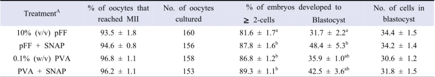
Immature oocytes were treated with SNAP at 0.1 µM in medium containing PVA and FF for the first 22 h of IVM. The results showed that SNAP treatment significantly increased cleavage (81.6% vs. 87.8%) and blastocyst formation (31.7% vs. 48.4%) only in PFF-supplemented maturation medium while not influenced in PVA-supplemented medium (86.8 and 89.3% of cleavage and 35.9% and 42.5% of blastocyst formation in control and SNAP treatment, respectively) (Table 1). Nuclear
TreatmentA % of oocytes that
reached MII
No. of oocytes cultured
% of embryos developed to No. of cells in blastocyst 2-cells Blastocyst 10% (v/v) pFF 93.5 ± 1.8 160 81.6 ± 1.7a 31.7 ± 2.2a 34.4 ± 1.5 pFF + SNAP 94.6 ± 0.8 156 87.8 ± 1.6b 48.4 ± 5.3b 34.2 ± 1.4 0.1% (w/v) PVA 96.8 ± 1.1 158 86.8 ± 1.2b 35.9 ± 1.0ab 30.6 ± 1.2 PVA + SNAP 96.2 ± 1.1 153 89.3 ± 1.1b 42.5 ± 3.6ab 31.8 ± 1.5 Five replicates.
AOocytes were untreated or treated with 0.1 µM SNAP for 0-22 h of in vitro maturation. abValues with different superscripts denote difference within the same column (p < 0.05).
Table 1. Effect of SNAP in a maturation medium containing pig follicular fluid (pFF) or polyvinyl alcohol (PVA) on embryonic development after parthenogenesis
maturation of oocytes and mean cell number of blastocysts were not influenced by the SNAP treatment.
2. Effect of SNAP treatment on nuclear status of oocytes at 11 and 22 h of IVM
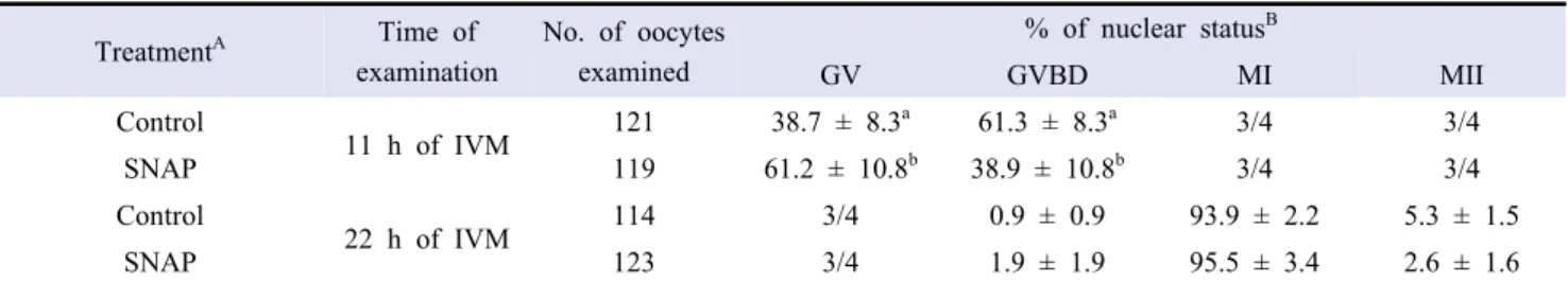
Effect of SNAP on nuclear progression during IVM was evaluated. It was revealed that SNAP significantly (p < 0.05) increased meiotic arrest at the GV stage (38.7 vs 61.2%) and significantly decreased the proportion of oocytes at the GVBD (61.3 vs 38.9%) at 11 h of IVM compared to control. However, nuclear status at 22 h of IVM was not influenced by the SNAP treatment (Table 2).
3. Effects of SNAP treatment at various doses during IVM on embryonic development after PA
To determine the optimal concentration of SNAP, oocytes were matured under 0.001, 0.1 and 10 µM SNAP for the first 22 h of IVM. It revealed that 0.1 μM SNAP significantly (p < 0.05) increased embryonic development to the blastocyst stage than control and other concentrations of SNAP (36.6-48.2 vs 54.2%). However, oocyte maturation, embryo cleavage, and cell number in blastocyst were not different among the tested groups (Table 3).
4. Effects of SNAP treatment at various stages of IVM on embryonic development after PA
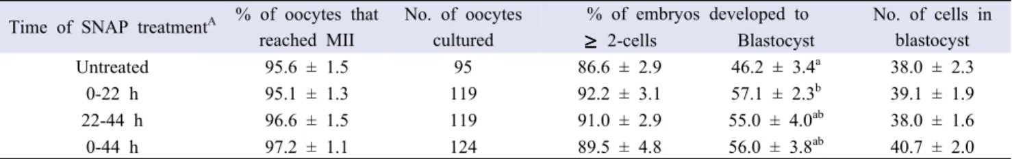
When oocytes were untreated or treated with SNAP for the first 22 h, the second 22 h, and whole period (44 h) of IVM, SNAP treatment for the first 22 h significantly improved (p < 0.05) blastocyst development compared to control, treatments for the second 22 h, and for whole IVM period (46.2 vs 57.1%). However, nuclear maturation, cleavage, and average cell per blastocyst were not altered in all groups (Table 4). 5. SNAP treatment with or without cysteine in a chemically
defined medium during IVM on embryonic development after PA
Antioxidant activity of SNAP was evaluated by comparing with cysteine. For this oocytes were treated with control, SNAP with or without cysteine in PVA-supplemented defined IVM medium. It was observed that SNAP with or without cysteine significantly (p < 0.05) improved cleavage and blastocyst formation compared to other treatments (34.4-37.5 vs 49.1-51.5%). However, there was no significant difference in maturation and cell per blastocyst among the groups tested (Table 5).
TreatmentA Time of examination No. of oocytes examined % of nuclear statusB GV GVBD MI MII Control 11 h of IVM 121 38.7 ± 8.3 a 61.3 ± 8.3a 3/4 3/4 SNAP 119 61.2 ± 10.8b 38.9 ± 10.8b 3/4 3/4 Control 22 h of IVM 114 3/4 0.9 ± 0.9 93.9 ± 2.2 5.3 ± 1.5 SNAP 123 3/4 1.9 ± 1.9 95.5 ± 3.4 2.6 ± 1.6 Three replicates.
AOocytes were treated with 0.1 µM SNAP for 0-22 h of IVM in medium containing 10% (v/v) porcine follicular fluid. BGV, germinal vesicle; GVBD, GV break down; MI, metaphase I; MII, metaphase II.
abValues with different superscripts denote difference within the same column (p < 0.05).
Table 2. Effect of SNAP treatment on nuclear status oocytes at 11 and 22 h of in vitro maturation (IVM)
SNAP (µM) treatmentA % of oocytes that
reached MII
No. of oocytes cultured
% of embryos developed to No. of cells in blastocyst 2-cells Blastocyst Control (0) 93.1 ± 2.4 123 81.9 ± 4.2 36.6 ± 4.1a 35.9 ± 2.2 0.001 92.8 ± 1.6 127 86.3 ± 4.2 48.2 ± 2.9ab 37.7 ± 2.0 0.1 92.9 ± 1.3 116 90.8 ± 2.6 54.2 ± 6.5b 37.7 ± 2.2 10 92.5 ± 2.8 121 90.6 ± 3.4 36.6 ± 5.1a 34.8 ± 2.1 Four replicates.
AOocytes were treated with SNAP for 0-22 h of IVM in medium containing 10% (v/v) porcine follicular fluid. abValues with different superscripts denote difference within the same column (p < 0.05).
6. Effect of SNAP treatment on GSH content, ROS level and mitochondrial oxidative activity of oocytes after IVM This experiment was performed to detect the GSH, ROS and mitochondrial oxidative activity of oocytes after SNAP treatment during IVM. SNAP significantly increased the GSH level while decreased the ROS level and mitochondrial oxidative activity compared to control (p < 0.05; Table 6).
7. Effect of SNAP treatment during 0-22 h of IVM on embryonic development after SCNT
Effect of SNAP treatment during IVM on in vitro development of SCNT pig embryos was determined. The result indicated that SNAP had no effect on oocyte-cell fusion, embryo cleavage, and total cell number in blastocyst after SCNT while did show a significant (p < 0.05) effect on blastocyst formation when SNAP-treated oocytes were used as cytoplasts for SCNT (26.6 vs 37.0%) (Table 7).
TreatmentA No. of oocytes
examined GSH content (pixel/oocyte) No. of oocytes examined ROS level (pixel/oocyte) No. of oocytes examined Mitochondrial oxidative activity (pixel/oocyte) Control 58 1.00 ± 0.07a 40 1.00 ± 0.08a 56 1.00 ± 0.05a SNAP 58 1.23 ± 0.05b 36 0.79 ± 0.06b 58 0.87 ± 0.03b
AOocytes were treated with SNAP for 0-22 h of IVM in medium containing 10% (v/v) porcine follicular fluid. abValues with different superscripts denote difference within the same column (p < 0.05).
Table 6. Effect of SNAP treatment during in vitro maturation (IVM) on glutathione (GSH), reactive oxygen species (ROS), and mitochondrial oxidative activity in IVM oocytes
Time of SNAP treatmentA % of oocytes that
reached MII
No. of oocytes cultured
% of embryos developed to No. of cells in blastocyst 2-cells Blastocyst Untreated 95.6 ± 1.5 95 86.6 ± 2.9 46.2 ± 3.4a 38.0 ± 2.3 0-22 h 95.1 ± 1.3 119 92.2 ± 3.1 57.1 ± 2.3b 39.1 ± 1.9 22-44 h 96.6 ± 1.5 119 91.0 ± 2.9 55.0 ± 4.0ab 38.0 ± 1.6 0-44 h 97.2 ± 1.1 124 89.5 ± 4.8 56.0 ± 3.8ab 40.7 ± 2.0 Four replicates.
AOocytes were treated with 0.1 µM SNAP at various stages of IVM in medium containing 10% (v/v) porcine follicular fluid. abValues with different superscripts denote difference within the same column (p < 0.05).
Table 4. Effect of SNAP treatment at various stages of in vitro maturation (IVM) on embryonic development after parthenogenesis
TreatmentA % of oocytes
that reached MII
No. of oocytes cultured
% of embryos developed to No. of cells in blastocyst
Cysteine SNAP 2-cells Blastocyst
No No 95.7 ± 0.9 215 90.0 ± 2.1ab 34.4 ± 2.6a 34.9 ± 1.5
Yes No 95.9 ± 0.6 197 88.3 ± 1.4a 37.5 ± 2.0a 35.1 ± 1.2
No Yes 96.5 ± 1.3 202 93.9 ± 2.5b 51.5 ± 4.3b 37.8 ± 1.2
Yes Yes 96.5 ± 1.3 204 95.8 ± 1.1b 49.1 ± 2.0b 35.0 ± 1.2
Six replicates.
AOocytes were treated with SNAP for 0-22 h of IVM in medium containing 0.1% (w/v) polyvinyl alcohol. abValues with different superscripts denote difference within the same column (p < 0.05).
Table 5. Effect of SNAP treatment with or without cysteine during in vitro maturation (IVM) on embryonic development after parthenogenesis
TreatmentA % of oocytes fused No. of oocytes
cultured
% of embryos developed to No. of cells in blastocyst
2-cells Blastocyst
Control 77.1 ± 6.5 157 87.0 ± 2.0 26.6 ± 2.0a 39.7 ± 2.3
SNAP 75.9 ± 4.1 163 79.7 ± 7.7 37.0 ± 2.4b 39.6 ± 2.1
Four replicates.
AOocytes were treated with SNAP for 0-22 h of IVM in medium containing 10% (v/v) porcine follicular fluid. abValues with different superscripts denote difference within the same column (p < 0.05).
DISCUSSION
NO is present in the follicular fluid (Basini et al., 2014). To determine the effect of exogenous NO donor during IVM, oocytes were matured in medium supplemented with PFF or in a defined medium with PVA. SNAP in both PFF- and PVA-supplemented media significantly improved blastocyst development after PA, while there was no difference nuclear maturation, embryonic development, and blastocyst cell number between PFF and PVA controls. Thus, it was performed a next experiment to determine the optimal concentration for pigs using PFF-supplemented medium. Immature oocytes were exposed for the first 22 h of IVM and following without SNAP at 0.001, 0.1 and 10 µM concentration. It showed a significant increase in blastocyst development at 0.1 µM SNAP after PA. This result was agreed with a previous study in bovine (Katia et al., 2014).
NO has been revealed a biphasic function on oocyte meiosis in mice depending on its concentration. Abbasi et al. (2012) have suggested that the key of the meiotic arrest at the first meiotic division is the concentrations of cyclic adenosine-3' ,5'-monophosphate (cAMP) and cyclic guanosine monophosphate (cGMP) in the preovulatory follicle, since NO act as a cGMP modulator and a sensitive guanylcyclases (Bilodeau- Goeseels et al., 2007). Moreover, it can stimulate non-cGMP pathway like protein S-nitrosylation (SN) (Ji et al., 1999; Zhang & Hogg, 2005). Beside this, it has been shown that a high NO level halts meiotic progression, while a low NO level initiates the resumption of meiosis. In this study, the proportion of oocytes arrested at the GV stage was significant higher at 11 h of IVM in 0.1 µM SNAP treatment than in control, while nuclear progression to the GVBD, MI, MII stages were not influenced by the SNAP treatment for the first 22 h of IVM. It seems that a lower concentration of SNAP arrests the meiosis 11 h of treatment through the cAMP and cGMP pathway and this arrest is reversible. In contrast in bovine study, NO has not influenced meiotic resumption through cGMP pathway (Bilodeau- Goeseels et al., 2007). In addition, protein S-nitrosylation has been occurred in mouse embryo during IVC (Lee et al., 2013b).
Cysteine acts as an antioxidant during IVM of mammalian oocytes. When antioxidant effect of SNAP was compared with cysteine, SNAP with or without cysteine resulted in a higher embryonic development than no treatment and cysteine only. In addition, SNAP treatment significantly decreased the ROS level of oocytes. Although it was not clear how SNAP increased GSH and decreased ROS level, our results indicates that SNAP
acts as an antioxidant and alleviate the harmful action of ROS during cytoplasmic maturation of pig oocytes. Oocytes or embryos are inevitably prone to be exposed to oxidative stress during IVM and IVC. ROS can be produced due to handling of oocytes or embryos in vitro by being exposed to a high-oxygen atmosphere and activations such as electric stimulus for cell fusion or activation of SCNT oocytes and resultantly ROS level increases (Fujitani et al. 1997; Koo et al., 2008). High level of ROS influences lipid peroxidation and DNA fragmentation, and also effects RNA protein synthesis (Takahashi et al., 2000). These factors may impair the in vitro developmental competence and decrease the early embryonic death. In addition, ROS play a detrimental role in embryonic development though minimal level of ROS is needed for the better blastocyst development. Our finding was similar with the previous results that NO has a capacity to protect mammalian cells against oxidative stress (Kuo et al., 1996).
Mitochondria is an important cytoplasmic organelle for metabolism and respiration within a cell and has shown an influence on the oocyte maturation and embryonic development (Van Blerkom, 2004). Mitochondrial oxidative activity was significantly higher in control than SNAP-treated oocytes in this study. This result is in accordance with the previous results in human (Moawad et al., 2014) and pigs (Torner et al., 2004). Moawad et al. (2014) also confirmed that a high mitochondrial oxidative activity subsequently increased ROS level in oocytes. Beside this, cumulus apoptosis was influenced by the mitochondrial oxidative activity in pig (Torner et al., 2004). NO and NO donors can initiate S-nitrosylation of proteins and also accelerate S-nitrosylation in the endoplasmic reticulum and mitochondria. Protein S-nitrosylation controls the metabolic enzymes activity such as phosphatases, oxidoreductases, proteases and protein kinases in vitro and in vivo, as well as respiratory proteins, structural components and cytoskeletal, transcription factors, G proteins (Hess et al., 2005; Jaffrey et al., 2001). In this study, SNAP treatment during IVM effectively increased intra-oocyte GSH content, decreased ROS level, and also decreased the mitochondrial oxidative activity. This effect of SNAP might create better homeostatic microenvironments desirable for in vitro development of PA and SCNT embryos. In summary, it has been demonstrated in this study that exogenous NO donor SNAP treatment during the early stage of IVM improves embryonic development after PA and SCNT by maintaining the redox homeostasis as well as microenvironment of treated oocytes and thus stimulates early development of PA and SCNT embryos in pigs.
ACKNOWLEDGMENTS
This research was supported by Basic Science Research Program through the National Research Foundation of Korea (NRF) funded by the Ministry of Science, ICT and Future Planning (Grant No. 2015R1A2A2A01005490).
REFERENCES
Abbasi M, Akbari M, Amidi F, RagerdiKashani I, Mahmoudi R, Sobhani A, Takzare N, Pasbakhsh P, BarbarestaniM, Abolhassani F, Sato E. 2009. Nitric oxide acts through different signalling pathways in maturation of cumulus cell-enclosed mouse oocytes. DARU J. Pharm. Sci. 17: 8-52. Abe K, Matsuoka K, Inoue N, Taga M, Kato T. 1999. Messenger
RNA of neuronal nitric oxide synthase is expressed and possibly functions in mouse oocytes and embryos during preimplantation development. Biomedical Research 20: 61-65. Barroso RP, Osuamkpe C, Nagamani M, Yallampalli C. 1998.
Nitric oxide inhibits development of embryos and implantation in mice. Mol. Hum. Reprod. 4: 503-507.
Basini G, Bianchi F, Bussolati S, Baioni L, Ramoni R, Grolli S, Conti V, Bianchi F, Grasselli F. 2012. Atrazine disrupts steroidogenesis, VEGF and NO production in swine granulosa cells. Ecotoxicol. Environ. Saf. 85: 59-63. Bergandi L, Basso G, Evangelista F, Canosa S, Dalmasso P,
Aldieri E, Revelli A, Benedetto C, Ghigo D. 2014. Inducible nitric oxide synthase and heme oxygenase 1 are expressed in human cumulus cells and may be used as biomarkers of oocyte competence. Reprod. Sci. 21: 1370-1377.
Bilodeau-Goeseels S. 2007. Effects of manipulating the nitric oxide/cyclic GMP pathway on bovine oocyte meiotic resumption in vitro. Theriogenology 68: 693-701.
Bu S, Xia G, Tao Y, Lei L, Zhou B. 2003. Dual effects of nitric oxide on meiotic maturation of mouse cumulus cell-enclosed oocytes in vitro. Mol. Cell. Endocrinol. 207: 21-30. Bu S, Xie H, Tao Y, Wang J, Xia G. 2004. Nitric oxide
influences the maturation of cumulus cell-enclosed mouse oocytes cultured in spontaneous maturation medium and hypoxanthine-supplemented medium through different signaling pathways. Mol. Cell. Endocrinol. 223: 85-93.
Chmelikova E, Sedmıkova M, Petr J, Kott T, Lanska V, Tumova L, Tichovska H, Jeseta M. 2009. Expression and localization of nitric oxide synthase isoforms during porcine
oocyte growth and acquisition of meiotic competence. Czech. J. Anim. Sci. 54:137 149.
Curnow EC, Ryan JP, Saunders DM, Hayes ES. 2010. Developmental potential of bovine oocytes following IVM in the presence of glutathione ethyl ester. Reprod. Fertil. Dev. 22: 597-605.
Fujitani Y, Kasai K, Ohtani S, Nishimura K, Yamada M, Utsumi K. 1997. Effect of oxygen concentration and free radicals on in vitro development of in vitro produced bovine embryos. J. Anim. Sci. 75: 483 489.
Furnus CC, de Matos DG, Picco S, Garcia PP, Inda AM, Mattioli G, Errecalde AL. 2008. Metabolic requirements associated with GSH synthesis during in vitro maturation of cattle oocytes. Anim. Reprod. Sci. 109: 88-99.
Goud PT, Goud AP, Najafi T, Gonik B, Diamond MP, Saed GM, Zhang X, Abu-Soud HM. 2014. Direct real-time measurement of intra-oocyte nitric oxide concentration in vivo. PLoS One 9: e98720.
Hess DT, Matsumoto A, Kim SO, Marshall HE, Stamler JS. 2005. Protein S-nitrosylation: purview and parameters. Nat. Rev. Mol. Cell. Biol. 6: 150-166.
Hunter RH, Polge C. 1966. Maturation of follicular oocytes in the pig after injection of human chorionic gonadotrophin. J. Reprod. Fertil. 12: 525-531.
Jablonka-Shariff A, Olson LM. 1998. The role of nitric oxide in oocyte meiotic maturation and ovulation: meiotic abnormalities of endothelial nitric oxide synthase knock-out mouse oocytes. Endocrinology 139: 2944-2954.
Jablonka-Shariff A, Olson LM. 2000. Nitric oxide is essential for optimal meiotic maturation of murine cumulus-oocyte complexes in vitro. Mol. Reprod. Dev. 55: 412-421. Jaffrey SR, Erdjument-Bromage H, Ferris CD, Tempst P,
Snyder SH. 2001. Protein S-nitrosylation: a physiological signal for neuronal nitric oxide. Nat. Cell Biol. 3: 193-197. Ji Y, Akerboom TP, Sies H, Thomas JA.1999. S-nitrosylation
and S-glutathiolation of protein sulfhydryls by S-nitroso glutathione. Arch. Biochem. Biophys. 362: 67-78.
Katia RLS, Pedro RLP, Ligia GM, Marcos RC, Cláudia LVL. 2014. Effect of nitric oxide on the cyclic guanosine monophosphate (cGMP) pathway during meiosis resumption in bovine oocytes. Theriogenology, 81: 556-564
Koo OJ, Jang G, Kwon DK, Kang JT, Kwon OS, Park HJ, Kang SK, Lee BC. 2008. Electrical activation induces reactive oxygen species in porcine embryos. Theriogenology 70: 1111 1118. Kuo RC, Baxter GT, Thompson SH, Stricker SA, Patton C,
Bonaventura J, Epel D. 2000. NO is necessary and sufficient for egg activation at fertilization. Nature 406: 633-636. Kuo PC, Abe KY, Schroeder RA. 1996. Interleukin-1-induced
nitric oxide production modulates glutathione synthesis in cultured rat hepatocytes. Am. J. Physiol., 271: 851-862. Lee J, You J, Lee GS, Hyun SH, Lee E. 2013a. Pig oocytes
with a large perivitelline space matured in vitro show greater developmental competence after parthenogenesis and somatic cell nuclear transfer. Mol. Reprod. Dev. 80:753-762. Lee TH, Lee MS, Huang CC, Tsao HM, Lin PM, Ho HN, Shew
JY, Yang YS. 2013b. Nitric oxide modulates mitochondrial activity and apoptosis through protein S-nitrosylation for preimplantation embryo development. Journal of assisted reproduction and genetics 30: 1063-1072.
Lee Y, Lee H, Park B, Elahi F, Lee J, Lee ST, Park CK, Hyun SH, Lee E. 2016. Alpha-linolenic acid treatment during oocyte maturation enhances embryonic development by influencing mitogen-activated protein kinase activity and intraoocyte glutathione content in pigs. J. Anim. Sci. 94: 3255-3263. Li Q, Miao DQ, Zhou P, Wu YG, Gao D, Wei DL, Cui W,
Tan JH. 2011. Glucose metabolism in mouse cumulus cells prevents oocyte aging by maintaining both energy supply and the intracellular redox potential. Biol. Reprod. 84: 1111-1118.
Luberda Z. 2005. The role of glutathione in mammalian gametes. Reprod. Biol. 5: 5-17.
Matta SG, Caldas-Bussiere MC, Viana KS, Faes MR, Paes de Carvalho CS, Dias BL, Quirino CR. 2009. Effect of inhibition of synthesis of inducible nitric oxide synthase-derived nitric oxide by aminoguanidine on the in vitro maturation of oocyte- cumulus complexes of cattle. Anim. Reprod. Sci. 111: 189-201. Mizutani E, Ohta H, Kishigami S, Van Thuan N, Hikichi T,
Wakayama S, Kosaka M, Sato E, Wakayama T. 2006. Developmental ability of cloned embryos from neural stem cells. Reproduction 132: 849-857.
Moawad AR, Xu B, Tan SL, Taketo T. 2014. L-carnitine supplementation during vitrification of mouse germinal vesicle stage-oocytes and their subsequent in vitro maturation improves meiotic spindle configuration and mitochondrial distribution in metaphase II oocytes. Hum. Reprod. 29: 2256-2268. Nakamura Y, Yamagata Y, Sugino N, Takayama H. Kato H.
2002. Nitric oxide inhibits oocyte meiotic maturation. Biol. Reprod. 67: 1588-1592.
Pires PR, Santos NP, Adona PR, Natori MM, Schwarz KR, de Bem TH, Leal CL. 2009. Endothelial and inducible nitric
oxide synthases in oocytes of cattle. Animal reproduction science 116: 233-243.
Sakatani M, Suda I, Oki T, Kobayashi S, Kobayashi S, Takahashi M. 2007. Effects of purple sweet potato anthocyanins on development and intracellular redox status of bovine preimplantation embryos exposed to heat shock. J. Reprod. Dev. 53: 605-614.
Schwarz KR, Pires PR, Adona PR, Camara de Bem TH, Leal CL. 2008. Influence of nitric oxide during maturation on bovine oocyte meiosis and embryo development in vitro. Reprod. Fertil. Dev. 20: 529-536.
Schwarz KR, Pires PR, Mesquita LG, Chiaratti MR, Leal CL. 2014. Effect of nitric oxide on the cyclic guanosine monophosphate (cGMP) pathway during meiosis resumption in bovine oocytes. Theriogenology 81: 556-564.
Sela-Abramovich S, Edry I, Galiani D, Nevo N, Dekel N. 2006. Disruption of gap junctional communication within the ovarian follicle induces oocyte maturation. Endocrinology 147: 2280-2286.
Sengoku K, Takuma N, Horikawa M, Tsuchiya K, Komori H, Sharifa D, Tamate K, Ishikawa M. 2001. Requirement of nitric oxide for murine oocyte maturation, embryo development, and trophoblast outgrowth in vitro. Molecular reproduction and development 58: 262-268.
Somfai T, Ozawa M, Noguchi J, Kaneko H, Kuriani Karja NW, Farhudin M, Dinnyes A, Nagai T, Kikuchi K. 2007. Developmental competence of in vitro-fertilized porcine oocytes after in vitro maturation and solid surface vitrification: effect of cryopreservation on oocyte antioxidative system and cell cycle stage. Cryobiology 55: 115-126.
Song K. Hyun SH, Shin T, Lee E. 2009. Post-activation treatment with demecolcine improves development of somatic cell nuclear transfer embryos in pigs by modifying the remodeling of donor nuclei. Mol. Reprod. Dev. 76: 611-619.
Takahashi M, Keicho K, Takahashi H, Ogawa H, Schultz RM, Okano A. 2000. Effect of oxidative stress on development and DNA damage in in vitro cultured bovine embryos by comet assay. Theriogenology 54: 137-145.
Tao Y, Xie H, Hong H, Chen X, Jang J, Xia G. 2005. Effects of nitric oxide synthase inhibitors on porcine oocyte meiotic maturation. Zygote 13: 1-9.
Torner H, Brüssow KP, Alm H, Ratky J, Pöhland R, Tuchscherer A, Kanitz W. 2004. Mitochondrial aggregation n patterns and activity in porcine oocytes and apoptosis in surrounding cumulus cells depends on the stage of pre-ovulatory
maturation, Theriogenology 61: 1675-1689.
Van Blerkom J. 2008. Mitochondria as regulatory forces in oocytes, preimplantation embryos and stem cells. Reprod. Biomed. Online 16: 553-569.
Vargas F, Moreno JM, Wangensteen R, Rodriguez-Gomez I, Garcia-Estan J. 2007. The endocrine system in chronic nitric oxide deficiency. European Journal of Endocrinology 156: 1-12. Walker SC, Shin T, Zaunbrecher GM, Romano JE, Johnson
GA, Bazer FW, Piedrahita JA. 2002. A highly efficient method for porcine cloning by nuclear transfer using in vitro-matured oocytes. Cloning Stem cells 4: 105-112. Whitworth KM, Li R, Spate LD, Wax DM, Rieke A, Whyte JJ,
Manandhar G, Sutovsky M, Green JA, Sutovsky P, Prather
RS. 2009. Method of oocyte activation affects cloning efficiency in pigs. Mol. Reprod. Dev. 76: 490-500. Yoshioka K, Suzuki C, Tanaka A, Anas IM, Iwamura S. 2002.
Birth of piglets derived from porcine zygotes cultured in a chemically defined medium. Biol. Reprod. 66: 112-119. You J, Lee J, Hyun SH, Lee E. 2012. L-carnitine treatment
during oocyte maturation improves in vitro development of cloned pig embryos by influencing intracellular glutathione synthesis and embryonic gene expression. Theriogenology 78: 235-243.
Zhang Y, Hogg N. 2005. S-Nitrosothiols: cellular formation and transport. Free Radic. Biol. Med. 38: 831-838.