• 검색 결과가 없습니다.

Effect of L-Glutathione Treatment during Somatic Cell Nuclear Transfer Procedures on the Subsequent Embryonic Development and DNA Methylation Status of Cloned Bovine Embryos

N/A
N/A
Protected

Academic year: 2021

Share "Effect of L-Glutathione Treatment during Somatic Cell Nuclear Transfer Procedures on the Subsequent Embryonic Development and DNA Methylation Status of Cloned Bovine Embryos"

Copied!
6
0
0

로드 중.... (전체 텍스트 보기)

전체 글

(1)

Effect of L-Glutathione Treatment during Somatic Cell Nuclear Transfer Procedures on the Subsequent Embryonic Development and DNA Methylation

Status of Cloned Bovine Embryos

Hyo-Kyung Bae

1

, Nam-Sik Yoon

1

, In-Sun Hwang

1

, Choon-Keun Park

2

, Boo-Keun Yang

2

and Hee-Tae Cheong

1,

1

College of Veterinary Medicine and Institute of Veterinary Science, Kangwon National University, Chuncheon 200-701, Korea

2

College of Animal Life Science, Kangwon National University, Chuncheon 200-701, Korea

ABSTRACT

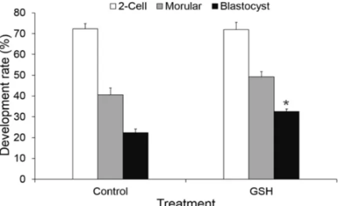
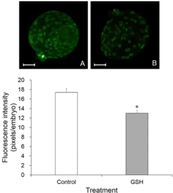
We investigate the effect of L-glutathione (GSH), an antioxidant, treatment during the somatic cell nuclear transfer (SCNT) procedures on the in vitro development and DNA methylation status of bovine SCNT embryos. Bovine in vitro matured (IVM) oocytes were enucleated and electrofused with a donor cell, then activated by a combination of Ca-ionophore and 6-dimethylaminopurine. The recipient oocytes or reconstituted oocytes were treated with 50 μM GSH during these SCNT procedures from enucleation to activation treatment. The SCNT embryos were cultured for 7 days to evaluate the in vitro development, apoptosis and DNA methylation in blastocysts. The apoptosis was measured by TUNEL assay and caspase-3 activity assay. Methylated DNA of SCNT embryos at the blastocyst stages was detected using a 5-methylcytidine (5-MeC) antibody. The developmental rate to the blastocyst stage was significantly higher (P<0.05) in GSH treatment group (32.5±1.2%, 78/235) than that of non-treated control SCNT embryos (22.3±1.8%, 50/224). TUNEL assay revealed that the numbers of apoptotic cells in GSH treatment group (2.3±0.4%) were signi- ficantly lower (P<0.05) than that of control (3.8±0.6%). Relative caspase-3 activity of GSH treated group was 0.8±0.06 fold compared to that of control. DNA methylation status of blastocysts in GSH treatment group (13.1±0.5, pixels/

embryo) was significantly lower (P<0.05) than that of control (17.4±0.9, pixels/embryo). These results suggest that antioxidant GSH treatment during SCNT procedures can improve the embryonic development and reduce the apoptosis and DNA methylation level of bovine SCNT embryos, which may enhance the nuclear reprogramming of bovine SCNT embryos.

(Key words : somatic cell nuclear transfer, GSH, in vitro development, apoptosis, DNA-methylation)

*

This study was supported by 2014 Research Grant from Kangwon National University (No. C1010767-01-01).

Correspondence : [email protected]

INTRODUCTION

The physical cellular stress derived from micromanipulation is regarded as one of the most critical factors affecting the low efficiency of somatic cell nuclear transfer (SCNT) embryos generating excessive reactive oxygen species (ROS) (Hwang et al., 2012). The excessive level of ROS can alter the cellular molecules, which may result in serious damages of the mito- chondrial alterations, DNA fragmentation, abnormal metabolism, apoptosis, embryo cell block and epigenetic abnormality (Hal- liwell and Aruoma, 1991; Raha and Robinson, 2000; Kitagawa et al., 2004; Rhoads et al., 2006).

Antioxidant enzymes such as L-ascorbic acid (Vit. C, Tate- moto et al., 2004), α-tocopherol (Vit. E, Olson and Seidel,

2000), β-mercaptoethanol (β-ME, Nedambale et al., 2006),

GSH (Salmen et al., 2005) and superoxide dismutase (SOD,

Tatemoto et al., 2004) have been used for reduction of ROS

in various species. Out of these, GSH is intracellular free thiol

compound, and protects cells from ROS toxicity and regulates

the intracellular redox balance (Lafleur et al., 1994). Intracel-

lular level of GSH was known to be an important factor that

influences oocyte maturation and the subsequent development

of embryos after IVF or SCNT (Yoshida et al., 1993). Recently,

we reported that 50 μM GSH during manipulation procedures

reduced the ROS generation in bovine parthenogenetic and

SCNT embryos (Bae et al., 2012). Moreover, the antioxidant

treatment during SCNT procedures could inhibit the mitochon-

drial and DNA damages at the one-cell stage of bovine SCNT

(2)

embryos (Kim et al., 2012).

The epigenetic reprogramming of donor cells was needed to successful cloning by SCNT. DNA methylation level of normal embryos during preimplantation development is relatively low.

However, DNA methylation patterns of SCNT embryos are not fully demethylated, the level of DNA methylation in SCNT embryos is higher than normal embryos and is similar to somatic cells (Kang et al., 2001). Thus, this hypermethylation status in SCNT embryos is regarded one of the reasons for abnormal gene expression and low cloning efficiency (Arms- trong et al., 2006).

The present study was conducted to examine the effect of an antioxidant, GSH treatment during SCNT procedures on the in vitro development, apoptosis and DNA methylation status of cloned bovine embryos.

MATERIALS AND METHODS

1. In Vitro Maturation of Oocytes

Bovine ovaries were obtained from a local slaughterhouse and transported to the laboratory at 30 ∼35℃. Bovine cumulus- oocyte complexes (COCs) were aspirated from ovary follicles (2 ∼7 mm diameter), and washed in Tyrode's lactate (TL)-Hepes buffer containing 0.1% (w/v) polyvinyl alcohol (PVA; Sigma- Aldrich, St. Louis, MO, USA). The COCs with compact se- veral layers of cumulus cells and homogeneous ooplasm were selected for in vitro maturation (IVM). Approximately 50 ∼ 100 COCs were cultured in 500 μl of Tissue Culture Medium 199 (TCM-199; Gibco, Grand Island, NY, USA) supplemented with 10% FBS (Gibco), 0.2 mM Na-pyruvate (Sigma-Aldrich), 50 μg/ml gentamicin (Sigma-Aldrich), 0.02 IU/ml FSH (Sigma- Aldrich) and 1 μg/ml 17β-estradiol (Sigma-Aldrich) at 39 ℃, 5%

CO

2

in air for 20 ∼22h..

2. Preparation of Somatic Cells

Bovine somatic cells were obtained from an ear tissue of Korean native cow (Hanwoo). Cells were cultured in Dulbecco's modified Eagle's medium (DMEM; Gibco) supplemented with 10% FBS (Gibco) and 1% antibiotic (Sigma-Aldrich) at 39 ℃, 5% CO

2

in air. The cells were passaged 2 ∼5 times, and then frozen by using DMEM supplemented with 10% dimetylsul- foxide (DMSO; Sigma-Aldrich) and stored in liquid nitrogen.

Before SCNT, cells were thawed and cultured in DMEM supplemented with 10% FBS and 1% antibiotic until they rea-

ched confluence to synchronize the cell cycleat the G0/G1 phase at 39 ℃, 5% CO

2

in air. Cells were trypsinized with 0.05%

trypsin-EDTA (Gibco) and centrifuged at 500×g for 5 min in TCM-199 (Gibco) supplemented with 3 mg/ml BSA (Sigma- Aldrich) (TCM-BSA). Cells were cultured in TCM-BSA con- taining 10 μg/ml phytohemagglutinin-P (PHA-P; Sigma-Aldrich) for 5 min prior to SCNT.

3. Nuclear Transfer, GSH Treatment and In Vitro Culture

The cumulus cells of IVM oocytes were removed by vor- texing in PBS (Gibco) supplemented with 0.1% hyaluronidase (Sigma-Aldrich) and 0.1% polyvinyl pyrrolidone (PVP; Sigma- Aldrich) for 3 min. Prior to enucleation, cumulus-free oocytes were cultured in TCM-BSA containing 0.4 μg/ml demecolcine (Sigma-Aldrich) for 40 min to extrude their metaphase II (MII) chromosome mass. The enucleation of oocytes was performed by removing the MII chromosome mass and first polar body using injection pipette in Hepes-buffered TCM-BSA containing 5 μg/ml cytochalasin B (Sigma-Aldrich). Then a donor cell was injected into the perivitelline space of an enucleated recipient oocyte. The reconstructed oocytes were placed between two wire electrodes (1 mm apart) of a fusion chamber overlaid with fu- sion medium (0.3 M mannitol, 0.1 mM CaCl

2

․2H

2

O, 0.1 mM MgCl

2

․6H

2

O and 0.5 mM Hepes). For the fusion, a single direct current (DC) pulse of 1.25 kV/cm was applied for 30 μs using a BTX Electro Cell Manipulator 200 (BTX, San Diago, CA, USA). After fusion treatment, there constructed oocytes were cultured in TCM-BSA for 40 min and checked for fusion.

The fused embryos were further activated by 10 μ M Ca-iono- phore (A23187; Sigma-Aldrich) for 5 min and followed by an incubation in CR1aa supplemented with 3 mg/ml BSA and 2 mM 6-dimethylaminopurine (DMAP; Sigma-Aldrich) for 3 h.

For GSH treatment group, 50 μM GSH (Sigma-Aldrich) was added to each medium for micromanipulation, fusion, holding and activation culture. The above concentrations of antioxidants were selected by our previous experiment (Bae et al., 2012).

After activation, SCNT embryos were cultured in CR1aa (Ro- senkrans and First, 1994) supplemented with 3 mg/ml BSA (Sigma-Aldrich) for 2 days and then cultured in CR1aa supple- mented with 10% FBS (Gibco) for further 5 days at 39 ℃, 5%

CO

2

in air.

4. TUNEL Assay

Terminal deoxynucleotidyl transferase dUTP nick end la-

(3)

beling (TUNEL) assay was perfoemed using In Situ Cell Death Detection Kit (TMR red, Roche, Mannhaim, Germany). The SCNT blastocysts were fixed with 4% (v/v) paraformaldehyde (Sigma-Aldrich) in PBS for 1 h at 4 ℃. Fixed blastocysts were permeabilized by incubation in 0.5% Triton X-100 (Sigma- Aldrich) for 30 min at room temperature (RT), followed by incubation with TUNEL mixture (TMR red in a ratio of 1:9) for 1 h at 37 ℃ in the dark and by incubation with 0.5% Triton X-100 for 5 min at RT. Embryos were then stained with 40 μg/mL Hoechst 33342 (Sigma-Aldrich) for 30 min at RT in the dark and mounted on a slide glass with mounting solution (Vec- tashield, Vector Laboratories, CA, USA). Slides were analyzed for TUNEL positive staining under a fluorescence microscope (BX-40, Olympus, Tokyo, Japan). Experiments were repeated at least 4 times using total 20 ∼25 blastocysts in each group.

5. Caspase-3 Activity Assay

Intracellular caspase-3 activities in blastocysts were analyzed using the Caspase-3 Colorimetric Activity Assay Kit (CHEMI- CON, Billerica, MA, USA). Briefly, 3 blastocysts from each group were transferred to a microcentrifuge tube containing 50 μL of 1× lysis buffer and lysed for 15 min on ice. Lysates were centrifuged at 500 ×g for 5 min and the supernatants were incubated with assay mixture (5× assay buffer 20 μl, distilled water 20 μl and caspase-3 substrate 10 μl) for 2 h at 37 ℃ in the dark. The optical density (OD) values were obtained using a microplate reader (Power Wave XS, BioTek, Winooski, VT, USA) at 405 nm, 37 ℃. Experiments were repeated 5 times using total 15 blastocysts in each group.

6. Immunofluorescence Staining of Methylated DNA

Methylated DNA of SCNT embryos at the blastocyst stage was detected using a 5-methylcytidine (5-MeC) antibody. Em- bryos were fixed with 4% (v/v) paraformaldehyde (Sigma- Aldrich) in PBS for 1 h at 4 ℃ and permeabilized with 0.5%

Triton X-100 (Sigma-Aldrich) for 15 min at RT. Embryos were then washed and further treated with 4 N HCl for 1 h at RT to denature DNA. After washing with PBS supplemented with 0.3% PVP (Sigma-Aldrich), embryos were treated with 3%

BSA in PBS (Sigma-Aldrich) for 1 h at RT to preclude unspe- cific binding of primary antibody. Embryos were incubated with primary antibody (monoclonal antibody 5-methylcitidine; Euro- genetec, BI-MECY-0100, Fremont, CA, USA) for 1 h at 37 ℃ for the immunostaining of methylated DNA. Embryos were then

incubated with Alexa Fluor 488 goat-anti-mouse IgG (Invitro- gen, A10680, Karlsruhe, Germany) for 1 h at 37 ℃ in the dark.

Stained embryos were mounted on a slide glass with PBS supplemented with 0.3% PVP (Sigma-Aldrich) and slides were analyzed for green fluorescence intensity under a fluorescence microscope (BX-40, Olympus). The fluorescence intensity of each blastocyst in images was measured using ImageJ software (National Institute of Mental Health, Bethesda, MD, USA).

Experiments were repeated 4 times using total 20 blastocysts in each group.

7. Statistical Analysis

Data were pooled and statistically expressed as mean ± standard error (SEM). Significant differences between data were analyzed by Student’s t-tests using Statistical Analysis System (SAS Institute, Inc., Cary, NC, USA).

RESULTS

1. In Vitro Development of SCNT Embryos

There were no significant differences between control and GSH treatment groups, and among treatment groups in the clea- vage rate (72.3 ± 2.5% vs. 71.9 ± 3.5%) and developmental rate to the morular stage (40.6 ± 3.2% vs. 49.3 ± 2.5%). However, the developmental rates to the blastocyst stage were significantly higher (P<0.05) in GSH treatment groups (32.5 ± 1.2%, 78/235) than that of control (22.3 ± 1.8%, 50/224) (Fig. 1). There was no difference in blastocyst cell number between both groups (data not shown).

Fig 1. In vitro development of SCNT embryos. Experiments were

repeated 10 times in each treatment group. Percentage data

are presented as mean ± S.E. * Significantly different from the

SCNT control within the same development stage (P<0.05).

(4)

2. Apoptosis

TUNEL assay revealed that the percentage of apoptotic cells in blastocysts from GSH treatment group (2.3 ± 0.4%, 49/2096 cells) were significantly lower (P<0.05) than that of control (3.8 ± 0.6%, 73/1,912 cells, Fig. 2). Although there was no difference, the relative intracellular caspase-3 activity of GSH treatment group at the blastocyst stage was 0.79 ± 0.06 fold compared to control (defined as 1 fold) (Fig. 3).

3. DNA Methylation Status

DNA methylation level at the blastocyst stage in GSH treat- ment groups (13.1 ± 0.5, pixels/embryo) were significantly lower (P<0.05) than that of control (17.4 ± 0.9, pixels/embryo) (Fig. 4).

Fig. 2. TUNEL assay of the blastocyst stage SCNT embryos. Data are presented as mean ± S.E. * Significantly different from the SCNT control (P<0.05).

Fig. 3. The relative intracellular caspase-3 activity of SCNT embryos.

Datum in GSH is presented as mean ± SE.

Fig. 4. DNA methylation of SCNT embryos at the blastocyst stage.

(The upper part) Fluorescence images of SCNT embryos stained with 5-MeC. (A) SCNT control embryo, (B) GSH- treated SCNT embryo. Scale bar = 50 μm. (The lower part) DNA methylation status of SCNT embryos at the blastocyst stage. Data are presented as mean ± S.E. * Significantly different from the SCNT control (P<0.05).

DISCUSSION

ROS such as hydrogen peroxide (H

2

O

2

), superoxide anions (O

2—

·) and hydroxyl radicals (OH·), are produced in cells during the normal aerobic metabolism. A suitable amount of ROS are necessary to maintain the normal cell function (Nasr-Esfahani and Johnson, 1991; Guerin et al., 2001), and maintained by the balance between the rate of production and scavenging by intracellular antioxidant systems (Cui et al., 2012). However, if ROS are not effectively removed from cells, the high level of ROS in the cells can induce oxidative stress, which may damage the mitochondrial function, nuclear DNA and viability of embryos (Halliwell and Aruoma, 1991).

The cellular stresses during SCNT procedures such as enu-

cleation, cell injection, fusion and activation, could generate the

excessive ROS (Hwang et al., 2012). ROS can induce the

several dysfunctions including mitochondria and DNA damages

and subsequently inhibit the reprogramming of SCNT embryos

(5)

(Hwang et al., 2013). However, the cellular stress of embryos during micromanipulation procedures has been overlooked until now.

Many researchers have been used various methods such as the application of low O

2

tension (Iwamoto et al., 2005) and addition of antioxidants (Ali et al., 2003) to prevent the exce- ssive ROS. The addition of antioxidants in culture medium such as Vit. C, Vit. E, β-ME, GSH and SOD enhanced the antioxidant systems within embryo and embryonic development (Olson and Seidel, 2000; Orsi and Leese, 2001). However, there has been limited information about the effect of antioxidant treatment during micromanipulation procedures. Thus, in the present study, we focused the antioxidant treatment during SCNT procedures, but not during in vitro culture period, which is the different point compared to other previous study. In our previous studies, we determined the optimal concentrations of GSH in the bovine parthenogenetic embryos, and we confirmed that GSH treatment during SCNT procedures is effective to inhibit the ROS generation in bovine SCNT embryos (Bae et al., 2012). Moreover, the antioxidant treatment during SCNT procedures could inhibit the mitochondrial and DNA damages of bovine SCNT embryos (Kim et al., 2012).

The blastocyst stage is a crucial period during preimplanta- tion development, and quality of blastocysts portends their ability to implant (Fabian et al., 2005). Apoptosis is one of the main factors determining the quality of blastocyst. Apoptosis in blastocyst might lead to either early embryonic death or fetal anomalies (Brill et al., 1999). In this study, TUNEL assay result showed that GSH treatment reduced the number of apoptotic cells in blastocysts compared to SCNT control. The caspase-3 activity, however, was similar in both control and GSH-treated groups, which was different from TUNEL result.

The reason for this difference is unclear, but might be due to the small number of blastocysts used for caspase-3 assay rather than for TUNEL assay.

DNA methylation status is an indicator of the occurrence of appropriate epigenetic reprogramming in SCNT embryos. Many studies reported that genomic hypermethylation in cloned em- bryos induce transcriptional abnormalities, (Bourćhis et al., 2001; Dean et al., 2001; Kang et al., 2001). DNA methylation status in SCNT blastocysts was significantly higher than IVF blastocysts (Yamanaka et al., 2011). Similarly, the present results showed that the level of DNA methylation at the blas- tocyst stage in SCNT control group was hypermethylated.

GSH treatment during the SCNT procedures could reduce the DNA methylation level of SCNT bovine embryos. It was su- ggested that the protection of cells from cellular damage during manipulation procedures by an antioxidant, GSH, resulted in more normal methylation in SCNT embryos during in vitro development.

In conclusion, the present study shows that an antioxidant, GSH treatment during SCNT procedures could improve the embryonic development and reduce the apoptosis and DNA methylation level of bovine SCNT embryos, which may enhance the nuclear reprogramming of bovine SCNT embryos and im- prove the efficiency of SCNT.

REFERENCES

Ali AA, Bilodeau JF and Sirard MA. 2003. Antioxidant requi- rements for bovine oocytes varies during in vitro maturation, fertilization and development. Theriogenology 59:939-949.

Armstrong L, Lako M, Dean W and Stojkovic M. 2006. Epi- genetic modification is central to genome reprogramming in somatic cell nuclear transfer. Stem Cells. 24:805-814.

Bae HK, Kim JY, Hwang IS, Park CK, Yang BK and Cheong HT. 2012. Inhibition of reactive oxygen species generation by antioxidant treatments during bovine somatic cell nuclear transfer. Reprod. Dev. Biol. 36:115-120.

Bourćhis D, Le Bourhis D, Patin D, Niveleau A, Comizzoli P, Renard JP and Viegas-Pequignot E. 2001. Delayed and incomplete reprogramming of chromosome methylation pa- tterns in bovine cloned embryos. Curr. Biol. 11:1542-1546.

Brill A, Torchinsky A, Carp H and Toder V. 1999. The role of apoptosis in normal and abnormal embryonic development.

Reprod. Genet. 16:512-519.

Cui H, Kong Y and Zhang H. 2012. Oxidative stress, mitochon- drial dysfunction, and aging. J. Signal Transduct. 646354:1- 13.

Dean W, Santos F, Stojkovic M, Zakhartchenko V, Walter J, Wolf E and Reik W. 2001. Conservation of methylation reprogramming in mammalian development: Aberrant repro- gramming in cloned embryos. Proc. Natl. Acad. Sci. USA.

98:13734-13738.

Fabian D, Koppel J and Maddox-Hyttel P. 2005. Apoptotic processes during mammalian preimplantation development.

Theriogenology 64:221-231.

Guerin P, Mouatassim SE and Menezo Y. 2001. Oxidative

(6)

stress and protection against reactive oxygen species in the pre-implantation embryo and its surroundings. Hum. Re- prod. Update 7:175-189.

Halliwell B and Aruoma OI. 1991. DNA damage by oxygen derived species. Its mechanism and measurement in mam- malian systems. FEBS. Lett. 281:9-19.

Hwang IS, Bae HK and Cheong HT. 2013. Mitochondrial and DNA damages in bovine somatic cell nuclear transfer em- bryos. J. Vet. Sci. 14:235-240.

Hwang IS, Bae HK, Park CK, Yang BK and Cheong HT. 2012.

Generation of reactive oxygen species in bovine somatic cell nuclear transfer embryos during micromanipulation pro- cedures. Reprod. Dev. Biol. 36:49-53.

Iwamoto M, Onishi A, Fuchimoto D, Somfai T, Takeda K, Tagami T, Hanada H, Noguchi J, Kaneko H, Nagai T and Kikuchi K. 2005. Low oxygen tension during in vitro matu- ration of porcine follicular oocytes improves parthenogenetic activation and subsequent development to the blastocyst stage. Theriogenology 63:1277-1289.

Kang YK, Koo DB, Park JS, Choi YH, Chung AS, Lee KK and Han YM. 2001. Aberrant methylation of donor genome in cloned bovine embryos. Nat. Genet. 28:173-177.

Kim JY, Bae HK, Hwang IS, Park CK, Yang BK and Cheong HT. 2012. Inhibition of mitochondria and DNA damages of cloned bovine embryos by antioxidant treatment during so- matic cell nuclear transfer procedures. Reprod. Dev. Biol.

36(Suppl.):125 (abstr.).

Kitagawa Y, Suzuki K, Yoneda A and Watanabe T. 2004. Effect of oxygen concentration and antioxidants on the in vitro developmental ability, production of reactive oxygen species (ROS), and DNA fragmentation in porcine embryos. The- riogenology 62:1186-1197.

Lafleur MV, Hoorweg JJ, Joenje H, Westmijze EJ and Retel J. 1994. The ambivalent role of glutathione in the protection of DNA against singlet oxygen. Free Radic. Res. 21:9-17.

Nasr-Esfahani MM and Johnson MH. 1991. The origin of re- active oxygen species in mouse embryos cultured in vitro.

Development 113:551-560.

Nedambale TL, Du F, Yang X and Tian XC. 2006. Higher survival rate of vitrified and thawed in vitro produced

bovine blastocysts following culture in defined medium su- pplemented with beta-mercapto ethanol. Anim. Reprod. Sci.

93:61-75.

Olson SE and Seidel GE Jr. 2000. Culture of in vitro-produced bovine embryos with vitamin E improves development in vitro and after transfer to recipients. Biol. Reprod. 62:248- 252.

Orsi NM and Leese HJ. 2001. Protection against reactive oxygen species during mouse preimplantation embryo development:

role of EDTA, oxygen tension, catalase, superoxide dismu- tase and pyruvate. Mol. Reprod. Dev. 59:44-53.

Raha S and Robinson BH. 2000. Mitochondria, oxygen free radicals, disease and ageing. Trends Biochem. Sci. 25:502- 508.

Rhoads DM, Umbach AL, Subbaiah CC and Siedow JN. 2006.

Mitochondrial reactive oxygen species. Contribution to oxi- dative stress and interorganellar signaling. Plant Physiol.

141:357-366.

Salmen JJ, Skufca F, Matt A, Gushansky G, Mason A and Gardiner CS. 2005. Role of glutathione in reproductive tract secretions on mouse preimplantation embryo development.

Biol. Reprod. 73:308-314.

Tatemoto H, Muto N, Sunagawa I, Shinjo A and Nakada T.

2004. Protection of porcine oocytes against cell damage caused by oxidative stress during in vitro maturation: role of superoxide dismutase activity in porcine follicular fluid.

Biol. Reprod. 71:1150-1157.

Yamanaka K, Sakatani M, Kubota K, Balboula AZ, Sawai K and Takahashi M. 2011. Effects of downregulating DNA methyltansferase 1 transcript by RNA interference on DNA methylation status of the satellite I region and in vitro de- velopment of bovine somatic cell nuclear transfer embryos.

Reprod. Dev. 57:393-402.

Yoshida M, Ishigaki K, Nagai T, Chikyu M and Pursel VG.

1993. Glutathione concentration during maturation and after fertilization in pig oocytes: Relevance to the ability of oo- cytes to form male pronucleus. Biol. Reprod. 49:89-94.

Received Octber 28, 2014, Revised November 28, 2014,

Accepted December 7, 2014

수치

Fig  1.  In  vitro  development  of  SCNT  embryos.  Experiments  were  repeated  10  times  in  each  treatment  group
Fig.  2.  TUNEL  assay  of  the  blastocyst  stage  SCNT  embryos.  Data  are  presented  as  mean  ±  S.E

참조

관련 문서