Effect of Glycine and Various Osmolarities of Culture
Medium on In Vitro Development of Parthenogenesis and
Somatic Cell Nuclear Transfer Embryos in Pigs
Joohyeong Lee1,2,*, Yongjin Lee1,*, Hae Hong Jung1, Seung Tae Lee3,
Geun-Shik Lee1,2 and Eunsong Lee1,2,
1Laboratory of Theriogenology, College of Veterinary Medicine, Kangwon National University, Gangwon 24341, Korea
2Institute of Veterinary Science, Kangwon National University, Gangwon 24341, Korea 3Division of Applied Animal Science, College of Animal Life Science, Kangwon National
University, Gangwon 24341, Korea
Abstract
The osmolarity of a medium that is commonly used for in vitro culture (IVC) of oocytes and embryos is lower than that of oviductal fluid in pigs. In vivo oocytes and embryos can resist high osmolarities to some extent due to the presence of organic osmolytes such as glycine and alanine. These amino acids act as a protective shield to maintain the shape and viability in high osmotic environments. The aim of this study was to determine the effects of glycine or/and alanine in medium with two different osmolarities (280 and 320 mOsm) during IVC on embryonic development after parthenogenesis (PA) and somatic cell nuclear transfer (SCNT) in pigs. To this end, IVC was divided into two stages; the 0-2 and 3-7 days of IVC. In each stage, embryos were cultured in medium with 280, 320, or 360 mOsm and their combinations with or without glycine or/and alanine according to the experimental design.
Treatment groups were termed as, for example, "T(osmolarity of a medium used in 0-2 days of IVC)-(osmolarity of a medium used in 3-7 days of IVC)" T280-280 was served as control. When PA embryos were cultured in medium with various osmolarities, T320-280 showed a significantly higher blastocyst formation (29.0%) than control (22.2%) and T360-360 groups (6.9%). Glycine treatment in T320-280 significantly increased blastocyst formation (50.4%) compared to T320-280 only (36.5%) while no synergistic was observed after treatment with glycine and alanine together in T320-280 (45.7%). In contrast to PA embryonic development, the stimulating effect by the culture in T320-280 was not observed in SCNT blastocyst development (27.6% and 23.7% in T280-280 and T320-280, respectively) whereas the number of inner cell mass cells was significantly increased in T320-280 (6.1 cells vs. 9.6 cells). Glycine treatment significantly improved blastocyst formation of SCNT embryos in both T280-280 (27.6% vs. 38.0%) and T320-280 (23.7% vs. 35.3%). Our results demonstrate that IVC in T320-280 and treatment with glycine improves blastocyst formation of PA and SCNT embryos in pigs.
Received 7 December 2018 Revised 13 December 2018
Accepted 21 December 2018 Key Words : Osmotic stress, Glycine, Embryonic development, Somatic cell nuclear transfer, Pigs
* Joohyeong Lee and Yongjin Lee contributed equally to this work. Correspondence: Eunsong Lee (ORCID: 0000-0001-9654-7788)
INTRODUCTION
In vitro production (IVP) of pig embryos is a valuable tool that aids development of assisted reproductive technologies. However, the developmental competence and quality of IVP embryos derived from somatic cell nuclear transfer (SCNT) are still low compared to those of in vivo-derived embryos (Gil et al., 2010; Dang-Nguyen et al., 2011). The low developmental competence of IVP pig embryos is partially due to the suboptimal culture conditions for embryonic development in vitro (Funahashi and Day, 1997).
The nutritional and energy requirements of developing embryos are associated with biochemical and morphological changes occurring during the oocyte maturation and embryonic development. Several reports have identified pyruvate, lactate and glucose as essential nutrients and important energy sources for early preimplantation development of mammalian embryos (Brinster, 1974; Brown and Whittingham, 1992). The addition of amino acids to a culture medium promotes in vitro embryonic development in a variety of mammalian species (Gardner and Lane, 1993; Bavister and Arlotto, 1990; Lee et al., 2004; Suzuki and Yoshioka, 2006). Some amino acids are present at higher concentrations in the female reproductive tract fluid and in embryos than in plasma (Schultz et al., 1981). In particular, glycine is the most abundant amino acid in porcine oviduct and uterine fluid (Iritani et al., 1974; Li et al., 2007). The presence of glycine at high concentrations in the natural milieu implies this amino acid may have some important functions in oocyte maturation, fertilization, and early embryonic development. In addition to glycine, alanine is one of the free amino acids detected at a high concentration in mammalian reproductive tract (Rousseau et al., 1994). Our previous study demonstrated that supplementation of a culture medium with glycine and alanine synergistically improved in vitro development of in vitro fertilization (IVF) embryos and also increased blastocyst cell number in the bovine (Lee and Fukui, 1996).
In vitro culture (IVC) medium is one of the major critical factors that affects in vitro development of pig embryos (Miyoshi and Mizobe, 2014). Optimized IVC systems to support embryonic development can be applied for increasing the efficacy of assisted reproductive technologies such as IVF and SCNT by improving developmental potential of IVP embryos. Many studies have been carried out to understand and establish an optimized IVC system to support early development of pig embryos in vitro. Common strategies to acquire an optimized IVC medium have been to supplement IVC medium with vitamins, hormones, and growth factors (Kere et al., 2013). Interestingly, the osmolarity of a
culture medium commonly used and known to be effective for IVC of mammalian oocytes and embryos are lower than that of oviductal fluid which ranged between 250~280 mOsm (Liu and Foote, 1995; Liu and Foote, 1996). Several previous studies reported that IVC medium with a higher osmolarity than 300 mOsm showed detrimental effect on the development of mammalian embryos (Van Winkle et al., 1990; Hadi et al., 2005). However, this detrimental outcome could be overcome using potential organic osmolytes such as taurine (Harris et al., 2005), hypotaurine (Dawson and Baltz, 1997), glutamine (Dawson and Baltz, 1997), and others which are known to protect mammal embryos against high osmolarity due to the raised NaCl concentration in medium during the embryo culture. Moreover, supplementation with organic osmolytes such as glycine (Dawson and Baltz, 1997) and alanine (Hammer and Baltz, 2003) that are non-essential amino acids, allows in vitro oocytes and embryos to tolerate such a high physiological osmolarity (320 mOsm). These amino acids act as a protective shield to maintain the shape and survive in high osmotic environment (Li et al., 2007). Recently, Li et al. (2007) reported that the concentrations and composition of free amino acids in the female pig reproductive tract are very different from the formulation of porcine zygote medium (PZM)-3. On Day 3 after the onset of estrus, the total concentration of free amino acids in oviductal fluid was found to be four times higher than that in PZM-3. Remarkably, the concentration of glycine at that stage was 41 times higher in the fluid than in PZM-3. Hence, in this study we examined effects of glycine or/and alanine in IVC medium on the parthenogenesis (PA) or SCNT embryonic development after supplementing them to IVC medium of two different osmolarities (320 and 280 mOsm) during the various stages (0-2 and 3-7 days) of IVC in pigs.
MATERIALS AND METHODS
1. Culture media and reagents
All chemicals and reagents were purchased from Sigma-Aldrich (St. Louis, MO, USA), otherwise specified. Medium-199 (M-199; Invitrogen, Grand Island, NY, USA) was used as the base medium for in vitro maturation (IVM) of oocytes. The base M-199 was supplemented with 10% (v/v) porcine follicular fluid, 75 μg/mL kanamycin, 0.6 mM cysteine, 15 ng/mL epidermal growth factor, 0.91 mM pyruvate, and 1 μg/mL insulin. The IVC medium was used nonessential amino acid removed PZM (Yoshioka et al., 2002) + 0.1% polyvinyl alcohol (PVA) for
embryo development after PA and SCNT, it contains 5.5 mM glucose, 0.34 mM trisodium citrate, 2.77 mM myo-inositol, and 10 μM β-mercaptoethanol (You et al., 2012). According to experimental design, 4.1 mM glycine or/and 1.2 mM alanine were added to IVC medium. Each concentration of glycine and alanine was set based on the concentration of free amino acids in the porcine oviductal fluid (Li et al., 2007).
2. Oocytes maturation
Prepubertal gilt ovaries were obtained from a local slaughterhouse and delivered to the laboratory in warm physiological saline. The cumulus-oocyte-complexes (COCs) were subsequently aspirated from the superficial follicles sized between 3 8 mm in diameter. COCs with multiple layers of compact cumulus cells and uniform ooplasm were selected and washed three times in HEPES-buffered Tyrode's medium (TLH) containing 0.05% (w/v) PVA. The COCs were then shifted to 500 μL of IVM medium supplemented with 10 IU/mL hCG (Intervet International BV, Boxmeer, Holland) and 80 μg/mL FSH (Antrin R-10; Kyoritsu Seiyaku, Tokyo, Japan) in each of a four-well multi-dish (Nunc, Roskilde, Denmark). COCs were matured at 39°C with 5% CO2 at maximum humidity for 22 h, then washed three
times in fresh hormone-free IVM medium, and further cultured in hormone-free IVM medium for 22 h or 20 h for PA or SCNT, respectively.
3. Preparation of nuclei donor cells
Porcine fetal fibroblasts were seeded into four-well culture dishes. Then, it was grown using Dulbecco's modified Eagle medium with F-12 nutrient mixture (Invitrogen, Grand Island, NY, USA) supplemented with 10% (v/v) fetal bovine serum from a single batch until a complete monolayer of cells are formed. By contact inhibition for 72 to 96 h, donor cells were synchronized at the G0/G1 stage of the cell cycle. Cells of the same passage (three to seven) were used in each replicate for the various treatments. Before nuclear transfer, a suspension of single cells was prepared by resuspending the cells in TLH supplemented with 0.4% (w/v) bovine serum albumin (BSA) and trypsinizing the cultured cells.
4. Somatic cell nuclear transfer and parthenogenetic activation After completing IVM for 41 h, the cumulus cells of COCs were dispersed by pipetting in the presence of 0.1% hyaluronidase. Only those oocytes which have first polar bodies and uniform
ooplasm were selected and stained with 5 μg/mL Hoechst 33342 for 15 min. Then, oocytes were washed twice in fresh manipulation medium, transferred into a drop of this media containing 7.5 μ g/mL cytochalasin B, and overlaid with warm mineral oil. Enucleation was later performed using a 17-μm beveled glass pipette (Humagen, Charlottesville, VA, USA) by aspirating the first polar body, along with a small amount of surrounding cytoplasm. The expelled cytoplasm was then surveyed by epifluorescence microscopy (TE300; Nikon, Tokyo, Japan) to verify whether the nuclear material had been removed. Prior to injecting a single disaggregated donor cell into the perivitelline space of the enucleated oocytes, oocyte-cell couplets were placed on a 1-mm fusion chamber overlaid with 1 mL of 280 mM mannitol solution containing 0.05 mM MgCl2 and 0.001 mM CaCl2, as
previously described. Membrane fusion was induced by applying an alternating current field of 2 V cycling at 1 MHz for 2 seconds, followed by two pulses of 170 V/mm direct current for 30 μsec using a cell fusion generator (LF101; NepaGene, Chiba, Japan). After the evaluation of fusion under a stereomicroscope, oocytes were then incubated for 1 h in TLH-BSA. Immediately after incubation, the reconstructed oocytes were activated with two pulses of 120 V/mm direct current for 60 μsec in a 280 mM mannitol solution containing 0.05 mM MgCl2 and 0.1 mM
CaCl2. For PA, fused oocytes were activated using a pulse
sequence identical to that used to activate SCNT oocytes. 5. Post-activation treatment and embryo culture
Subsequently after electrical activation, the PA and SCNT embryos were treated with 5 μg/mL cytochalasin B and 0.4 μg/mL demecolcine combined with 1.9 mM 6-dimethylaminopurine in IVC medium for 4 h, respectively. The SCNT and PA embryos were washed three times in fresh IVC medium, transferred into 30 μL IVC medium droplets under mineral oil, and then cultured at 39°C in a humidified atmosphere of 5% CO2, 5% O2, and
90% N2 for 7 days. Cleavage and blastocyst formation were
evaluated on Days 2 and 7, respectively. The day of SCNT or PA was designated as Day 0.
6. Differential staining of the inner cell mass (ICM) and trophectoderm (TE)
After the formation of blastocyst, the total cell counts in blastocysts were performed using following procedures. Firstly, blastocysts were treated with 0.25% pronase (w/v) for 5 mins to remove the zona pellucida. After rinsing in PZM-3, zona free blastocysts
were stained with 5 μg/mL Hoechst 33342 for 1h. After rinsing in PZM-3, the blastocysts were treated with 0.04% (v/v) Triton X-100 for 3 mins followed by treatment with 0.005% (w/v) propidium iodide for 10 mins. After rinsing in PZM-3, stained blastocysts were mounted on glass slides under a cover slip and examined under an inverted microscope (Nikon Corp.) equipped with epifluorescence. The ICM nuclei labeled with Hoechst 33342 appeared blue and TE cell nuclei labeled with both Hoechst 33342 and propidium iodide appeared pink.
7. Experimental design
IVC was divided into two stages of 0-2 and 3-7 days through the entire study. Treatment groups were termed as follows; T(osmolarity of a medium used in 0-2 days)-(osmolarity of a medium used in 3-7 days of IVC), for example, T280-280, T320-280, and so on. T280-280 was served as control. In Experiment 1, the effect of various osmolarities of a culture medium on embryonic development after PA were examined. PA embryos were exposed to medium during the first 2 days and then further cultured for 5 days in IVC medium, respectively, with 280, 320 or 360 mOsm. The effect of alanine or/and glycine treatment in IVC medium of 280 and 320 mOsm on embryonic development after PA were examined in Experiment 2. PA embryos were cultured in medium without or with 1.2 mM alanine or/and 4.1 mM glycine during IVC. In Experiment 3, the effect of glycine in IVC medium with various osmolarities on the development to the blastocyst stage and mean cell number of inner cell mass and trophectoderm of SCNT embryos were investigated.
8. Statistical analysis
All statistical analyses were performed using the Statistical Analysis System (version 9.3; SAS Institute, Cary, NC, USA). The data were analyzed using the general linear model procedure
followed by the least significant difference mean separation procedure when treatments differed at p < 0.05. The percentage data were subjected to arcsine transformation before analysis to maintain homogeneity of variance. The results are expressed as the mean ± standard error of the mean.
RESULTS
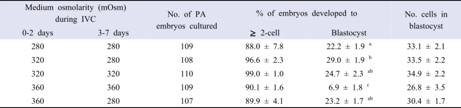
1. Effect of various osmolarities of medium during IVC on embryonic development after PA
PA embryos were cultured in medium with various osmolarities during IVC. As shown in Table 1, there were no significant differences in the embryo cleavage (88.0-99.0%) and mean number of cells in blastocysts (26.8-34.9 cells/blastocyst) among treatment groups. T320-280 (29.0%) showed a higher (p < 0.05) blastocyst formation than control (22.2%) and T360-360 groups (6.9%) while T360-360 showed significantly decreased blastocyst formation compared to all other treatment groups (24.7% and 23.2% in T320-320 and T360-320, respectively).
2. Effect of alanine or/and glycine in IVC medium with various osmolarities on embryonic development after PA The effect of alanine or/and glycine treatment during IVC in medium with various osmolarities on PA embryonic development was evaluated. Treatment with alanine and glycine in T280-280 did not show a significant effect on blastocyst formation (26.1, 28.4, and 27.9% in control, control + alanine, and control + glycine, respectively) and mean cell number of blastocyst (31.4, 28.5, and 31.4 cells/blastocyst in control, control + alanine, and control + glycine, respectively) (Table 2). When PA embryos were cultured in T320-280, glycine treatment with (45.7%) or without alanine (50.4%) significantly (p < 0.05) increased blastocyst
Medium osmolarity (mOsm)
during IVC No. of PA
embryos cultured
% of embryos developed to No. cells in blastocyst
0-2 days 3-7 days 2-cell Blastocyst
280 280 109 88.0 ± 7.8 22.2 ± 1.9 a 33.1 ± 2.1 320 280 108 96.6 ± 2.3 29.0 ± 1.9 b 33.5 ± 2.2 320 320 110 99.0 ± 1.0 24.7 ± 2.3 ab 34.9 ± 2.2 360 360 109 90.1 ± 1.6 6.9 ± 1.8 c 26.8 ± 3.5 360 280 107 89.9 ± 4.1 23.2 ± 1.7 ab 30.4 ± 1.7 Four replicates.
a-cValues in the same column with different superscript letters are different (p < 0.05).
formation compared to T320-280 and T320-280 + alanine (36.5 and 38.7%, respectively) while no synergistic effect of alanine and glycine supplementation was observed.
3. Effect of glycine in IVC medium with various osmolarities on embryonic development and cell number of ICM and TE after SCNT
Effect of IVC in T320-280 and glycine supplementation on SCNT embryonic development was determined. Glycine supplementation in T320-280 showed a significantly higher (p < 0.05) cleavage (94.2%) than control (85.6% in T280-280) but cleavage was not different from that in control + glycine (93.7%) and T320-280 (88.8%). Glycine supplemented to both in T280-280 (38.0%) and T320-280 (35.3%) significantly (p < 0.05) increased blastocyst formation compared to no supplementation (27.6% and 23.7% in T280-280 and T320-280, respectively) while mean cell numbers in blastocyst were not differed among all treatment groups (Table
3). When SCNT blastocysts were examined for their cell number of ICM and TE, the number of ICM cells in T320-280 + glycine (9.6 cells) was significantly higher than that in T280-280 + glycine (6.1 cells) while not different from those in T280-280 and T320-280 (7.3 and 6.4 cells, respectively). Glycine supplementation in T280-280 showed a decreased proportion of ICM cells to total cells compared to no supplementation (17.8% vs. 27.2%).
DISCUSSION
Many studies have been performed using IVM oocytes to produce IVF and SCNT pig embryos. However, low developmental potential of IVP pig embryos compared to in vivo embryos is a major issue that needs to be solved. For this reason, many researchers have been trying to improve the developmental competence of IVP embryos by creating an optimized IVM and Medium osmolarity (mOsm)
during IVC Amino acid No. of PA embryos cultured
% of embryos developing to No. of cells in blastocyst
0-2 days 3-7 days 2-cells Blastocyst
280 280 None 121 89.6 ± 3.6 26.1 ± 1.8 a 31.4 ± 1.5 280 280 ALA 122 89.8 ± 2.4 28.4 ± 2.5 a 28.5 ± 1.2 280 280 GLY 122 86.3 ± 2.2 27.9 ± 0.7 a 31.4 ± 1.4 320 280 None 122 89.0 ± 2.5 36.5 ± 1.6 b 30.9 ± 1.6 320 280 ALA 122 89.1 ± 0.9 38.7 ± 1.9 b 29.9 ± 1.1 320 280 GLY 122 90.0 ± 3.6 50.4 ± 3.4 c 30.5 ± 1.0 320 280 ALA + GLY 121 92.0 ± 1.9 45.7 ± 1.6 c 31.7 ± 1.2 Four replicates.
ALA, 1.2 mM alanine; GLY, 4.1 mM glycine.
a-cValues in the same column with different superscript letters are different (p < 0.05).
Table 2. Effect of alanine (ALA) or/and glycine (GLY) in in vitro culture (IVC) medium with various osmolarities during IVC on embryonic development after parthenogenesis (PA)
Medium osmolarity (mOsm)
during IVC Glycine No. of SCNT
embryos cultured
% of embryos developing to No. of cells in blastocyst
0-2 days 3-7 days 2-cells Blastocyst
280 280 - 161 85.6 ± 2.8 b 27.6 ± 1.6 ac 31.9 ± 1.5
280 280 + 159 93.7 ± 1.6 ab 38.0 ± 1.6 b 35.5 ± 1.7
320 280 - 159 88.8 ± 3.0 ab 23.7 ± 4.1 c 34.7 ± 1.7
320 280 + 158 94.2 ± 3.4 a 35.3 ± 3.8 ab 33.7 ± 1.5
*Four replicates.
a-cValues in the same column with different superscript letters are different (p < 0.05).
Table 3. Effect of glycine in in vitro culture (IVC) medium with various osmolarities on embryonic development after somatic cell nuclear transfer (SCNT)
IVC systems. Oocyte maturation and embryonic development in vitro are greatly influenced by the osmolarity of a culture medium (Collins and Baltz, 1999). In addition, the optimal osmolarity of a medium for early pig embryonic development depends on the developmental stage of embryos. Interestingly, most effective osmolarity of IVC medium for culture of mammalian embryos was lower than the physiological osmolarity (Liu and Foote, 1995; Liu and Foote, 1996). PZM-3 (Yoshioka et al., 2008; Li et al., 2007) is the most common culture medium used for culture of pig embryos produced by IVF, PA and SCNT. The osmolarity of PZM-3 is ranged from 260 to 280 mOsm that is lower than the physiological osmolarity of 318 to 321 mOsm of pig oviductal fluid. In this study, PA and SCNT embryos were exposed to medium having a similar osmolarity with that in female reproductive tract. The results of the present study showed that the high osmolarity of a culture medium (320 mOsm) induced by increasing NaCl concentration during the first 2 days of IVC improved preimplantation development of PA embryos compared to medium with 280 mOsm. Similar results were obtained when the osmotic pressure was increased from 273 to 318 mOsm by adding sucrose as well as NaCl (Miyoshi and Mizobe, 2014). In previous studies, culturing pig embryos in a higher osmolality (320-330 mOsm) first, and then transferring into a lower osmolality (250-280 mOsm) in time appeared to be beneficial for pig embryonic development (Li et al., 2007; Miyoshi and Mizobe, 2014). Our results are consistent with these previous findings.
The main discovery from this study is that glycine treatment during IVC can support the developmental competence of PA and SCNT embryos. In this study, the effects of glycine or/and alanine supplemented to IVC medium of 320 or 280 mOsm on in vitro development of PA and SCNT pig embryos were examined. Unexpectedly, glycine or/and alanine did not show any significant improvement in embryonic development when supplemented to
IVC medium with 280 mOsm. On the other hand, supplementing glycine in a high osmolarity (320 mOsm) environment during the first 2 days of IVC further improved the percentage of embryos developed to the blastocyst than by just providing a high osmolarity environment. The improved developmental competence of PA embryos was mainly due to the supplementation of glycine, which might act as a protective shield to maintain the shape and survive better in high osmolarity environment (Li et al., 2007). Alanine is also well known for its protective effect against high osmolarity in mouse embryonic development (Hammer and Baltz, 2003). However, alanine supplementation to IVC medium did not show any stimulating effect on embryonic development to the blastocyst stage. It was possible that the differences in developmental physiology including metabolic requirements between mouse and pig embryos might have influenced the function of alanine and thus showed different response to the alanine treatment during IVC.
In contrast to PA embryos, in vitro developmental competence of SCNT embryos was not improved by the culture in high osmolarity medium during the first 2 days of IVC. On the other hand, the glycine treatment improved the developmental competence of SCNT embryos in normal osmotic (T280-280) as well as in high osmotic (T320-280) conditions in this study. There are numerous steps in the process of embryo production via SCNT than PA. In the SCNT process including physical enucleation and electrical cell fusion. Moreover, SCNT oocytes are exposed to more oxidative stresses than PA oocytes. SCNT oocytes undergoes nuclear remodeling and reprogramming of newly introduced somatic cell nucleus that was different from those occurring in PA embryos. The different response to the glycine treatment of SCNT from PA embryos might be attributed to the difference in embryo metabolism and developmental nature between PA and SCNT embryos.
Evaluating the quality of blastocysts obtained in vitro is essential process to improve the efficiency of assisted reproductive biotechnologies. Medium osmolarity (mOsm)
during IVC Glycine No. of SCNT
blastocysts examined
Cell number % of inner cell mass cells/total cells
0-2 day 3-7 day Inner cell mass Trophectoderm
280 280 - 10 7.3 ± 1.2 ab 19.9 ± 2.1 27.2 ± 4.9 a
280 280 + 15 6.1 ± 1.0 b 26.2 ± 2.4 17.8 ± 2.2 b
320 280 - 8 6.4 ± 0.8 ab 26.9 ± 4.4 20.8 ± 2.9 ab
320 280 + 13 9.6 ± 0.5 a 26.5 ± 3.4 26.0 ± 2.5 a
a,bValues in the same column with different superscript letters are different (p < 0.05).
Table 4. Mean numbers of inner cell mass and trophectoderm cells in somatic cell nuclear transfer (SCNT) blastocysts derived from the in vitro culture (IVC) under the treatment with glycine in medium with various osmolarites
One of the quality evaluation methods is to count the total cell number of blastocysts and the proportion of ICM to total cell numbers by differential staining (Kim et al., 2004). Previous studies reported that total cell number of blastocyst similar to that of in vivo-derived blastocysts can be regarded as a valuable marker to assess the viability of IVP embryos (De la Fuenta and King, 1997; Soom et al., 1997). In this study, it was observed that the mean number of ICM and the proportion of ICM to total cell number of SCNT blastocysts were not altered by the glycine treatment and the culture in medium with various osmolarities during IVC. Interestingly, glycine supplementation in T320-280 showed a significant increase in the number of ICM cells as well as in the ration of ICM to total cells compared to glycine supplementation in T280-280 indicating embryo quality was improved by the glycine. In summary, our results demonstrated that the two-step culture system established in this study using a medium with 320 mOsm for the first 2 days and 280 mOsm medium for further 5 days of IVC improved embryonic development after PA in pigs. In addition, glycine supplementation in this two-step culture system improved SCNT embryonic development and influenced the quality of blastocyst by increasing the ICM cells and the proportion of ICM cells to total cells.
ACKNOWLEDGMENTS
This research was supported by Basic Science Research Program through the National Research Foundation of Korea (NRF) funded by the Ministry of Education (Grant No. 2016R1D1A1B03934064).
REFERENCES
Bavister BD, Arlotto T. 1990. Influence of single amino acids on the development of hamster one-cell embryos in vitro. Mol Reprod Dev 25:45-51.
Brinster R. 1974. Embryo Development. J Anim Sci 38:1003-1012. Brown JJ, Whittingham DG. 1992. The dynamic provision of
different energy substrates improves development of one-cell random-bred mouse embryos in vitro. J Reprod Fertil 95:503-511.
Collins JL, Baltz JM. 1999. Estimates of mouse oviductal fluid tonicity based on osmotic responses of embryos. Biol Reprod 60:1188-1193.
Dang-Nguyen TQ, Somfai T, Haraguchi S, Kikuchi K, Tajima
A, Kanai Y, Nagai T. 2011. In vitro production of porcine embryos: current status, future perspectives and alternative applications. Anim Sci J 82:374-382.
Dawson KM, Baltz JM. 1997. Organic osmolytes and embryos: substrates of the Gly and β transport systems protect mouse zygotes against the effects of raised osmolarity. Biol Reprod 56:1550-1558.
De la Fuente R, King WA. 1997. Use of chemically defined system for the direct comparison of inner cell mass and trophectoderm distribution in murine, porcine and bovine embryos. Zygote 5:309-320.
Funahashi H, Day BN. 1997. Advances in in vitro production of pig embryos. J Reprod Fertil Suppl 52:271-283. Gardner DK, Lane M. 1993. Amino acids and ammonium regulate
mouse embryo development in culture. Biol Reprod 48:377-385. Gil M, Cuello C, Parrilla I, Vazquez J, Roca J, Martinez E. 2010.
Advances in swine in vitro embryo production technologies. Reprod Domest Anim 45:40-48.
Hadi T, Hammer M, Algire C, Richards T, Baltz JM. 2005. Similar effects of osmolarity, glucose, and phosphate on cleavage past the 2-cell stage in mouse embryos from outbred and F1 hybrid females. Biol Reprod 72:179-187.
Hammer M, Baltz JM. 2003. β-Alanine but not taurine can function as an organic osmolyte in preimplantation mouse embryos cultured from fertilized eggs. Molecular Reproduction and Development: Incorporating Gamete Research 66:153-161. Harris SE, Gopichandran N, Picton HM, Leese HJ, Orsi NM.
2005. Nutrient concentrations in murine follicular fluid and the female reproductive tract. Theriogenology 64:992-1006. Iritani A, Sato E, Nishikawa Y. 1974. Secretion rates and chemical
composition of oviduct and uterine fluids in sows. J Anim Sci 39:582-588.
Kere M, Siriboon C, Lo N, Nguyen NT, Ju J. 2013. Ascorbic acid improves the developmental competence of porcine oocytes after parthenogenetic activation and somatic cell nuclear transplantation. J Reprod Dev 59:78-84.
Kim H, Lee G, Hyun S, Lee S, Nam D, Jeong Y, Kim S, Kang S, Lee B, Hwang W. 2004. Improved in vitro development of porcine embryos with different energy substrates and serum. Theriogenology 61:1381-1393.
Lee ES, Fukui Y, Lee BC, Lim JM, Hwang WS. 2004. Promoting effect of amino acids added to a chemically defined medium on blastocyst formation and blastomere proliferation of bovine embryos cultured in vitro. Anim Reprod Sci 84:257-267. Lee E, Fukui Y. 1996. Synergistic effect of alanine and glycine
on bovine embryos cultured in a chemically defined medium and amino acid uptake by in vitro-produced bovine morulae and blastocysts. Biol Reprod 55:1383-1389.
Li R, Whitworth K, Lai L, Wax D, Spate L, Murphy CN, Rieke A, Isom C, Hao Y, Zhong Z. 2007. Concentration and composition of free amino acids and osmolalities of porcine oviductal and uterine fluid and their effects on development of porcine IVF embryos. Mol Reprod Dev 74:1228-1235.
Li R, Whitworth K, Lai L, Wax D, Spate L, Murphy CN, Rieke A, Isom C, Hao Y, Zhong Z. 2007. Concentration and composition of free amino acids and osmolalities of porcine oviductal and uterine fluid and their effects on development of porcine IVF embryos. Mol Reprod Dev 74:1228-1235.
Liu Z, Foote RH. 1996. Sodium chloride, osmolyte, and osmolarity effects on blastocyst formation in bovine embryos produced by in vitro fertilization (IVF) and cultured in simple serum-free media. J Assist Reprod Genet 13:562-568.
Liu Z, Foote RH. 1995. Development of bovine embryos in KSOM with added superoxide dismutase and taurine and with five and twenty percent 02. Biol Reprod 53:786-790.
Miyoshi K, Mizobe Y. 2014. Osmolarity-and stage-dependent effects of glycine on parthenogenetic development of pig oocytes. J Reprod Dev 60:349-354.
Rousseau J. 1993. Role of the female genital tract in the transport and survival of gametes and fertilized egg. Reproduction in mammals and man.
Schultz GA, Kaye PL, McKay DJ, Johnson MH. 1981. Endogenous amino acid pool sizes in mouse eggs and preimplantation embryos. J Reprod Fertil 61:387-393.
Soom AV, Boerjan ML, Bols PE, Vanroose G, Lein A, Coryn M, Kruif Ad. 1997. Timing of compaction and inner cell allocation in bovine embryos produced in vivo after superovulation. Biol Reprod 57:1041-1049.
Suzuki C, Yoshioka K. 2006. Effects of amino acid supplements and replacement of polyvinyl alcohol with bovine serum albumin in porcine zygote medium. Reprod Fertil Dev 18:789-795.
Van Winkle LJ, Haghighat N, Campione AL. 1990. Glycine protects preimplantation mouse conceptuses from a detrimental effect on development of the inorganic ions in oviductal fluid. J Exp Zool 253:215-219.
Yoshioka K, Suzuki C, Onishi A. 2008. Defined system for in vitro production of porcine embryos using a single basic medium. J Reprod Dev 54:208-213.
Yoshioka K, Suzuki C, Tanaka A, Anas IM, Iwamura S. 2002. Birth of piglets derived from porcine zygotes cultured in a chemically defined medium. Biol Reprod 66:112-119. You J, Lee J, Hyun S, Lee E. 2012. L-carnitine treatment during
oocyte maturation improves in vitro development of cloned pig embryos by influencing intracellular glutathione synthesis and embryonic gene expression. Theriogenology 78:235-243.