• 검색 결과가 없습니다.

Study on Chemicals for Post-activation in Porcine Somatic Cell Nuclear Transfer

N/A
N/A
Protected

Academic year: 2021

Share "Study on Chemicals for Post-activation in Porcine Somatic Cell Nuclear Transfer"

Copied!
6
0
0

로드 중.... (전체 텍스트 보기)

전체 글

(1)

http://dx.doi.org/10.12750/JET.2016.31.2.131

INTRODUCTION

Since animal cloning achieved a first success, somatic cell nuclear transfer (SCNT) present ideas for application in many research areas such as regenerative medicine Park et al. 2012.

And animal cloning techniques showed wide variety of applicability for the manufacturing of transgenic animals for research in human Lee et al. 2010. Especially In the pig, transgenic techniques has a potential value for the generation of human diseases model and organs for xenotransplantation Martinez Diaz et al. 2003.

However, SCNT embryos has very poor survival ability and an nasty efficiency of cloned offspring production. Therefore, improvement of the growth of SCNT embryos to the blastocyst stage was needed Martinez Diaz et al. 2003.

SCNT is composed of various stages, including oocytes

collection and IVM and preparation of donor cells, nuclear transfer, cell fusion and activation of reconstructed oocytes, and embryo culture Song et al. 2009. Effectiveness of NT in pigs is influenced by many factors, and post-activation treatment is one of the critical steps that directly impact the developmental capability of SCNT embryos Im et al. 2006.

Because SCNT is not a natural process, some side effects like low calcium concentration and loss of nuclear chromosomes happen. So postactivation process needed to cover these problems.

Firstly because sperms are not enough in NT, activation process is necessary and is generally imitated by enhancing oocyte’s intracellular calcium to resume the metaphase II meiosis Im et al. 2006. In order that activating NT embryos, electric pulse has been commonly used, although the electric pulse makes a single temporary rise in intracellular calcium

Study on Chemicals for Post-activation in Porcine Somatic Cell Nuclear Transfer

Kyuhong Min

1

, Seungwon Na

1

, Euncheol Lee

1

, Ghangyong Kim

1,2

, Youngkwang Yu

1

, Pantu Kumar Roy

1,2

, Xun Fang

1,2

, MB Salih

1

, Jongki Cho

1,2,*

1

College of Veterinary Medicine, Chungnam National University, Daejeon 34134, Republic of Korea.

2
Climate Change Disease Control Center, BrainKorea21 plus, Daejeon 34134, Republic of Korea

ABSTRACT

Since the first success of animal cloning, somatic cell nuclear transfer presented various ideas in many research areas such as regenerative medicine. However, SCNT embryos has poor survival rate. Therefore, numerous researches carried out to enhance the developmental capability of porcine nuclear transfer embryos. Cytochalasin B, demecolcine, latrunculin A, cycloheximide and 6-dimethylaminopurine are efficient chemicals treated in post-activation procedure to increase the efficiency of SCNT. This review study is aim to investigate the effects of these chemicals applied to post-activation in porcine SCNT. Cytochalasin B, demecolcine, latrunculin A are cytoskeletal manuplators inhibit extrusion of pseudo-polar body. Cytochalasin B and demecolcine showed considerably higher blastocyst formation proportion (26-28%) compared to when they are not treated (16%). And when latrunculin A was treated for postactivation, blastocyst formation proportion was increased in SCNT embryos exposed to LA (38%) than those in control (14%). On the other hand, cycloheximide and 6-dimethylaminopurine are protein synthesis and kinase inhibitors. And they help to maintain Ca

2+

fluctuation in oocytes. Cleavage and blastocyst rates of NT embryos were increased when they were exposed to CHX (16.9% and 5.4% with no CHX).And 6-DMAP also showed higher blastocyst formation (21.5% compared to 15.7%, control). Although all these chemicals have different mechanisms, they showed developmental competence enhancement in NT embryos. However, there are only few studies comparing each chemical’s post-activation effect. Therefore, further research and study should be conducted to find optimal chemical for improving the efficiency of SCNT.

(Key words: embryo, porcine, postactivation, somatic cell nuclear transfer)

* Correspondence: Jongki Cho

Tel: +82-42-821-6788

E-mail: cjki@cnu.ac.kr

(2)

concentration Wang et al. 1998. Though the single transient can continue the second meiosis, it is not enough to facilitate high rates of further development. So chemicals elevate intracellular calcium, such as A23187, and protein synthesis inhibitors, such as cycloheximide (CHX) and phosphorylation inhibitors like 6-dimethylaminopurine (6-DMAP), have been used in combination to activate pig NT embryos Martinez Diaz et al. 2003(, Im et al. 2007).

Secondly, when the reconstructed oocytes from SCNT activated by electric or chemical stimuli, they tend to export a part of the somatic cell chromosomes in the pseudo-polar body (PPB) form Lai et al. 2002. Nuclear aneuploidy can be down to losing nuclear chromosomes of the reconstructed oocytes, which lead to washout in normal embryonic development to delivery and decreases production efficiency of cloned animals in the end Song et al. 2009. So, some chemicals like cytochalasin, demecolcine, and latrunculin A have been used to suppress PPB emission from the reconstructed oocytes (Himaki et al. 2012).

Cytochalasin B, demecolcine, latrunculin A, cycloheximide, 6-DMAP have been used for post-activation to increase the efficiency of SCNT. In this study, it was investigated for the influence of these chemicals on porcine SCNT embryos development.

Cytochalasin B

Cytochalasin B (CytoB), which chemical formula is C

29

H

37

NO

5

, is a cell-permeable mycotoxin (Figure 1). And its name comes from the Greek cytos (cell) and chalasis (relaxation) Scherlach et al. 2010. It is known that Substoichiometric CytoB suppresses both the interaction of actin filaments and the speed of actin polymerization in solution MacLean-Fletcher and Pollard 1980.

The main mechanism of CytoB is the suppression of actin filament polymerization by binding to the fast-growing (barbed) end of F-actin filaments Theodoropoulos et al. 1994.

Also, it suppresses cytoplasmic division by preventing the formation of contractile microfilaments. And it restrains cell movement and causes nuclear extrusion Copeland 1974.

Due to CytoB’s above-mentioned properties, it is often used in theriogenologic research. CytoB suppresses pseudopolarbody (PPB) extrusion, disrupts spindle structure, inhibits oocyte maturation, and causes formation of diploid oocytes Bai et al.

2011. And CytoB have been used in SCNT. Because CytoB has ability to depolymerize actin filaments, it has been extensively

used to inhibit the extrusion of PB in SCNT oocytes after activation Song et al. 2009.

In the SCNT experiment of Song et al’s (2009), 5 mg/ml cytoB and 0.4 mg/ml demecolcine was used in the post-activation. And in the result, treatment with CytoB and/or demecolcine (DC) (26–28%) was higher than in the controls (16%) in blastocyst formation. And the number of oocytes forming a single pronucleus (PN) was higher when they were treated with Dc or CytoB + Dc (86%, each) than control conditions (44%) or treated with CytoB (63%).

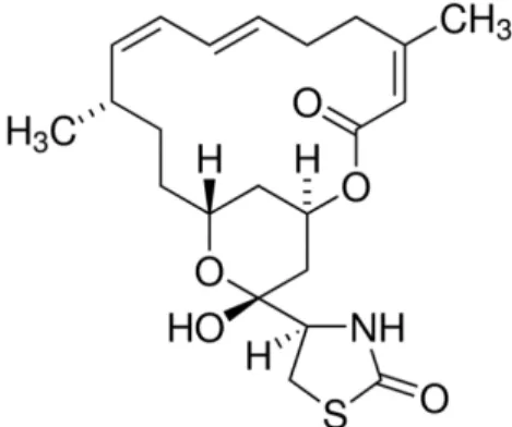
Figure 1. Cytochalasin B. The Cytochalasins (Greek cytos, cell;

chalasis, relaxation) are a family of mycotoxin. These fungal toxins are related by chemical structure. Cytochalasin B has highly substituted hydrogenated isoindole ring, and it is fused a macrocyclic ring. This fungal toxins also has many unusual,amusing, and characteristic effects on the animal cell.

(http://www.sigmaaldrich.com/catalog/product/sigma/c676 2?lang=ko&region=KR)

Demecolcine (DC)

Stemed from plants of the genus Colchicum, Colchicine has long been known to be a strong inhibitor of cell division by its impacts on spindle microtubule assembly (Figure 2) Dustin 1974. And demecolcine (DC), which chemical formula is C

21

H

25

NO

5

, is a closely concered with colchicine. However, it has less toxicity. Because of its ability to depolymerizing microtubules and limiting microtubule formation, it arrests cells in metaphase Rieder and Palazzo 1992.

Like CytoB, DC also widely applied to oocytes and SCNT

embryos. Demecolcine causes the enucleation of oocytes and

porcine metaphase II–arrested oocytes. In addition, presumably

by its ability to induce the formation of a single pronucleus

(PN) and improving DNA ploidy, postactivation treatment with

demecolcine sequentially supports in vivo development to delivery

in pig SCNT embryos Lee et al. 2010.

(3)

In the SCNT experiment of Lee et al’s (2010), embryos were postactivation treated with 0.4 mg/mL DC or 2 mg/mL nocodazole in IVC medium for 4 h. Blastocyst formation of SCNT oocytes was higher after post-activation treatment with DC (29%) and nocodazole (30%) than control oocytes (16%).

The rate of oocytes that formed pronucleus was increased when treated with nocodazole and DC (82%, 86% each) than under the control conditions (66%). Both nocodazole and DC suppressed pseudo-polar body extrusion, and the rate of embryos with diploid chromosomes was increased after treated with nocodazole and DC (86%, 77% each) than under control conditions (58%).

And, in the SCNT experiment of Song et al’s (2009), concentration of used chemicals were 5 mg/ml (CytoB) and 0.4 mg/ml (DC). Recipients of embryos treated with DC had a increased portion of pregnancies lead to delivery (50% for DC-, compared to 37.5% for control), and they showed increased production of piglets (12 from DC-treated, compared to 7 from control).

Figure 2. Demecolcine is secondary amino compound which is (S)-colchicine where the N-acetyl group is replaced by an N-methyl group. It can be Isolated from the autumn crocus, Colchicum autumnale. And it has lesser toxicity than colchicine and it depolymerizes microtubules;

blocks mitosis at metaphase.

(http://www.sigmaaldrich.com/catalog/product/sigma/d73 85?lang=ko&region=KR)

Latrunculin A

A toxin purified from the Latrunculia magnifica, Latrunculin A(LA) is a family of the cytoskeletal inhibitors, which prevent the polymerization of actin (Figure 3) Cooper 1987. The main mechanism of LA is as in the following. LA impacts on the polymerization of actin, a main component of the cytoskeleton, in a manner consistent with formation of a complex with G-actin Wakatsuki et al. 2001. LA can bind specifically to G-actin and has no effect to prevent the F-actin

Yarmola et al. 2000.

Recently, some research suggested that postactivation treatment with LA was efficient in enhancing the developmental competence of pig embryos Himaki et al. 2010. And Himaki et al. (2012) reported that post-activation treatment with LA showed higher embryonic development of SCNT oocytes than in CytoB.

LA indicates a similar impact with CytoB at a low concentration and the influence of LA on actin are 10~100 times more stronger than cytochalasins Wakatsuki et al. 2001.

Thus, LA may have weaker cytotoxicity when it is applied for post-activation treatment of reconstructed oocytes.

In the SCNT experiment of Himaki et al’s (2012), SCNT embryos were postactivation treated with 0, 0,5 or 1uM LA and cultured. Blastocyst formation proportion was higly increased in SCNT embryos exposed to 0.5uM LA (38%) than those in control (14%). When embryos exposed to 0.5uM were transferred into 2 recipient, and they made production of piglets.

Figure 3. Latrunculin A is a toxin derived from the red sea sponge Latrunculia magnifica. Latrunculin A effects in vitro in the morphological change of the actin polymerization. It inhibits actin polymerization with different way than cytochalasin. It hinders microfilament-mediated processes.

(http://www.sigmaaldrich.com/catalog/product/sigma/l5163

?lang=ko&region=KR)

Cycloheximide

Cycloheximide (CHX), which chemical formula is C

15

H

23

NO

4,

is an inhibitor of protein biosynthesis in yeast s, derived from Streptomyces griseus (Figure 4) Baliga et al.

1969. The laboratory chemical agent that widely used to

suppress protein synthesis, it is indicated that CHX blocks

eukaryotic translation. It attaches to the ribosome and

suppresses eEF2-mediated translocation Schneider-Poetsch et

(4)

al. 2010.

Protein synthesis inhibition by CHX decreases the cyclin and accordingly reduce MPF Im et al. 2006. Thus, CHX was thought to be efficient to cause activation of porcine metaphase II oocytes when combined with electric stimulation Martinez Diaz et al. 2003.

In the SCNT experiment of Martinez Diaz et al.’s (2003), NT embryos were treated with 10 ug/ml of CHX or without CHX before evaluating the in vitro development to the blastocyst stage. Although there was no difference in the fusion rate between the two groups (No CHX, with CHX), cleavage and blastocyst rates of NT embryos were increased when they were exposed to CHX (16.9% when CHX treatment whereas 5.4% with no CHX).

Figure 4. Cycloheximide (CHX) is an antibiotic which is achieved by S. griseus. Its main function is translation inhibition in eukaryotes, and it results in cell growth arrest and cell death. CHX is widely applied for selection of CHX-resistant strains of yeast and fungi. And it is also used in protein synthesis inhibition for detection of short-lived proteins and super-induction of protein expression.

(http://www.sigmaaldrich.com/catalog/product/sigma/c769 8?lang=ko&region=KR)

6-DMAP

6-dimethylaminopurine(6-DMAP) is a puromycin analogue, which chemical formula is C

7

H

9

N

5.

As the protein kinase inhibitor, 6-DMAP inactivates the catalytic subunit of MPF, p34cdc2 kinase, by inactivating mitogen-activated protein kinase (MAPK) (Figure 5) Gordo et al. 2001Liu et al. 1998.

6-DMAP is widely used in somatic cell nuclear transfer research. And lately cloning mammals with 6-DMAP has done successfully Katoh et al. 2004. It has been shown to reduce

levels of active MPF Susko-Parrish et al. 1994 and enhance pronuclear formation Szollosi et al. 1993 in incubation of activated mammalian oocytes using 6-DMAP.

Thus, 6-DMAP has been applied to improve the maturation of oocytes and capability of embryos. And especially 6-DMAP can be used in post-activation in SCNT procedure. 6-DMAP could activate oocytes by suppressing phosphotyrosine dephosphorylation as well as changing Ca

2+

in oocytes. It is indicated that activation by 6-DMAP could keep the fluctuation line of Ca

2+

level and enhance following development of porcine NT embryos Im et al. 2007.

In the SCNT experiment of Im et al.’s (2006), fused oocytes were treated to additional chemical activation by exposing to 2 mM 6-DMAP for 3 h or 10 mg/ml, CHX for 6 h. Cleavage rates were not considerably different among treatments (66.4%

– 68.3%, each). However, oocytes activated with chemicals showed higher increased development to the blastocyst stage compared to E (electrically activated control) alone (DMAP : 21.5%, CHX : 23.4% vs. E : 15.7%, respectively)

Figure 5. 6-Dimethylaminopurine (6-DMAP) is analogue of puromycins and it is a member of the class of organic compounds known as 6-alkylaminopurines. These compounds have an alkylamine group which is attached at the 6-position of a purine. 6-DMAP is a inhibitor of protein kinase and cyclin-dependent kinase used in parthenogenesis and meiosis studies.

(http://www.sigmaaldrich.com/catalog/product/sigma/d2 629?lang=ko&region=KR)

Conclusion

There are some kinds of chemicals for postactivation on

porcine SCNT. And they can be categorized into two groups

by mechanism of postactivation. The one is the inhibiting

extrusion of nuclear nucleosomes, and cytoB, DC, and LA

were included in this group. And they are cytoskeletal

modifier.

(5)

CytoB and DC showed considerably higher blastocyst formation proportion when they are treated (26-28) than they are not treated (16). However there was no significant difference between when they were treated with each other.

And in pronucleus formation, when CytoB with DC showed enhancement compared to using CytoB alone. And this is maybe due to DC has higher ability to formation of pronucleus. And when LA was treated for postactivation, it was indicated that LA is efficient in enhancing developmental competence of SCNT, and LA had ability to develop to deliver. So in the face of fact that LA has less cytotoxicity than cytoB, LA showed replaceability to the cytoB. However, further studies about comparison between cytoB and LA should be carried out. The other including CHX and 6-DMAP was to enhance the calcium concentration of oocyte. CHX, which inhibit protein synthesis, activate NT embryo by reducing MPF. CHX rarely effect fusion rate. However, cleavage and blastocyst rates of NT embryos were increased when they were exposed to CHX (16.9% and 5.4% with no CHX) 6-DMAP changes Ca2+ in oocytes and inhibits dephosphorylation. And in the former study, blastocyst formation of 6-DMAP : 21.5% was higher than control (15.7%), and has no significant difference with CHX (23.4%).

Therefore, they showed the enhancement of developmental capability of SCNT embryos.

In summary, there are many chemicals that can be applied to postactivation of porcine SCNT, and they had different mechanisms of activation. However, they all showed developmental capability improvement in SCNT embryos.

Including above-mentioned treatments, research and study should be continued to find optimal chemical for improving the efficiency of SCNT.

ACKNOWLEDGEMENTS

This research was supported by a grant of the CNU research grants 2014.

REFERENCES

Bai C, Liu H, Liu Y, Wu X, Cheng L, Bou S, Li GP. 2011.

Diploid oocyte formation and tetraploid embryo development induced by cytochalasin B in bovine. Cell Reprogram 13(1):37-45.

Baliga BS, Pronczuk AW, Munro HN. 1969. Mechanism of cycloheximide inhibition of protein synthesis in a cell-free system prepared from rat liver. J Biol Chem 244(16):4480-4489.

Cooper JA. 1987. Effects of cytochalasin and phalloidin on actin. J Cell Biol 105(4):1473-1478.

Copeland M. 1974. The cellular response to cytochalasin B: a critical overview. Cytologia (Tokyo) 39(4):709-727.

Dustin P. 1974. Some recent advances in the study of microtubules and microtubule poisons. Arch Biol (Liege) 85(2):263-288.

Gordo AC, He CL, Smith S, Fissore RA. 2001. Mitogen activated protein kinase plays a significant role in metaphase II arrest, spindle morphology, and maintenance of maturation promoting factor activity in bovine oocytes.

Mol Reprod Dev 59(1):106-114.

Himaki T, Mizobe Y, Tsuda K, Suetomo M, Yamakuchi H, Miyoshi K, Takao S, Yoshida M. 2012. Effect of postactivation treatment with latrunculin A on in vitro and in vivo development of cloned embryos derived from kidney fibroblasts of an aged Clawn miniature boar. J Reprod Dev 58(4):398-403.

Himaki T, Mori H, Mizobe Y, Miyoshi K, Sato M, Takao S, Yoshida M. 2010. Latrunculin A dramatically improves the developmental capacity of nuclear transfer embryos derived from gene-modified clawn miniature pig cells. Cell Reprogram 12(2):127-131.

Im GS, Samuel M, Lai L, Hao Y, Prather RS. 2007.

Development and calcium level changes in pre-implantation porcine nuclear transfer embryos activated with 6-DMAP after fusion. Mol Reprod Dev 74(9):1158-1164.

Im GS, Seo JS, Hwang IS, Kim DH, Kim SW, Yang BC, Yang BS, Lai L, Prather RS. 2006. Development and apoptosis of pre-implantation porcine nuclear transfer embryos activated with different combination of chemicals.

Mol Reprod Dev 73(9):1094-1101.

Katoh M, Araki A, Ogura T, Valdivia RP. 2004.

6-Dimethylaminopurine (6-DMAP), which is used to produce most cloned animals, is mutagenic in Salmonella typhimurium TA1535. Mutat Res 560(2):199-201.

Lai L, Kolber-Simonds D, Park KW, Cheong HT, Greenstein JL, Im GS, Samuel M, Bonk A, Rieke A, Day BN, Murphy CN, Carter DB, Hawley RJ, Prather RS. 2002.

Production of alpha-1,3-galactosyltransferase knockout pigs

(6)

by nuclear transfer cloning. Science 295(5557):1089-1092.

Lee J, You J, Kim J, Hyun SH, Lee E. 2010. Postactivation treatment with nocodazole maintains normal nuclear ploidy of cloned pig embryos by increasing nuclear retention and formation of single pronucleus. Theriogenology 73(4):429- 436.

Liu L, Ju JC, Yang X. 1998. Parthenogenetic development and protein patterns of newly matured bovine oocytes after chemical activation. Mol Reprod Dev 49(3):298-307.

MacLean-Fletcher S, Pollard TD. 1980. Mechanism of action of cytochalasin B on actin. Cell 20(2):329-341.

Martinez Diaz MA, Suzuki M, Kagawa M, Ikeda K, Takahashi Y. 2003. Effects of cycloheximide treatment on in-vitro development of porcine parthenotes and somatic cell nuclear transfer embryos. Jpn J Vet Res 50(4):147-155.

Park SJ, Park HJ, Koo OJ, Choi WJ, Moon JH, Kwon DK, Kang JT, Kim S, Choi JY, Jang G, Lee BC. 2012.

Oxamflatin improves developmental competence of porcine somatic cell nuclear transfer embryos. Cell Reprogram 14(5):398-406.

Rieder CL, Palazzo RE. 1992. Colcemid and the mitotic cycle.

J Cell Sci 102 ( Pt 3):387-392.

Scherlach K, Boettger D, Remme N, Hertweck C. 2010. The chemistry and biology of cytochalasans. Nat Prod Rep 27(6):869-886.

Schneider-Poetsch T, Ju J, Eyler DE, Dang Y, Bhat S, Merrick WC, Green R, Shen B, Liu JO. 2010. Inhibition of eukaryotic translation elongation by cycloheximide and lactimidomycin. Nat Chem Biol 6(3):209-217.

Song K, Hyun SH, Shin T, Lee E. 2009. Post-activation treatment with demecolcine improves development of somatic cell nuclear transfer embryos in pigs by modifying

the remodeling of donor nuclei. Mol Reprod Dev 76(7):611-619.

Susko-Parrish JL, Leibfried-Rutledge ML, Northey DL, Schutzkus V, First NL. 1994. Inhibition of protein kinases after an induced calcium transient causes transition of bovine oocytes to embryonic cycles without meiotic completion. Dev Biol 166(2):729-739.

Szollosi MS, Kubiak JZ, Debey P, de Pennart H, Szollosi D, Maro B. 1993. Inhibition of protein kinases by 6-dimethylaminopurine accelerates the transition to interphase in activated mouse oocytes. J Cell Sci 104 ( Pt 3):861-872.

Theodoropoulos PA, Gravanis A, Tsapara A, Margioris AN, Papadogiorgaki E, Galanopoulos V, Stournaras C. 1994.

Cytochalasin B may shorten actin filaments by a mechanism independent of barbed end capping. Biochem Pharmacol 47(10):1875-1881.

Wakatsuki T, Schwab B, Thompson NC, Elson EL. 2001.

Effects of cytochalasin D and latrunculin B on mechanical properties of cells. J Cell Sci 114(Pt 5):1025-1036.

Wang WH, Abeydeera LR, Prather RS, Day BN. 1998.

Functional analysis of activation of porcine oocytes by spermatozoa, calcium ionophore, and electrical pulse. Mol Reprod Dev 51(3):346-353.

Yarmola EG, Somasundaram T, Boring TA, Spector I, Bubb MR. 2000. Actin-latrunculin A structure and function.

Differential modulation of actin-binding protein function by latrunculin A. J Biol Chem 275(36):28120-28127.

Received June 24, 2016, Revised June 28, 2016,

Accepted June 28, 2016

수치

Figure 2. Demecolcine is secondary amino compound which is  (S)-colchicine where the N-acetyl group is replaced by  an  N-methyl group

참조

관련 문서

In the SEM & EDS analysis of the solid-residue, it was discovered that dissolution on the mineral surface increased much more when the microwave

In the POLARIS-2 and POLARIS-3 studies 14 , it was not determined that SOF/VEL/VOX for 8 weeks was non-inferior to SOF/VEL for 12 weeks for the treatment of non-cirrhotic

pollutants exposed to the gravel was increased to 10 times of the normal concentration, the oxygen uptake rate, indicating microbial activity of the gravel

In the present study, consistent to that report, up-regulation of VEGF mRNA was observed in the colon cancer cell with the acquired resistance to 5-FU and this

The purpose of this study was to examine the effects of a swimming program for 12 weeks on health-related physical fitness and growth hormone in

Among the sub-factors of self-esteem, positive, male was higher than female, and on the contrary, negative In this study, females were higher than males,

In order to derive a new fuzzy controller, a fixed design-parameter fuzzy PID controller was surveyed in view of deriving a control law based on the

In the late nineteenth century, although the proportion of ether was increased and that of chloroform decreased due to the raised awareness of the high