R E S E A R C H Open Access
Change of the airway space in mandibular prognathism after bimaxillary surgery
involving maxillary posterior impaction
Woo-Young Lee, Young-Wook Park * , Kwang-Jun Kwon and Seong-Gon Kim
Abstract
Background: The purpose of this retrospective study was to develop a two- and three-dimensional analysis of the airway using cone-beam computed tomography (CBCT) and to determine whether the airway space would be changed in mandibular prognathism after bimaxillary surgery involving maxillary posterior impaction.
Methods: Patients requiring orthognathic surgery from 2012 to 2014 were recruited for this study. CBCT scans were obtained at three points: preoperatively (T0), immediate postoperatively (T1), and after 6 months postoperatively (T2).
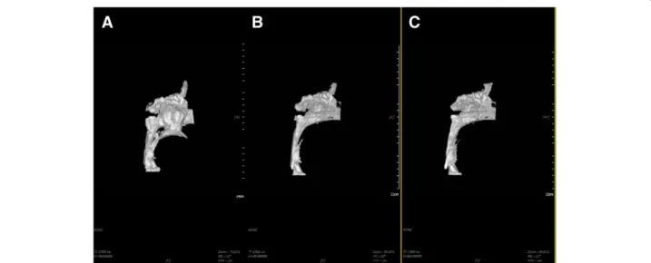
The nasopharynx, oropharynx, and hypopharynx were measured on the CBCT scan for each patient in a repeatable manner. With the midsagittal plane, linear measurements in the middle of each were obtained. For the CBCT, volumetric measurements of each and total airway were obtained.
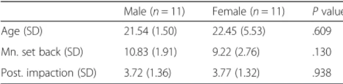
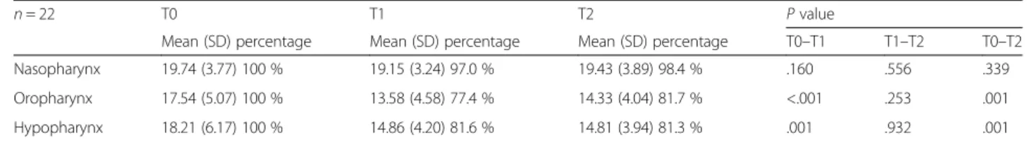
Results: A total of 22 consecutive patients (11 men and 11 women) were included in the present study. The total volume was significantly reduced ( p < .001). However, the change of the diameter and volume of the nasopharynx was not statistically significant ( p = .160, p = .137, respectively). In the oropharynx, the change of both the diameter and volume showed statistical significance between preoperatively and immediate postoperatively ( p < .001, p = .001, respectively) and also preoperatively and after 6 months postoperatively ( p = .001, p = .010, respectively). In the
hypopharynx, the change of both the diameter and volume showed statistical significance between preoperatively and immediate postoperatively ( p = .001, p < .001, respectively) and also preoperatively and after 6 months postoperatively ( p = .001, p < .001, respectively).
Conclusions: The bimaxillary surgery involving maxillary posterior impaction can reduce the volume of airway in the patients of mandibular prognathism. Although total airway volume was reduced significantly, the changes in the volume and diameter of the nasopharynx were not statistically significant. The maxillary posterior impaction affects on the nasopharyngeal airway minimally.
Keywords: Orthognathic surgery, Airway management, Cone-beam computed tomography
Background
Typically, class III malocclusion patients represent a combination of facial malformations in the skeletal and dental alveolar bone [1]. Most of these patients can exhibit mandibular prognathism and maxillary retro- gnathism. For the establishment of the patient ’s optimal occlusion and profile, it is necessary to perform orthog- nathic surgery, especially bilateral sagittal split ramus osteotomy (SSRO) or vertical ramus osteotomy (VRO)
[2]. However, some patients required both maxillary and mandibular surgery, the so-called bimaxillary orthog- nathic surgery [3]. Clockwise rotation of the maxilla (posterior impaction) can achieve correction of acute nasolabial angle and stabilization of the occlusal plane.
But this surgical movement of the maxilla may reduce the volume of airway space [4]. In the case of the class III malocclusion patients performed with orthognathic surgery, it can change the position of the hyoid bone and tongue, and the base of the tongue moves to the posterior, which will increase the contact surface between the soft palate and tongue [5, 6]. As a result, the pharyngeal airway space
* Correspondence: [email protected]
Department of Oral and Maxillofacial Surgery, College of Dentistry, Gangneung-Wonju National University, 7 Jukheon-gil, Gangneung, Gangwondo 210-702, South Korea
© 2016 The Author(s). Open Access This article is distributed under the terms of the Creative Commons Attribution 4.0
International License (http://creativecommons.org/licenses/by/4.0/), which permits unrestricted use, distribution, and
reproduction in any medium, provided you give appropriate credit to the original author(s) and the source, provide a link to
the Creative Commons license, and indicate if changes were made.
is narrowed [7]. Up to date, there are many reports of air- way reduction in mandibular set back surgery but rare in maxillary posterior impaction.
In these patients, cone-beam computed tomography (CBCT) has been attracting attention for the three-dimen- sional assessment of the airway, an important role in the process of diagnosis in place of the conventional CT [8, 9].
CBCT is equipped with a fast scanning equipment and low exposure to radiation as compared to conventional CT [9, 10]. As it can observe on various directions for evaluation of three-dimensional airway reconstruction, it can be suit- able for use as a measuring apparatus for a change in the air- way between preoperative and postoperative procedure [11].
The purpose of this retrospective study was to develop a two and three-dimensional analysis of the airway using CBCT and to determine whether the difference in the air- way volume would be changed with posterior impaction of maxilla in orthognathic surgery.
Methods Patient analysis
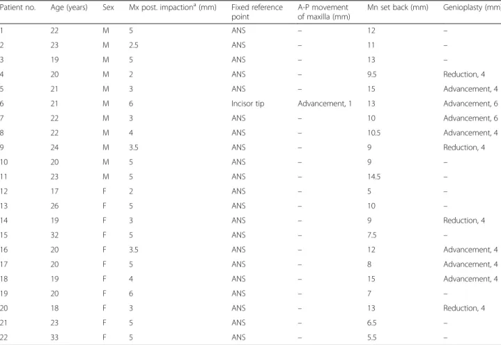
From January 2012 to January 2014, a selected group of 22 patients with skeletal class III malocclusion who have
complete records were chosen (Tables 1 and 2). Patients with systemic surgical contraindications were excluded from the study. The operations have been performed by the experi- enced surgeon, and anesthesia was administered by an experi- enced anesthetist. After surgery, most patients remained in the hospital for 7 days. A standard regimen of antibiotics was administered for 7 days according to clinical conditions. All patients were admitted 1 day before the surgery. A prophylac- tic antibiotic treatment (augmentin or cefazolin) was routinely given with the induction of anesthesia. NSAID (ketoro- lac tromethamine) was given for postoperative anal- gesia. Corticosteroid was given for 3 days via tapering process. All patients were done with intermaxillary fixation by elastic rubber to fit in occlusal stent. When they would be discharged, intermaxillary fixation was removed, and rubber guiding was done.
Radiographic analysis
The radiologic information was taken by cone-beam computed tomography (Alphard, Asahi Roentgen Co., Kyoto, Japan) which was used in the Gangneung-Wonju National University dental hospital, Department of Oral and Maxillofacial Radiology for patient radiographic
Table 1 Patients information
Patient no. Age (years) Sex Mx post. impaction
a(mm) Fixed reference point
A-P movement of maxilla (mm)
Mn set back (mm) Genioplasty (mm)
1 22 M 5 ANS – 12 –
2 23 M 2.5 ANS – 11 –
3 19 M 5 ANS – 13 –
4 20 M 2 ANS – 9.5 Reduction, 4
5 21 M 3 ANS – 15 Advancement, 4
6 21 M 6 Incisor tip Advancement, 1 13 Advancement, 6
7 22 M 3 ANS – 10 Advancement, 6
8 22 M 4 ANS – 10.5 Advancement, 4
9 24 M 3.5 ANS – 9 Reduction, 4
10 20 M 5 ANS – 9 –
11 23 M 5 ANS – 14.5 –
12 17 F 2 ANS – 5 –
13 26 F 5 ANS – 10 –
14 19 F 3 ANS – 9 Reduction, 4
15 32 F 5 ANS – 7.5 –
16 20 F 3.5 ANS – 12 Advancement, 4
17 20 F 5 ANS – 8 Advancement, 4
18 19 F 4 ANS – 15 Advancement, 4
19 20 F 6 ANS – 7 –
20 18 F 3 ANS – 13 Reduction, 4
21 23 F 5 ANS – 6.5 –
22 33 F 5 ANS – 5.5 –
Abbreviations: Mx maxillary, Mn mandibular, Post posterior, ANS anterior nasal spine, A-P anteroposterior
a