465
DOI: 10.4046/trd.2010.69.6.465
ISSN: 1738-3536(Print)/2005-6184(Online) Tuberc Respir Dis 2010;69:465-468
CopyrightⒸ2010. The Korean Academy of Tuberculosis and Respiratory Diseases. All rights reserved.
Erlotinib-Related Spontaneous Pneumothorax in Patient with Primary Lung Cancer
Hae-Seong Nam, M.D., Hyeon-Jeong Lee, M.D., Min Su Kim, M.D., Sung Wook Park, M.D., Sang-Hoon Jeon, M.D., Jae Hwa Cho, M.D., Seung Min Kwak, M.D., Hong Lyeol Lee, M.D., Jeong-Seon Ryu, M.D.
Division of Pulmonary and Critical Care Medicine, Department of Internal Medicine, Inha University Hospital, Inha University School of Medicine, Incheon, Korea
Spontaneous pneumothorax (SPTx) associated with primary lung cancer is quite rare, but has been reported as the initial presentation or a complication of disease progression. Moreover, chemotherapy-related SPTx in primary lung cancer occurs at a very low frequency, accounting for less than 0.05% of all cases. Here, we report the first case of erlotinib-related SPTx in a patient with advanced lung adenocarcinoma in Korea. After 3 cycles of cisplatin-based chemotherapy as first-line therapy, erlotinib was administered as second-line treatment. Asymp- tomatic SPTx accompanied by a significant decrease in tumor size was observed in the left lung 7 weeks later.
The patient received continuous administration of erlotinib, without additional treatment. This case showed that SPTx can occur in patients with primary lung cancer receiving erlotinib, and asymptomatic chemotherapy-related SPTx in primary lung cancer may not require therapeutic intervention.
Key Words: Pneumothorax; Lung neoplasm; erlotinib
Address for correspondence: Jeong-Seon Ryu, M.D.
Division of Pulmonary, Department of Internal Medicine, Center for Lung Cancer, Inha University Hospital, Inha University School of Medicine, 7-206, Shinheung-dong 3-ga, Jung-gu, Incheon 400-711, Korea
Phone: 82-32-890-3738, Fax: 82-32-882-6578 E-mail: [email protected]
Received: Aug. 12, 2010 Accepted: Oct. 1, 2010
Introduction
Spontaneous pneumothorax (SPTx) is a relatively rare clinical presentation in patients with primary lung cancer. It has been reported mainly as the initial pre- sentation of lung cancer or a complication of disease progression1,2. Especially, chemotherapy-related SPTx in primary lung cancer have been reported by sporadic cases2-4. Here, we describe the first case of a patient with adenocarcinoma of the lung who presented with SPTx after erlotinib administration, the first reported in Korea.
Case Report
An otherwise healthy 48-year-old Korean woman was admitted to our hospital with a 4-month history of dry cough and dyspnea. She had never smoked.
The results of physical examination and laboratory tests were normal. No endobronchial lesions were not- ed on bronchoscopic examination. Polymerase chain re- action and microscopic examinations (acid-fast bacillus smear and culture) with bronchial washing or sputum were negative for Mycobacterium tuberculosis. A chest computed tomography (CT) scan showed dif- fuse consolidation in the left upper lobe of the lung, consolidation and ground glass opacities (GGOs) in the left lower lobe, and tiny consolidations and GGOs in the right lower lobe. Pathology results from a specimen obtained by CT-guided fine-needle biopsy of the con- solidation in the left lower lobe showed a well-differ- entiated adenocarcinoma. The other primary lesion was not found by positron emission tomography-CT (PET- CT) scan. Brain magnetic resonance imaging revealed multiple brain metastases. She was administered three
Case Report
HS Nam et al: Erlotinib-related spontaneous pneumothorax
466
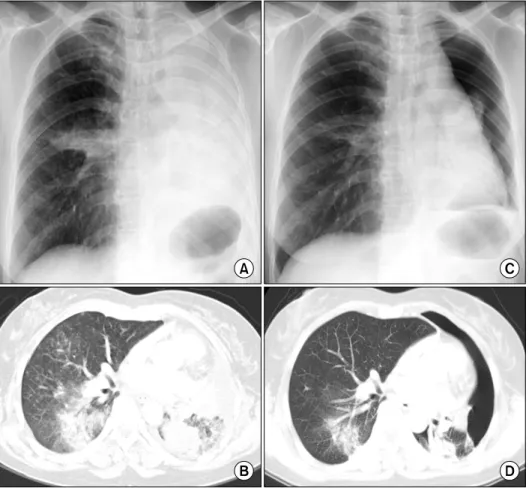
Figure 1. Chest X-ray and computed tomography were performed before treatment with erlotinib (A, B). After 7 weeks of erlotinib administration, a spontaneous pneumothorax was first confirmed in the left lung; the consolidations and ground glass opacities in the lung showed marked reductions in size with erlotinib treatment (C, D).
Figure 2. Nine weeks after spontaneous pneumothorax, the left pneumo- thorax showed spontaneous improvement in chest X-ray (A) and com- puted tomography (B).
Tuberculosis and Respiratory Diseases Vol. 69. No. 6, Dec. 2010
467 cycles of cisplatin-based doublets chemotherapy, which
showed no effect. After three cycles of cisplatin-based doublets chemotherapy, a chest X-ray (Figure 1A) re- vealed total opacification of the left lung and right mid- dle lobe infiltration. A chest CT scan (Figure 1B) showed this to be secondary to diffuse consolidations and GGOs in the left lung, and small multiple nodules and consolidations in the right lower lobe. As she had never smoked and tumor biopsy specimens showed a deletion in exon 19 of the epidermal growth factor re- ceptor (EGFR), second-line chemotherapy with erlotinib (daily 150 mg) was begun.
After 7 weeks, she was asymptomatic at a routine checkup at our outpatient clinic, but her chest X-ray (Figure 1C) demonstrated left SPTx. She reported no history of trauma. Chest CT (Figure 1D) indicated that the consolidations or GGOs in the lung had been mark- edly reduced in size by erlotinib without severe side ef- fects, except for the SPTx. Thus, the SPTx was observed without any therapeutic intervention. Nine weeks later, chest X-ray (Figure 2A) and CT (Figure 2B) taken at the outpatient clinic revealed spontaneous improvement of the left pneumothorax, even though erlotinib admin- istration had been continued.
Discussion
SPTx associated with malignant pulmonary neoplasm is rare. It has been reported as a first sign of lung in- volvement for certain tumors or as a complication aris- ing from radiotherapy or chemotherapy in patients suf- fering from a variety of malignancies5-8. In general, che- motherapy-related SPTx has been reported in chemo- sensitive tumors, particularly sarcomas or germ cell tu- mors, with multiple lung metastases6-8. However, SPTx associated with primary lung cancer occurs at a very low frequency. Lai et al.2 reported that 18 (0.32%) of 5,567 patients with primary lung cancer had SPTx as a complication, and only two (0.04%) of these cases oc- curred after chemotherapy. Rarely, sporadic cases of SPTx have been reported associated with chemotherapy in primary lung cancer2-4. To the best of our knowledge,
this is the first case of erlotinib-related SPTx in a patient with primary lung cancer in Korea.
The mechanism of chemotherapy-related SPTx is largely elusive; however, various mechanisms have been proposed4,6,8. For example, rapid tumor lysis or tumor tissue necrosis due to chemotherapy may directly induce the formation of fistulas. Alternatively, there may be underlying subpleural bullae or blebs that rupture after chemotherapy.
Erlotinib is a small-molecule tyrosine kinase inhibitor directed against the EGFR. The presence of an EGFR mutation or of clinical factors such as adenocarcinoma or never having smoked is associated with the response to erlotinib9. Taken together with the temporal relation- ship between erlotinib administration and radiological regression of the tumors, this was likely the etiology in the patient described here, as she had no history of smoking, was positive for EGFR mutation, had no pre- vious history of lung disease, and lacked any other rele- vant history. Apoptosis of subpleural tumor cells, in- duced by tyrosine kinase inhibitors such as erlotinib or gefitinib that are directed against EGFR10, probably in- duces the formation of bronchopleural fistulas. There- fore, we postulated that erlotinib induced rapid tumor lysis and necrosis of peripherally located tumor tissues, leading to SPTx.
Generally, treatment of chemotherapy-related SPTx involves closed chest tube insertion8. In addition, chem- ical pleurodesis or surgical intervention may be consid- ered in patients with recurrent and/or bilateral chemo- therapy-related SPTx6,7. However, some chemotherapy- related SPTx in patients with primary lung cancer, par- ticularly those responding to chemotherapy for lung cancer, improve spontaneously, without any therapeutic intervention, while continuing chemotherapy3,4. The present case showed also that asymptomatic SPTx ac- companied by significant decrease in the size of the tu- mor resolved spontaneously, without any therapeutic in- tervention, during continued erlotinib administration.
These cases suggest that the tissue repair process can continue despite chemotherapy for lung cancer. Further studies are needed to understand the cause of sponta-
HS Nam et al: Erlotinib-related spontaneous pneumothorax
468
neously resolving chemotherapy-related SPTx.
In summary, this case showed that SPTx can occur in patients with primary lung cancer receiving erlotinib.
Asymptomatic chemotherapy-related SPTx in primary lung cancer serves as a reminder to clinicians that it may spontaneously improve with no therapeutic inter- vention, and that it may represent an indirect measure of response to chemotherapy.
Acknowledgements
This study was supported by INHA UNIVERSITY Research Grant (INHA-40900-01).
References
1. Steinhäuslin CA, Cuttat JF. Spontaneous pneumothorax:
a complication of lung cancer? Chest 1985;88:709-13.
2. Lai RS, Perng RP, Chang SC. Primary lung cancer com- plicated with pneumothorax. Jpn J Clin Oncol 1992;
22:194-7.
3. O'Connor BM, Ziegler P, Spaulding MB. Spontaneous pneumothorax in small cell lung cancer. Chest 1992;
102:628-9.
4. Mori M, Nakagawa M, Fujikawa T, Iwasaki T, Kawa- mura T, Namba Y, et al. Simultaneous bilateral sponta-
neous pneumothorax observed during the administ- ration of gefitinib for lung adenocarcinoma with multi- ple lung metastases. Intern Med 2005;44:862-4.
5. Dines DE, Cortese DA, Brennan MD, Hahn RG, Payne WS. Malignant pulmonary neoplasms predisposing to spontaneous pneumothorax. Mayo Clin Proc 1973;48:
541-4.
6. Biran H, Dgani R, Wasserman JP, Weissberg D, Shani A. Pneumothorax following induction chemotherapy in patients with lung metastases: a case report and liter- ature review. Ann Oncol 1992;3:297-300.
7. Fenlon HM, Carney D, Breatnach E. Case report: bi- lateral recurrent tension pneumothorax complicating combination chemotherapy for soft tissue sarcoma. Clin Radiol 1996;51:302-4.
8. Stein ME, Shklar Z, Drumea K, Goralnik L, Ben-Arieh Y, Haim N. Chemotherapy-induced spontaneous pneu- mothorax in a patient with bulky mediastinal lympho- ma: a rare oncologic emergency. Oncology 1997;54:
15-8.
9. Tsao MS, Sakurada A, Cutz JC, Zhu CQ, Kamel-Reid S, Squire J, et al. Erlotinib in lung cancer - molecular and clinical predictors of outcome. N Engl J Med 2005;353:133-44.
10. Moyer JD, Barbacci EG, Iwata KK, Arnold L, Boman B, Cunningham A, et al. Induction of apoptosis and cell cycle arrest by CP-358,774, an inhibitor of epi- dermal growth factor receptor tyrosine kinase. Cancer Res 1997;57:4838-48.