Yonsei Med J http://www.eymj.org Volume 52 Number 4 July 2011 695
Case Report
DOI 10.3349/ymj.2011.52.4.695pISSN: 0513-5796, eISSN: 1976-2437 Yonsei Med J 52(4):695-698, 2011
Bowel Perforation after Erlotinib Treatment in a Patient with Non-Small Cell Lung Cancer
Yun-Hong Cheon,
1,2,3Moon Jin Kim,
1,2,3Min Gyu Kang,
1,2,3Hee Jin Kim,
1,2,3Sang Su Lee,
1,2,3Cha Young Kim,
1,2,3Dae-Hong Jeon,
1,2,3Yu Eun Kim,
1,2,3and Gyeong-Won Lee
1,2,31Department of Internal Medicine, School of Medicine, Gyeongsang National University, Jinju;
2Gyeongsang Institute of Health Science, Jinju; 3Gyeongnam Regional Cancer Center, Jinju, Korea.
Received: February 9, 2010 Revised: February 26, 2010 Accepted: March 3, 2010
Corresponding author: Dr. Gyeong-Won Lee, Division of Oncology-Hematology, Department of Internal Medicine, School of Medicine,
Gyeongsang National University, 79 Gangnam-ro, Jinju 660-702, Korea.
Tel: 82-55-750-8066, Fax: 82-55-758-9122 E-mail: [email protected]
∙ The authors have no financial conflicts of interest.
© Copyright:
Yonsei University College of Medicine 2011 This is an Open Access article distributed under the terms of the Creative Commons Attribution Non- Commercial License (http://creativecommons.org/
licenses/by-nc/3.0) which permits unrestricted non- commercial use, distribution, and reproduction in any medium, provided the original work is properly cited.
Erlotinib is accepted as a standard second-line chemotherapeutic agent in patients with non-small cell lung cancer who are refractory or resistant to first-line plati- num-based chemotherapy. There has been no previous report of bowel perforation with or without gastrointestinal metastases related to erlotinib in patients with non- small cell lung cancer. The exact mechanism of bowel perforation in patients who received erlotinib remains unclear. In this report, we report the first case of entero- cutaneous fistula in a female patient with metastatic non-small cell lung cancer 9 months, following medication with erlotinib as second-line chemotherapy.
Key Words: Lung neoplasm, intestinal fistula, erlotinib
INTRODUCTION
Chemotherapy is a mainstay treatment for advanced or metastatic non-small cell lung cancer (NSCLC).1 Erlotinib, an epidermal growth factor receptor-tyrosine ki- nase inhibitor (EGFR-TKI), has been shown to be effective for Asian patients with favorable clinical features, such as female gender, never or non-smoker, and ade- nocarcinoma.2 The most common adverse effect is skin rash. However, other ad- verse effects are relatively minor and manageable.2-6
Among several targeted agents with different mechanisms of action, patients treated with vascular endothelial growth factor (VEGF)-targeted agents, such as be- vacizumab, develop hypertension, thrombosis, gastrointestinal bleeding, and bowel perforation as uncommon adverse effects.7-11 However, there has been no previous report of gastrointestinal perforation or fistula related to erlotinib administration.
Here, we report a case of enterocutaneous fistula in a patient with NSCLC after the ninth course of erlotinib treatment as a second-line palliative chemotherapy.
CASE REPORT
A 66-year-old female was diagnosed with metastatic non-small cell adenocarcino- ma of the lung [NSCLC; cT4N0M1 for metastases of the lung to the contralateral
Yun-Hong Cheon, et al.
Yonsei Med J http://www.eymj.org Volume 52 Number 4 July 2011 696
of bone metastasis was detected in the bone biopsy speci- men, which was compatible with a benign compression frac- ture related to severe osteoporosis. Additionally, ibuprofen (200 mg three times daily) was added to relieve low back pain for 7 days after the procedures.
Nine months after starting second-line chemotherapy with erlotinib, the patient was admitted with purulent yellowish sputum, cough, and mild shortness of breath without abdom- inal symptoms. Chest CT scan demonstrated decreased size of the primary lung mass, which was still evaluated as a par- tial response, with focal, patchy consolidation and peribron- chial thickening in the left lung field, clinically compatible with pneumonia.
On physical examination, a coarse breathing sound with a crackle, especially in the left lung field, was detected, and an enterocutaneous fistula with leakage of fecal material was found in the previous sigmoid colon herniation through the abdominal wall defect (Fig. 1A and B). Nevertheless, no sign of strangulation, including tenderness or rebound tenderness, was observed. Fistulography also demonstrated passage of contrast media into the sigmoid colon without another fistula (Fig. 2). Further administration of erlotinib was withheld after documentation of the enterocutaneous fistula.
After resolution of pneumonia via empirical antibiotic treatment for 14 days, a scheduled sigmoid colon resection and primary anastomosis were performed at the Depart- ment of General Surgery. Surgical pathologic findings in- cluded submucosal edema and serosal hemorrhage of the perforated sigmoid colon, consistent with enterocutaneous fistula and no metastatic tumor foci in the sigmoid colon (Fig. 3A and B). Ten days postoperatively, the patient was discharged with complete recovery from the enterocutane- ous fistula. She was alive and well on regular follow-up at lung and pleura on 18F-fluorodeoxyglucose positron emis-
sion tomography computed tomography (FDG-PET CT) scan] in January 2008. The patient had a history of explor- atory laparotomy and a total hysterectomy, due to acute ab- domen about 30 years earlier. Physical examination re- vealed sigmoid colon herniation through an abdominal wall defect, with no sign or symptom of strangulation. Although surgical management of the herniated colon loop and clo- sure of the abdominal wall were strongly recommended, she refused further surgical treatment, because there had been no symptom, such as abdominal pain or bowel habit abnormality associated with the sigmoid colon herniation, over the previous 30 years.
She was treated with a first-line chemotherapy, including cisplatin (70 mg/m2, day 1) and docetaxel (70 mg/m2, day 1) every 3 weeks for 6 cycles from January 2008 to July 2008. Following completion of 6 cycles of first-line chemo- therapy, a chest CT scan showed a partial response, accord- ing to the Response Evaluation Criteria in Solid Tumors (RECIST).
In October 2008, second-line chemotherapy with erlo- tinib was started because a follow-up chest CT scan re- vealed regrowth of the primary lung mass in the left upper lobe and increased malignant pleural effusion. A subse- quent chest CT scan showed persistent partial response at the first, third, fifth, and seventh month following erlotinib administration.
In November 2008 (seventh month following erlotinib administration), the patient complained of low back pain after a fall at home, and a compression fracture of the fifth lumbar vertebra was detected on a simple spine X-ray and spine magnetic resonance image (MRI). Percutaneous ver- tebroplasty and concomitant bone biopsy were performed at the Department of Neurosurgery. However, no evidence
Fig. 2. Fistulography shows well passage of contrast media into the sigmoid colon without another fistu.
Fig. 1. (A and B) White arrow indicates an enterocutaneous fistula in the sigmoid colon herniation through the abdominal wall defect.
A B
Bowel Perforation Following Erlotinib
Yonsei Med J http://www.eymj.org Volume 52 Number 4 July 2011 697
to bowel perforation regardless of their mechanism of ac- tion, especially in patients with visceral metastases caused by tumor regression and necrosis, our patient showed no ev- idence of bowel metastasis, according to radiologic findings at initial diagnosis or during administration of erlotinib, or pathologic findings of the post-operative resected bowel specimen.
In conclusion, physicians should be aware of the rare, but possible, adverse effect of bowel perforation or fistula fol- lowing administration of erlotinib, even in NSCLC patients without visceral metastases, and erlotinib administration should be discontinued when patients develop gastrointesti- nal perforation.
REFERENCES
1. Non-Small Cell Lung Cancer Collaborative Group. Chemothera- py and supportive care versus supportive care alone for advanced non-small cell lung cancer. Cochrane Database Syst Rev 2010:
CD007309.
2. Uhm JE, Park BB, Ahn MJ, Lee J, Ahn JS, Kim SW, et al. Erlotinib monotherapy for stage IIIB/IV non-small cell lung cancer: a mul- ticenter trial by the Korean Cancer Study Group. J Thorac Oncol 2009;4:1136-43.
3. Dudek AZ, Kmak KL, Koopmeiners J, Keshtgarpour M. Skin rash and bronchoalveolar histology correlates with clinical benefit in patients treated with gefitinib as a therapy for previously treated advanced or metastatic non-small cell lung cancer. Lung Cancer 2006;51:89-96.
4. Mohamed MK, Ramalingam S, Lin Y, Gooding W, Belani CP.
Skin rash and good performance status predict improved survival with gefitinib in patients with advanced non-small cell lung can- cer. Ann Oncol 2005;16:780-5.
5. Hotta K, Kiura K, Ueoka H, Tabata M, Fujiwara K, Kozuki T, et al. Effect of gefitinib (‘Iressa’, ZD1839) on brain metastases in patients with advanced non-small-cell lung cancer. Lung Cancer 2004;46:255-61.
6. Kris MG, Natale RB, Herbst RS, Lynch TJ Jr, Prager D, Belani CP, et al. Efficacy of gefitinib, an inhibitor of the epidermal growth fac- tor receptor tyrosine kinase, in symptomatic patients with non-small cell lung cancer: a randomized trial. JAMA 2003;290:2149-58.
7. Reck M, von Pawel J, Zatloukal P, Ramlau R, Gorbounova V, Hirsh V, et al. Phase III trial of cisplatin plus gemcitabine with either placebo or bevacizumab as first-line therapy for nonsquamous non-small-cell lung cancer: AVAil. J Clin Oncol 2009;27:1227-34.
8. Di Costanzo F, Mazzoni F, Micol Mela M, Antonuzzo L, Chec- cacci D, Saggese M, et al. Bevacizumab in non-small cell lung cancer. Drugs 2008;68:737-46.
9. Johnson DH, Fehrenbacher L, Novotny WF, Herbst RS, Nemun- aitis JJ, Jablons DM, et al. Randomized phase II trial comparing bevacizumab plus carboplatin and paclitaxel with carboplatin and paclitaxel alone in previously untreated locally advanced or meta- static non-small-cell lung cancer. J Clin Oncol 2004;22:2184-91.
10. Cohen MH, Gootenberg J, Keegan P, Pazdur R. FDA drug ap- proval summary: bevacizumab plus FOLFOX4 as second-line
the outpatient clinic, with no evidence of lung cancer pro- gression.
DISCUSSION
To our knowledge, there is no previously reported case of enterocutaneous fistula without intestinal metastasis in a pa- tient with NSCLC who received erlotinib.
Hoffman-La Roche Ltd. announced that gastrointestinal perforation (including fatalities) has been reported in pa- tients receiving erlotinib, and the prescribing information in- dicates that patients receiving concomitant anti-angiogenic agents, corticosteroids, nonsteroidal anti-inflammatory drugs (NSAIDs), and/or taxane-based chemotherapy, or who have a prior history of peptic ulceration or diverticular disease, are at increased risk of gastrointestinal perforation.12
Our patient had risk factors related to gastrointestinal per- foration during medication with erlotinib. First, she had a history of previous abdominal surgery, which was eventual- ly complicated with sigmoid colon herniation through an abdominal wall defect. This condition may cause a continu- ous increase of the pressure on already weakened areas of the bowel, thus further predisposing patients to bowel perfo- rations or fistulae that re-epithelialize poorly during erlotinib administration.13,14 Second, NSAIDs were administered con- comitantly for 7 days following percutaneous vertebroplasty, which could block COX 1 and COX 2 enzyme activity, also leading to gastrointestinal perforations through the de- velopment of ulcers and poor ulcer healing.13,14 There is strong evidence that HER-1/EGFR and VEGF share a com- mon downstream signaling pathway both in vitro and in vivo, and they exert effects on tumor cells directly or indirectly.15-18
Although we suggest that potent cytotoxic agents can lead
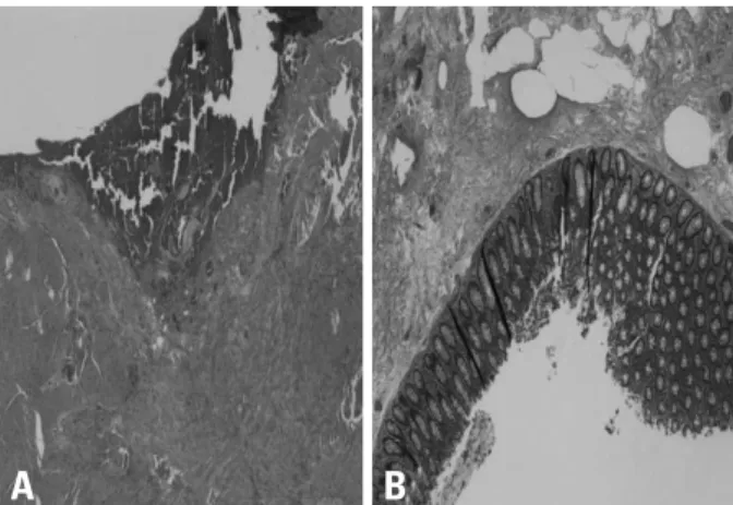
Fig. 3. (A and B) Submucosal edema and serosal hemorrhage of the perfo- rated sigmoid colon.
A B
Yun-Hong Cheon, et al.
Yonsei Med J http://www.eymj.org Volume 52 Number 4 July 2011 698
drogen-independent human prostate cancer growing in the bone of nude mice. Clin Cancer Res 2003;9:1200-10.
16. de Jong JS, van Diest PJ, van der Valk P, Baak JP. Expression of growth factors, growth-inhibiting factors, and their receptors in in- vasive breast cancer. II: Correlations with proliferation and angio- genesis. J Pathol 1998;184:53-7.
17. Petit AM, Rak J, Hung MC, Rockwell P, Goldstein N, Fendly B, et al. Neutralizing antibodies against epidermal growth factor and ErbB-2/neu receptor tyrosine kinases down-regulate vascular en- dothelial growth factor production by tumor cells in vitro and in vivo: angiogenic implications for signal transduction therapy of solid tumors. Am J Pathol 1997;151:1523-30.
18. Bruns CJ, Harbison MT, Davis DW, Portera CA, Tsan R, McCon- key DJ, et al. Epidermal growth factor receptor blockade with C225 plus gemcitabine results in regression of human pancreatic carcinoma growing orthotopically in nude mice by antiangiogenic mechanisms. Clin Cancer Res 2000;6:1936-48.
treatment of colorectal cancer. Oncologist 2007;12:356-61.
11. Saif MW, Elfiky A, Salem RR. Gastrointestinal perforation due to bevacizumab in colorectal cancer. Ann Surg Oncol 2007;14:1860-9.
12. Johnson JR, Cohen M, Sridhara R, Chen YF, Williams GM, Duan J, et al. Approval summary for erlotinib for treatment of patients with locally advanced or metastatic non-small cell lung cancer af- ter failure of at least one prior chemotherapy regimen. Clin Cancer Res 2005;11:6414-21.
13. Schellhaas E, Loddenkemper C, Schmittel A, Buhr HJ, Pohlen U.
Bowel perforation in non-small cell lung cancer after bevacizum- ab therapy. Invest New Drugs 2009;27:184-7.
14. Badgwell BD, Camp ER, Feig B, Wolff RA, Eng C, Ellis LM, et al.
Management of bevacizumab-associated bowel perforation: a case series and review of the literature. Ann Oncol 2008;19:577-82.
15. Kim SJ, Uehara H, Karashima T, Shepherd DL, Killion JJ, Fidler IJ. Blockade of epidermal growth factor receptor signaling in tu- mor cells and tumor-associated endothelial cells for therapy of an-