Tuberc Respir Dis 2009;67:331-337
CopyrightⒸ2009. The Korean Academy of Tuberculosis and Respiratory Diseases. All rights reserved.
폐외 결핵에서 전혈 인터페론 감마 측정법의 진단적 유용성
중앙대학교 의과대학 내과학교실
이혜민, 조성근, 강형구, 박성운, 이병욱, 이재희, 전은주, 최재철
The Usefulness of Whole-blood Interferon-gamma Release Assay for the Diagnosis of Extra-pulmonary Tuberculosis
Hye-Min Lee, M.D., Sung Gun Cho, M.D., Hyung Koo Kang, M.D., Sung Woon Park, M.D., Byung Ook Lee, M.D., Jae Hee Lee, M.D., Eun Ju Jeon, M.D., Jae Chol Choi, M.D.
Division of Pulmonary and Critical Care Medicine, Department of Internal Medicine, Chung-Ang University College of Medicine, Seoul, Korea
Background: The whole-blood interferon-gamma release assay (QuantiFERON-TB Gold [QFT-G]: Cellestis, Carnegie, Victoria, Australia) has been studied primarily for the use of diagnosing active pulmonary tuberculosis (TB) or latent TB. In the present study, the usefulness of QFT-G was evaluated for the diagnosis of extra-pulmonary tuberculosis (EP-TB).
Methods: From June 2006 to February 2009, we evaluated the usefulness of QFT-G in patients (n=65) suspected with EP-TB, retrospectively. The diagnostic sensitivity, specificity, positive predictive value (PPV), and negative predictive value (NPV) of the QFT-G assay were analyzed.
Results: EP-TB was diagnosed in 33 (51%) participants. The overall sensitivity, specificity, PPV, and NPV of the QFT-G assay for EP-TB were 78%, 79%, 81%, and 77%, respectively. Of the 33 with EP-TB, 14 (42%) were diagnosed with TB pleurisy, 7 (21%) with TB lymphadenitis, 7 (21%) with intestinal TB, and 5 (15%) with EP-TB in other sites. In subgroup analyses according by site of infection, the QFT-G showed 86% sensitivity, 64% specificity, and 78% NPV in TB pleurisy. On the other hand, the sensitivity, specificity, and NPV of the assay were 71%, 83%
and 71%, respectively in TB lymphadenitis, and 86%, 100% and 88%, respectively in intestinal TB. Among the patients with suspected alternative site EP-TB, the sensitivity, specificity, and NPV of the assay were 50%, 80%
and 67%, respectively.
Conclusion: The QFT-G assay showed moderate diagnostic accuracy in EP-TB. However, negative QFT-G assay does not exclude EP-TB because of the low NPV of this assay.
Key Words:
Mycobacterium tuberculosis
, Interferon-gamma, DiagnosisThis research was supported by the Chung-Ang University Research Grants in 2007.
Address for correspondence: Jae Chol Choi, M.D.
Division of Pulmonary and Critical Care Medicine, Depart- ment of Medicine, Chung-Ang University Medical Center, 65- 207, Hangangno 3-ga, Youngsan-gu, Seoul 140-757, Korea Phone: 82-2-748-9804, Fax: 82-2-749-9560
E-mail: [email protected] Received: Jul. 23, 2009 Accepted: Sep. 7, 2009
서 론
세계보건기구(World Health Organization)의 통계에 의
하면 2006년도에 세계 인구 중 920만명(10만명당 139명)
의 결핵 신고 신환자가 발생하고 있으며 이 중 170만명이
사망하였다
1. 우리나라의 2007년도 결핵 신고 신 환자 수
는 34,710명(연간 10만명당 71.6명)이었으며, 그 중 폐외
결핵 환자는 5,005명(연간 10만명당 10.3명)으로 보고되고
있다
2. 이처럼 폐외 결핵은 폐결핵에 비해 드물기는 하지
만 노령화, 사람면역결핍바이러스(human immunodefici-
ency virus, HIV) 감염 환자 및 면역억제제 사용 환자가 늘어남에 따라 관심이 증가하고 있다. 폐외 결핵은 폐 이외 의 모든 장기에 침범한 결핵으로써 흉막결핵이 가장 흔하 고, 림프절결핵이 두 번째이며, 복부결핵, 골관절결핵, 비 뇨기계 결핵, 신경계 결핵의 빈도로 보고되고 있다
3. 폐결핵은 객담검사와 영상의학적 검사 등의 간단한 방 법으로 진단되는데 비해 폐외 결핵은 환자의 분비물이나 조직을 얻기 위한 침습적인 방법이 필요한 경우가 많다.
또한, 임상양상이 비전형적인 경우가 많고, 검체의 항산균 도말 검사와 배양 검사의 민감도가 낮으며 결핵균 배양에 는 2∼7주 정도가 소요되어 진단에 어려움이 많다
4,5. 또 한, 결핵을 진단하는데 도움이 되는 투베르쿨린 피부검사 (tuberculin skin test, TST)는 우리나라처럼 bacilli Cal- mette-Guérin (BCG) 예방접종을 필수적으로 하는 경우 투베르쿨린 피부검사의 특이도에 대한 제한점이 있다
6,7. 2001년 이후 전혈 인터페론 감마 측정법(interferon- gamma releasing assay, IGRA)이 결핵을 진단하는 데 사 용되고 있다
8. 최근에는 활동성 폐결핵에서 유용성에 대 한 연구가 보고 되었으나
9-11, 폐외 결핵에서의 진단적 유 용성에 대한 연구는 많지 않으며, 폐외 결핵의 진단 시 IGRA의 민감도가 14%에서 80%까지 다양하게 보고되고 있다
6,7,10,12-14. 이에 본 연구에서는 폐외 결핵에서 IGRA의 진단적 유용성에 대해 알아보고자 하였다.
대상 및 방법
1. 대상 환자
2006년 6월부터 2009년 2월까지 중앙대학교병원에서 임상적으로 폐외 결핵이 의심되는 환자 중 IGRA를 시행 한 환자 65명을 후향적으로 분석하였다. 폐외 결핵은 감 염부위에 따라 흉막결핵, 장결핵, 림프절결핵, 신경계결 핵, 골관절결핵 및 기타결핵으로 나눠 살펴보았으며, 각각 폐외 결핵으로 의심되는 환자는 다음과 같이 정의하였다.
1) 흉막결핵은 흉부 방사선촬영에서 편측성의 흉막액이 있고 폐렴을 의심할 경결은 관찰 되지 않으며 흉막액의 세균 배양검사에서 음성인 경우에 흉막결핵을 의심하였 고, 2) 장결핵은 복통, 설사, 발열, 체중감소, 오심 등의 증상이 있고 대장내시경 소견에서 주로 회맹부를 침범하 는 궤양 또는 점막비후가 있는 경우 장결핵을 의심하였으 며, 3) 림프절결핵은 단단하고 경계가 명확한 무통성 림프 절종창이 특징인 경우 의심하였고, 4) 신경계결핵은 두통, 발열, 경부강직 등의 뇌막자극증상이 있으면서, 뇌척수액
검사에서 림프구 우세를 보이는 경우 신경계결핵을 의심 하였다. 5) 골관절 결핵, 간결핵 등의 기타 폐외 결핵으로 의심되는 경우는 전산화 단층 촬영(computed tomog- raphy, CT)과 같은 영상의학적 검사에서 결핵이 의심되었 던 환자를 대상으로 하였다.
이 환자 65명을 대상으로 결핵의 과거력이 있는 환자와 기저질환(당뇨병, 간경화, 만성신부전, 악성종양, HIV 감 염, 면역억제제를 최근 3개월 이내 복용한 경우) 동반 여 부에 대해 분석하였다. 폐외 결핵은 폐 이외의 부위에 감 염된 결핵으로 폐외 결핵의 진단은 1) 감염부위의 조직에 서 건락육아종(caseous granuloma)이 증명된 경우, 2) 조 직검사에서 항산막대균(acid-fast bacillus)이 발견된 경우, 3) 조직배양에서 결핵균이 증명된 경우, 4) 진단적 흉강천 자를 시행하고 흉막액이 삼출액이며, 아데노신 탈아미노효 소(adenosine deaminase, ADA) 40 U/L 이상과 림프구/
호중구 비가 0.75 이상일 때 진단하였다
1,15,16. 흉막결핵을 진단할 때 사용한 흉수 아데노신 탈아미노효소 활성도는 자동화기기로 분석하였다(model 7180; Hitachi, Tokyo, Japan). 이 연구는 중앙대학교 용산병원 의학연구심의위 원회 승인을 받았다.
2. IGRA
IGRA는 QuantiFERON-TB Gold (QuantiFERON-TB Gold [QFT-G]; Cellestis, Carnegie, Victoria, Australia)로 시행 하였다. QFT-G는 헤파린 처리된 용기에 환자의 전혈을 채취하여 배양용기에 각각 1 mL씩 분주하고 각 well에 결핵균 특이항원 early secreted antigenic target-6 kDa (ESAT-6)과 culture filtrate protein-10 kDa (CFP-10), 양성 대조 항원(mitogen) 및 음성 대조 항원(nil)을 첨가한 뒤 16∼24시간 배양한다. 배양 후 생성되는 인터페론 감마 를 효소면역 측정법(enzyme-linked immunosorbent as- say, ELISA)으로 측정하여 판독하였다. 인터페론 감마 농 도가 ESAT-6 또는 CFP-10에서 0.35 IU/mL 이상일 경우 양성으로 판정하고, 환자의 면역 상태와 관련된 양성 대조 항원이 0.5 IU/mL 이하인 경우에는 미정(indeterminate) 인 경우로 하였다
8,12.
3. 통계
QFT-G의 진단적 유용성은 민감도, 특이도, 양성예측
도, 음성예측도를 통하여 분석하였다. 양측검정 95%의 신
뢰수준은 Wilson score method을 사용하였다. 폐외 결핵
환자와 폐외 결핵이 아닌 환자군 간의 비교는 연속변수의
Table 1. Demographic and clinical characteristics of the patients with suspected extra-pulmonary tuberculosis
Characteristics
Final diagnosis
EP-TB (n=33) Not EP-TB (n=32) p-value
Age, median years (range) 40 (13∼86) 47 (17∼98) 0.397
Male/female 16/17 22/10 0.1
Duration of follow-up, month (range) 6 (1∼60) 6 (0.6∼60) 0.963
Prior history active TB 3 (9) 4 (13) 0.66
Immune-compromised conditions 7 (21) 8 (25) 0.719
Diabetes mellitus 5 (15) 3 (9)
Malignancy 1 (3) 2 (6)
Chronic renal failure 0 2 (6)
Liver cirrhosis 0 2 (6)
Immunosuppressant (≤3 months) 1 (3) 0
HIV infection 0 1 (3)
Data are no. (%) of patients, unless otherwise indicated.
EP-TB: extra-pulmonary tuberculosis; TB: tuberculosis; HIV: human immunodeficiency virus.
Table 2. Distribution of the site of infection with suspected extra-pulmonary tuberculosis
Site of infection
Final diagnosis
EP-TB (n=33) Not EP-TB (n=32)
Pleural 14 (42) 12 (38)
Gastrointestinal system 7 (21) 7 (22)
Lymph node 7 (21) 6 (19)
Central nervous system 0 6 (19)
Bone and/or joint 4 (12) 0
Liver 1 (3) 1 (3)
Data are no. (%) of patients, unless otherwise indicated.
EP-TB: extra-pulmonary tuberculosis.
경우 Mann-Whitney U-test를, 범주형 변수의 경우 Fish- er’s exact test를 시행하였다. p값이 0.05 미만일 때 통계 적으로 유의하다고 판정하였다. 통계분석은 SPSS for windows 12.0 (SPSS Inc., Chicago, IL, USA) 프로그램을 이용하였다.
결 과
1. 대상 환자의 특성
폐외 결핵이 의심되어 QFT-G를 시행한 환자는 65명이 었고 그 중 폐외 결핵 환자가 33명(51%), 폐외 결핵이 아 닌 환자가 32명(49%)이었다. 전체 환자의 나이의 중앙값 은 46세(범위 13∼98세)이고 남자가 38명(59%)이었으며, 중앙 추적관찰 기간은 6개월(범위 0.6∼60)이었다. 결핵 치료의 과거력이 있는 환자는 7명(11%)이었으며 기저질 환을 가진 환자는 중복된 질환을 포함하여 15명(23%)이 었다. 폐외 결핵 환자와 폐외 결핵이 아닌 환자군 사이에 나이, 성별, 추적관찰 기간, 결핵치료의 과거력, 면역억제 질환의 동반여부는 차이가 없었다(Table 1).
폐외 결핵으로 진단된 33명의 감염부위별 빈도는 흉막 결핵 14명(42%), 장결핵 7명(21%), 림프절결핵 7명(21%) 및 기타 폐외 결핵의 순서였다. 폐외 결핵이 아닌 32명의 감염부위별 질환은 흉막 질환 12명(38%) [농흉 2명, 부폐 렴성 흉수 3명, 악성 흉수 3명, 심부전에 동반된 흉막액 2명, 흉막유착 1명, 베게너씨 육아종(Wegener’s gran- ulomatosis) 1명], 장질환 7명(22%) [크론씨병(Crohn’s
disease) 5명, 장염 2명], 림프절 질환 6명(19%) [기쿠치씨 병(Kikuchi’s disease) 4명, 사르코이드증(sarcoidosis) 1 명, 림프종 1명], 기타 질환 7명[수막염 6명(19%), 간세포 성암(hepatocellular carcinoma)에 동반된 악성 복수 1명 (3%)]이었다(Table 2).
2. QuantiFERON-TB GOLD의 진단적 유용성
폐외 결핵이 의심되었던 환자에서 QFT-G양성인 환자
31명(48%) 중 폐외 결핵으로 진단된 환자는 25명(81%)이
었으며, QFT-G 음성인 환자 30명(46%) 중 폐외 결핵은
7명(23%)이었다. QFT-G결과가 미정(indeterminate)인 4
명(6%)을 제외하고, 폐외 결핵을 진단하는데 QFT-G의 민
감도, 특이도, 양성예측도, 음성예측도는 각각 78%, 79%,
Table 3. Results of the interferon gamma release assay among 65 patients with suspected extra-pulmonary tuberculosis, by patient population subgroup
Patients population
No. of subjects (%) Sensitivity Specificity PPV NPV
% (CI
95) % (CI
95) % (CI
95) % (CI
95)
EP-TB Not EP-TB
All patients 33 32 78 (60∼90) 79 (60∼91) 81 (62∼92) 77 (57∼89)
Pleural 14 (42) 12 (38) 86 (56∼97) 64 (32∼88) 75 (47∼92) 78 (40∼96)
Gastrointestinal 7 (21) 7 (21) 86 (42∼99) 100 (56∼100) 100 (52∼100) 88 (47∼99)
Lymph node 7 (21) 6 (19) 71 (30∼95) 83 (36∼99) 83 (36∼99) 71 (30∼95)
Others 5 (15) 7 (21) 50 (9∼91) 80 (30∼99) 67 (13∼98) 67 (24∼94)
EP-TB: extra-pulmonary tuberculosis; IGRA: interferon gamma release assay; PPV: positive predictive value; NPV: negative predictive value; CI
95: confidence interval 95%.
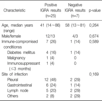
Table 4. Characteristics of patients who had verified cases of extra-pulmonary tuberculosis diagnosed
Positive Negative
Characteristic IGRA results IGRA results p-value
(n=25) (n=7)
Age, median years 41 (14∼86) 58 (13∼81) 0.264 (range)
Male/female 12/13 4/3 0.674
Immune-compromised 7 (26) 1 (14) 0.589 conditionss
Diabetes mellitus 4 (16) 1 (14)
Malignancy 1 (4) 0
Immunosuppressant 1 (4) 0 (≤3 months)
Site of infection 0.169
Pleural 12 (48) 2 (29)
Gastrointestinal 6 (24) 1 (14) Lymph node 5 (20) 2 (29)
Others 2 (8) 2 (29)
Data are no. (%) of patients, unless otherwise indicated.
IGRA: interferon gamma release assay.
81%, 77%이었다.
3. 감염부위별 QuantiFERON-TB GOLD의 진단적 유용성
흉막 결핵으로 의심된 환자 26명 중 16명(62%)에서 QFT-G 양성이었으며, 그 중 12명이 폐외 결핵으로 진단 되었다. QFT-G 검사가 음성이었던 9명(33%)의 환자 중 폐외 결핵은 2명이고, 미정 1명은 폐암에 동반된 악성흉 수로 진단되어, QFT-G의 민감도, 특이도, 양성예측도, 음 성예측도는 각각 86%, 64%, 75%, 78%이었다.
림프절 결핵으로 의심된 환자 13명 중 7명(54%)에서 림프절 결핵으로 진단되었다. QFT-G 양성인 6명(46%) 중 5명은 림프절 결핵으로 진단되었고, 1명은 기쿠치씨병으 로 진단되었다. QFT-G 음성인 환자 7명 중 2명(29%)이 림프절 결핵으로 진단되어 QFT-G의 민감도, 특이도, 양 성예측도, 음성예측도는 각각 71%, 83%, 83%, 71%였다.
장결핵으로 의심된 환자는 14명이었으며, 그 중 7명 (50%)이 장결핵으로 진단되었다. 장결핵이 의심된 환자 14명 중 QFT-G 양성 6명(43%)은 모두 장결핵으로 진단되 었고, QFT-G 음성인 환자 8명 중 1명이 장결핵으로 진단 되어 장결핵에서 QFT-G의 민감도, 특이도, 양성예측도, 음성예측도는 각각 86%, 100%, 100%, 88%였다.
기타 폐외 결핵 질환으로 의심된 경우는 신경계 결핵, 골관절결핵으로 분류되었으며 기타 폐외 결핵으로 의심 된 환자 12명 중 폐외 결핵으로 진단된 환자는 5명(42%), 다른 질환으로 진단된 환자는 7명(22%)이었다. 기타 폐외 결핵으로 의심되었던 환자 12명 중 QFT-G 양성인 환자 3명 중 2명(67%)이 폐외 결핵으로 진단되었으며, QFT-G 음성인 환자 6명 중 2명(33%)이 폐외 결핵으로 진단되었 고, 미정이 3명(25%)이었다. 기타 폐외 결핵에서 민감도,
특이도, 양성예측도, 음성예측도는 각각 50%, 80%, 67%, 67%였다(Table 3).
4. QuantiFERON-TB GOLD 위음성의 위험인자
폐외 결핵으로 진단된 33명 중 7명(21%)이 위음성의
소견을 보이고 있었다. QFT-G 양성인 환자와 QFT-G위음
성인 환자에서 나이, 성별, 면역억제 질환의 빈도 그리고
폐외 결핵의 감염부위는 통계학적으로 유의한 차이를 보
이지 않았다(Table 4).
고 찰
본 연구의 결과를 보면, QFT-G 검사법은 폐외 결핵이 의심되는 환자에서 중등도의 정확도를(민감도 78%, 특이 도 79%) 보임을 알 수 있었다. 하지만 77%의 음성예측도 를 보이고 있어, 이 검사법을 폐외 결핵을 배제하는 데 사용하는 것에는 제한점이 있음을 알 수 있다.
우리나라에서 결핵 신고 신환자 중 폐외 결핵은 14%
빈도로 보고되고 있다
2,16. 또한 스테로이드 등의 기존 면 역억제제 외에 종양괴사인자(tumor necrosis factor-α)에 대한 단일세포항체(monoclonal antibody) 제제가 류머티 즘, 크론씨병 등의 치료에 사용되면서 폐외 결핵의 빈도가 증가되고 있다
17. 따라서 이런 면역 억제 환자에서 다른 질환으로 오인되거나 진단이 늦어지면 치명적이거나 심 각한 후유증을 초래하기도 한다
18.
QFT-G 검사는 환자의 전혈과 결핵 특이적인 항원 (ESAT-6, CFP-10)을 이용하여 결핵에 감염되어 있는지를 검사하는 방법이다
19. 이 검사법은 투베르쿨린 피부검사와 달리 BCG 접종을 하는 군에서 위양성률이 낮은 장점 때문 에 우리나라와 같이 BCG 접종을 하는 나라에서도 결핵의 감염을 진단하는데 도움이 될 것으로 판단되고 있다
20,21. 최근에는 활동성 폐결핵에서 이 검사법의 유용성에 대한 보고들이 있다
10,22. 하지만, 잠복결핵과 현증결핵을 감별 할 수 없다는 근본적인 문제점으로 인하여 제한점이 있다.
그러나 IGRA와 TST를 병합하였을 때의 민감도가 99%로 높아서 두 검사 모두 음성이면 결핵을 배제하는 데에는 도움이 될 것으로 판단되고 있다
22.
IGRA의 폐외 결핵에 대한 보고는 하위 집단 분석을 통 한 연구가 대부분이었다
7,10,12,23. 최근에 Song 등
14은 100 명의 폐외 결핵 환자만을 대상으로 하여 진단율을 보고하 였다. 이 연구에서 민감도와 특이도는 69%와 82%로 중등 도의 정확도를 보였으나 78%의 낮은 음성예측도를 보이 고 있었다. IGRA가 잠복결핵과 현증결핵을 감별할 수 없 다는 문제점을 감안할 때 이 검사의 유용성은 결핵을 배제 할 수 있는가가 중요하다고 판단된다. 하지만, 이 결과를 바탕으로 생각하면 IGRA가 음성이라고 하더라도 결핵을 배제하기는 어려움을 알 수 있다. 그러나 이 연구는 다양 한 폐외 결핵 중 림프절 결핵과 척추결핵만을 대상으로 하였기 때문에 모든 폐외 결핵에 적용하기는 어려움이 있 을 수 있다. 따라서 본 연구에서는 다양한 형태의 폐외 결핵에서 이 검사의 정확성을 보고자 하였다. 본 연구의 결과를 살펴보면 QFT-G의 민감도는 78%, 음성예측도는
77%로 QFT-G 결과로 결핵을 예측하거나, 배제하는 것은 어려움을 알 수 있었다. 또한 감염부위로 나누어 살펴보 았을 때에도 음성예측도는 67∼88%로 감염부위에 상관 없이 낮은 음성예측도를 보이고 있었다. 최근 미국 질병 관리본부의 발표에 따르면 QFT-G 음성이 활동성 폐결핵 을 배제할 수 없다고 하였다
24. 본 연구 결과를 보아도 폐 외 결핵이 의심되는 환자에서 이 검사법을 이용하여 결핵 을 배제하기는 어려울 것으로 판단된다.
QFT-G 검사의 위음성은 면역억제 환자에서 흔하게 나 타난다고 알려져 있다
9,25. 본 연구에서도 위음성의 위험 인자를 알아보기 위하여 위음성과 양성인 군을 비교하여 보았으나 두 군 간에 면역 억제 환자의 빈도 그리고 폐외 결핵의 감염부위의 차이를 발견할 수는 없었다. 따라서 본 연구의 결과를 보면 면역 상태와 상관없이 일부의 환자 에서는 폐외 결핵에서도 위음성의 결과가 나타남을 알 수 있었고, 이러한 결과는 폐외 결핵의 진단에 있어서 면역상 태와 무관하게 QFT-G가 음성이라고 하더라도 폐외 결핵 을 배제해서는 안됨을 알 수 있었다.
폐외 결핵에서의 다양한 보고가 있지만 장결핵에 대한 결과는 많이 보고되지 않았다. 폐외 결핵 중 큰 비중을 차지하는 장결핵은 다른 장 질환과 비교할 때 증상이 큰 차이가 없다. 또한, 장결핵은 염증성 장질환과 내시경 소 견이 유사하여 감별이 어려운 경우가 많다. 장결핵과 크 론씨병을 감별하는 것이 중요한데 만약, 장결핵인데 크론 씨병으로 오진한 경우 면역억제 치료 후 장결핵이 전신 감염으로 악화될 가능성이 있기 때문에 정확한 진단이 필 수이다
26. 확진은 대장내시경 또는 조직학적 또는 조직검 사의 배양으로 진단할 수 있지만, 미생물학적 진단은 긴 잠복기로 진단에 어려우며, 조직검사에서도 감별이 어려 운 경우가 많이 있다. 본 연구에서 비록 환자 수가 많지는 않았으나 장결핵이 의심되었던 14명에서 음성 예측도는 88%로 내시경적 소견이 장결핵이 의심될 때 비록 QFT-G 검사가 음성이더라도 장결핵을 배제하기는 어려움을 알 수 있었으며, 특히 우리나라와 같이 잠복결핵의 빈도가 높은 경우에 낮은 민감도로 인하여 진단에 제한점이 있을 것으로 판단된다.
우리 연구에는 한계점이 있다. 첫 번째, 본 연구에서는
TST에 대한 비교 분석이 없다. TST와 IGRA를 같이 사용
하였을 때 결핵을 배제할 수 있다는 보고가 있듯이
22, 두
검사를 같이 시행하여 비교하여야 하나 후향적인 연구로
이에 대한 분석이 없다는 것이 가장 큰 제한점으로 생각된
다. 두 번째, 이 연구는 후향적인 연구로 모든 폐외 결핵이
의심되는 환자에서 QFT-G 검사가 시행되지 않았다. 따라 서 후향적 연구에 따른 선택적 비뚤림이 발생하였을 가능 성이 있다. 세 번째로, 본 연구에서는 전혈 인터페론 감마 검사로 QFT-G를 사용하였으며, T-SPOT.TB (Oxford Immunotec, Oxford, UK)이나 QuantiFERON-Gold In- Tube와 비교하지 않았으므로 본 연구 결과가 모든 IGRA 의 유용성을 반영하지는 않을 것으로 판단된다. 마지막으 로 작은 대상군 또한 중요한 제한점으로 판단된다. 하지 만, 이러한 제한점에도 불구하고 이번 연구는 다양한 폐외 결핵의 진단에 있어 QFT-G의 유용성을 본 논문으로 의미 가 있을 것으로 판단된다.
결론적으로, QFT-G는 폐외 결핵에서 중등도의 민감도 와 특이도를 보이고 있지만 폐외 결핵의 감염부위별로 67
∼88%의 음성 예측도로 인하여 QFT-G검사에서 음성의 결과를 보이더라도 폐외 결핵을 배제하기는 힘들 것으로 판단된다.
참 고 문 헌
1. World Health Organization. Global tuberculosis con- trol: surveillance, planning, financing, WHO/HTM/TB/
2008.393. Geneva: World Health Organization; 2008.
2. Korea Center for Disease Control and Prevention, Korean Institute of Tuberculosis. Annual report on the notified tuberculosis patients in Korea. Seoul: Korea Center for Disease Control and Prevention, Korean Institute of Tuberculosis; 2008.
3. Lew WJ. Tuberculosis surveillance system in Korea.
Tuberc Respir Dis 2000;48:298-307.
4. Steingart KR, Henry M, Laal S, Hopewell PC, Ramsay A, Menzies D, et al. A systematic review of commercial serological antibody detection tests for the diagnosis of extrapulmonary tuberculosis. Postgrad Med J 2007;83:
705-12.
5. Shim TS, Koh WJ, Yim JJ, Lew WJ. Diagnosis and treat- ment of latent tuberculosis infection in Korea. Tuberc Respir Dis 2004;57:101-8.
6. Pai M, Riley LW, Colford JM Jr. Interferon-gamma as- says in the immunodiagnosis of tuberculosis: a system- atic review. Lancet Infect Dis 2004;4:761-76.
7. Nishimura T, Hasegawa N, Mori M, Takebayashi T, Harada N, Higuchi K, et al. Accuracy of an interfer- on-gamma release assay to detect active pulmonary and extra-pulmonary tuberculosis. Int J Tuberc Lung Dis 2008;12:269-74.
8. Mazurek GH, LoBue PA, Daley CL, Bernardo J, Lardiza- bal AA, Bishai WR, et al. Comparison of a whole-blood interferon gamma assay with tuberculin skin testing for detecting latent Mycobacterium tuberculosis infection.
JAMA 2001;286:1740-7.
9. Menzies D, Pai M, Comstock G. Meta-analysis: new tests for the diagnosis of latent tuberculosis infection:
areas of uncertainty and recommendations for research.
Ann Intern Med 2007;146:340-54.
10. Dewan PK, Grinsdale J, Kawamura LM. Low sensitivity of a whole-blood interferon-gamma release assay for detection of active tuberculosis. Clin Infect Dis 2007;44:
69-73.
11. Mori T, Sakatani M, Yamagishi F, Takashima T, Kawabe Y, Nagao K, et al. Specific detection of tuberculosis in- fection: an interferon-gamma-based assay using new antigens. Am J Respir Crit Care Med 2004;170:59-64.
12. Ferrara G, Losi M, D'Amico R, Roversi P, Piro R, Meacci M, et al. Use in routine clinical practice of two commer- cial blood tests for diagnosis of infection with Mycobac- terium tuberculosis: a prospective study. Lancet 2006;
367:1328-34.
13. Munk ME, Arend SM, Brock I, Ottenhoff TH, Andersen P. Use of ESAT-6 and CFP-10 antigens for diagnosis of extrapulmonary tuberculosis. J Infect Dis 2001;183:175-6.
14. Song KH, Jeon JH, Park WB, Kim SH, Park KU, Kim NJ, et al. Usefulness of the whole-blood interferon- gamma release assay for diagnosis of extrapulmonary tuberculosis. Diagn Microbiol Infect Dis 2009;63:182-7.
15. Burgess LJ, Maritz FJ, Le Roux I, Taljaard JJ. Use of ad- enosine deaminase as a diagnostic tool for tuberculous pleurisy. Thorax 1995;50:672-4.
16. Park EB, Jeen YT, Ahn JH, Suh SJ, Lee SJ, Park NS, et al. Intestinal tuberculosis with a duodenal fistula.
Korean J Gastrointest Endosc 2007;35:346-50.
17. Lee YS, Jung JO, Hong JH, Seo YI, Eom KS, Jang SH, et al. Occurrence of tuberculous pleurisy associated with infliximab therapy. Korean J Med 2004;67:421-4.
18. Engin G, Acunas B, Acunas G, Tunaci M. Imaging of extrapulmonary tuberculosis. Radiographics 2000;20:
471-88.
19. Pai M, Gokhale K, Joshi R, Dogra S, Kalantri S, Mendir- atta DK, et al. Mycobacterium tuberculosis infection in health care workers in rural India: comparison of a whole-blood interferon gamma assay with tuberculin skin testing. JAMA 2005;293:2746-55.
20. Brock I, Weldingh K, Lillebaek T, Follmann F, Anders- en P. Comparison of tuberculin skin test and new spe- cific blood test in tuberculosis contacts. Am J Respir
Crit Care Med 2004;170:65-9.
21. Ewer K, Deeks J, Alvarez L, Bryant G, Waller S, Anders- en P, et al. Comparison of T-cell-based assay with tu- berculin skin test for diagnosis of Mycobacterium tuber- culosis infection in a school tuberculosis outbreak.
Lancet 2003;361:1168-73.
22. Dosanjh DP, Hinks TS, Innes JA, Deeks JJ, Pasvol G, Hackforth S, et al. Improved diagnostic evaluation of suspected tuberculosis. Ann Intern Med 2008;148:325- 36.
23. Pai M, Dheda K, Cunningham J, Scano F, O'Brien R.
T-cell assays for the diagnosis of latent tuberculosis in- fection: moving the research agenda forward. Lancet
Infect Dis 2007;7:428-38.
24. Mazurek GH, Jereb J, Lobue P, Iademarco MF, Met- chock B, Vernon A. Guidelines for using the Quanti- FERON-TB Gold test for detecting Mycobacterium tu- berculosis infection, United States. MMWR Recomm Rep 2005;54:49-55.
25. Kim SH, Song KH, Choi SJ, Kim HB, Kim NJ, Oh MD, et al. Diagnostic usefulness of a T-cell-based assay for extrapulmonary tuberculosis in immunocompromised patients. Am J Med 2009;122:189-95.
26. Golden MP, Vikram HR. Extrapulmonary tuberculosis:
an overview. Am Fam Physician 2005;72:1761-8.