© 2014 The Korean Ophthalmological Society
This is an Open Access article distributed under the terms of the Creative Commons Attribution Non-Commercial License (http://creativecommons.org/licenses /by-nc/3.0/) which permits unrestricted non-commercial use, distribution, and reproduction in any medium, provided the original work is properly cited.
Original Article
The Usefulness of Interferon-gamma Release Assay for Diagnosis of Tuberculosis-related Uveitis in Korea
Seong Joon Ahn1,2*, Ko Eun Kim1,3*, Se Joon Woo1, Kyu Hyung Park1
1Department of Ophthalmology, Seoul National University Bundang Hospital, Seoul National University College of Medicine, Seongnam, Korea
2Department of Ophthalmology, Armed Forces Capital Hospital, Seongnam, Korea
3Department of Ophthalmology, Seoul National University Hospital, Seoul National University College of Medicine, Seoul, Korea
Purpose: To evaluate the usefulness of the interferon-gamma release assay (IGRA) for diagnosing tuberculosis (TB)-related uveitis (TRU).
Methods: Records from 181 patients with ocular signs and symptoms suggestive of TRU and intraocular inflam- mation of unknown etiology were reviewed. All subjects underwent clinical and laboratory testing, including IGRA, to rule out presence of underlying disease. A diagnosis of presumed TRU was made based on an inter- nist’s TB diagnosis and a patient’s response to anti-TB therapy. Sensitivity, specificity, and positive predictive values of IGRA for TRU diagnosis were calculated. Clinical characteristics were compared between patients with positive and negative IGRA results.
Results: The sensitivity and specificity of IGRA for TRU were 100% and 72.0%, respectively. Mean age, per- centage of patients with retinal vasculitis, and the anatomic type of uveitis were significantly different between patients with positive and negative IGRA results (all p ≤ 0.001). Positive IGRA rates and false-positive rates were significantly different between age and anatomic type groups (both p = 0.001). The positive predictive value of the IGRA among patients with intraocular inflammation was high (70%) when all of younger age (≤40 years), posterior uveitis, and retinal vasculitis were present.
Conclusions: The IGRA is useful for diagnosing TRU in the Korean population, especially when it is used as a screening test. Clinical characteristics, including younger age (≤40 years), posterior uveitis, and retinal vasculi- tis in IGRA-positive patients, increase the likelihood of the patient having TRU.
Key Words: Age, Interferon-gamma release tests, Retinal vasculitis, Tuberculosis, Uveitis
Tuberculosis (TB) is the single most common cause of mor- bidity and mortality worldwide, but it is also the most cost-ef-
fective disease to treat [1-3]. TB is caused by Mycobacterium tuberculosis (MTB), which causes chronic and indolent sys- temic illness in multiple organs of the body [4]. Most patients with TB remain symptom-free, and illness occurs in only 3%
to 5% of infected people. The incidence of TB-related uveitis (TRU) in patients with intraocular inflammation varies be- tween 0.6% and 10.5%, depending on the country [4].
Cases of TRU are generally chronic and progress slowly.
As MTB requires oxygen to grow, it tends to remain in the
Received: May 22, 2013 Accepted: September 4, 2013
Corresponding Author: Se Joon Woo, MD. Department of Ophthalmolo- gy, Seoul National University Bundang Hospital, #82 Gumi-ro 173beon- gil, Bundang-gu, Seongnam 463-707, Korea. Tel: 82-31-787-7377, Fax: 82- 31-787-4057, E-mail: [email protected]
*These two authors contributed equally to this work.
choroid and ciliary body. However, it can be present in any part of the eye and thus, the condition presents in variable ways. TRU most commonly manifests as anterior uveitis, multifocal choroiditis, panuveitis, or vasculitis. The most accurate diagnostic tool to confirm TRU is the acid-fast smear/culture combination, followed by polymerase chain reaction of an ocular fluid sample. Unfortunately, obtain- ing an ocular fluid sample of sufficient volume is difficult in most cases and these tests are not highly specific. Al- though the current gold standard for TB diagnosis is a posi- tive tuberculin skin test (TST) combined with appropriate clinical findings, thi s test has somewhat limited accuracy.
The TST can have a high false-positive rate in Korea, which has an intermediate TB burden and a large number of Bacille Calmette-Guérin (BCG)-vaccinated individuals [2,5].
Among other diagnostic tests, the interferon-gamma re- lease assay (IGRA) has been reported to be useful for de- tecting TB infections [5,6]. Kang et al. [5] showed that 95.7% of Korean subjects had received the BCG vaccine, and that 51% and 4% of subjects had a positive TST and IGRA, respectively, even though the study group had no identifiable risk factors for TB. This result indicates that IGRA is a better TB test than the TST in Korea. Interest- ingly, Ang et al. [7] found that IGRA was more specific than TST, and the high TST false-positive rate in BCG-vac- cinated subjects favors the use of IGRA. Itty et al. [6] also suggested that IGRA is useful in detecting TRU, especially in immunocompromised or BCG-vaccinated patients.
However, the IGRA has a high cost, technical limita- tions, and a limited ability to differentiate between active and latent TB. Although several studies have shown the usefulness of IGRA in detecting TRU, few studies have investigated which patients should be tested with IGRA.
Clinical factors may affect IGRA results, including age (≥46 years) and TB history, both of which are independent risk factors for false-positive outcomes [8]. However, a re- cent study by Critselis et al. [9] showed that a positive IGRA result was not associated with age in children inves- tigated for latent TB infections.
The aim of the present study was to evaluate the usefulness of IGRA for diagnosing TRU in Korean patients with intraocular inflammation, and to assess the effect of clinical factors, including age, on IGRA results.
Clinical factors were compared between patients with and without positive IGRA results as well as between patients with and without presumed TRU. Furthermore, positive
IGRA rates were compared among subgroups created based on specific clinical factors. Lastly, associations be- tween clinical factors and IGRA results were assessed us- ing multiple regression analyses.
Materials and Methods
Patients
All aspects of this research protocol adhered to the te- nets of the Declaration of Helsinki. The institutional review board of Seoul National University Bundang Hos- pital approved this study and waived the need for informed consent. Included patients had clinical findings consistent with TRU [4] and intraocular inflammation of unknown infectious cause. The medical records of 246 consecutive patients with intraocular inflammation who visited Seoul National University Bundang Hospital between February 2010 and June 2012 were retrospectively reviewed. Among these patients, 238 had various types of intraocular inflam- mation and were screened with the IGRA. Additional lab- oratory tests were performed to identify possible causes of uveitis and included a complete blood count, serum chem- istry analysis, serology, urine analysis, chest and pelvis ra- diography, rheumatoid factor evaluations, antinuclear anti- body, and human leukocyte antigen-B27 levels. Fifty-three patients were excluded because of confirmed non-TB caus- es of intraocular inflammation, including toxoplasmosis, toxocariasis, and herpetic keratouveitis. An additional four patients were excluded because of indeterminate IGRA re- sults. Therefore, a total of 181 patients were included in the study analyses. Slit lamp examination, fundus examina- tion, optical coherence tomography, and fluorescein angi- ography were performed and all results were evaluated by two experienced retinal specialists (SJW and KHP).
Patients were asked about recent fevers, night sweats, pulmonary or extrapulmonary symptoms, and weight loss.
Additionally, any history of TB or immunosuppression was discussed and a thorough review of respiratory, gas- trointestinal, neurological, genitourinary, and musculoskel- etal systems was performed. Patients with suspected TB uveitis were referred to internists at Seoul National Uni- versity Bundang Hospital, who made independent TB di- agnoses using standard recommended guidelines [10]. Fol- lowing identification of a systemic TB infection and
exclusion of other uveitis causes, a diagnosis of presumed TRU (Fig. 1) was made. Patients with presumed TRU were treated with the standard daily four drug antimicrobial therapy cocktail (5 mg ⁄ kg isoniazid, 10 mg ⁄ kg rifampicin, 15 mg ⁄ kg ethambutol, and 25 mg ⁄ kg pyrazinamide) for 2 to 3 months followed by 4 to 10 months of daily 5 mg ⁄ kg isoniazid and 10-mg ⁄ kg rifampicin. In addition, 0.5-mg/kg oral prednisolone was administered daily for the first week of therapy and then tapered over 3 weeks. To support the TRU diagnosis, patients were examined 4 weeks after ini- tiating anti-TB therapy to confirm improvements in ocular inflammation and/or fundoscopic signs. Patients were not classified as having presumed TRU if remission was not achieved with antimicrobial therapy after 6 months.
Interferon-gamma release assay
The QuantiFERON-TB Gold blood test (Cellestis Limit- ed, Carnegie, VIC, Australia) was used for IGRA testing and is an in vitro laboratory diagnostic test used on whole blood specimens. It contains peptide cocktails that simu- late ESAT-6 and CFP-10 proteins, which are absent from all BCG strains and from most non-TB mycobacteria. The
test relies on the principle that previously sensitized T-cells produce interferon-gamma upon re-exposure [11-13].
Data analyses
Patients screened with the IGRA were divided into the following four groups, according to the primary location of ocular inflammation: anterior uveitis, intermediate uveitis, panuveitis, and posterior uveitis. Clinical features, including age, sex, anatomic type, and TB history, were compared be- tween patients with positive and negative IGRA results us- ing the Fisher’s exact or the Student’s t-test. Positive IGRA rates were calculated among age (group 1, 0 to 30 years;
group 2, 31 to 40 years; group 3, 41 to 50 years; group 4, 51 to 60 years; group 5, 61 to 80 years) and anatomic type sub- groups. Clinical features were compared between presumed TRU and non-TRU patients.
Continuous variables are presented as mean ± standard devia- tion throughout the manuscript. Statistical analyses were per- formed using the SPSS ver. 17.0 (SPSS Inc., Chicago, IL, USA).
Student’s t-tests and analyses of variance were used for continu- ous variables, while the chi-square test was used for dichotomous variables. Statistical significance was defined as a p-value <0.05.
Fig. 1. Diagnosis of presumed tuberculosis (TB)-related uveitis. (A) Fundus photograph (left) and early (middle) and late (right) phase fluorescein angiogram (FA) images from the right eye of an 18-year-old man at the initial clinical visit. Temporal retinal vasculitis with vascular sheathing, exudate, retinal hemorrhage, and vitreous haze were noted. The patient had a positive interferon-gamma release assay result and was referred to a pulmonologist, who made a diagnosis of pulmonary TB, confirmed with a positive Mycobacterium tubercu- losis culture and high-resolution computed tomography. (B) Fundus photograph and FA image of the right eye showing remission after scatter laser therapy and 6 months of anti-tuberculosis medication.
A
B
Results
Clinical characteristics of the 181 patients (99 men and 82 women) included in the analyses are presented in Table 1.
Mean patient age was 43.4 ± 16.2 years (range, 6 to 80 years). Among the 181 patients with intraocular inflamma- tion, 65 (35.9%) had a positive IGRA result. Additionally, 20 patients (11.0%) were diagnosed with presumed TRU because high resolution computed tomography (HRCT) re- sults or a positive acid-fast bacteria smear/MTB culture were suggestive of a pulmonary TB infection. All patients with presumed TRU had a positive IGRA test (IGRA sen- sitivity for presumed TRU = 100%) and 116 of 161 patients without presumed TRU had negative IGRA results (IGRA specificity for presumed TRU = 72.0%).
Patient age was significantly different between patients with positive and negative IGRA results (50.0 ± 13.5 years in positive patients, 39.7 ± 16.4 years in negative patients, p
< 0.001). Patients with positive and negative IGRA results also had significantly different anatomic types of inflam- mation. Remarkably, posterior uveitis was noted in 44.6%
of patients with a positive IGRA result, but in only 17.2%
of patients with a negative result. Furthermore, the propor- tion of patients with retinal vasculitis was significantly higher among patients with a positive IGRA result (35.4%) than among those with a negative IGRA result (7.8%, p <
0.001). Patient age was not significantly different between patients with (45.6 ± 14.6 years) and without (43.1 ± 16.4 years) presumed TRU (p = 0.531). However, posterior uve-
itis and retinal vasculitis were more frequently noted in patients with presumed TRU (75% and 21.1%, respectively) than in those with intraocular inflammation from other causes (60% and 12.4%, respectively, p < 0.001).
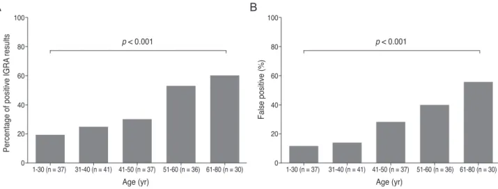
Data were examined and compared in each of the five age subgroups and the percentage of IGRA-positive results in each group is presented in Fig. 2. The percentage of pa- tients with positive IGRA results were 18.9%, 24.4%, 29.7%, 52.8%, and 60% in age groups 1, 2, 3, 4, and 5, re- spectively. The percentage of false-positive results were 11.8%, 13.9%, 27.8%, 39.3%, and 55.6%, in age groups 1, 2, 3, 4, and 5, respectively. Both true- and false-positive IGRA results were significantly different among age groups and were linearly correlated with age (i.e., true and false positive rate increased with age, p < 0.001, chi-square test for trend).
Positive IGRA result data were also analyzed among the four different inflammation location groups. Patients in the anterior uveitis, intermediate uveitis, posterior uveitis, and panuveitis groups had IGRA positive rates of 26.7%, 26.5%, 59.2%, and 30.4%, respectively (Fig. 3). More pa- tients with retinal vasculitis (71.9%) had positive IGRA test results than patients without retinal vasculitis (28.2%, p <
0.001). False positives occurred in 15.0%, 0.0%, 51.7%, and 28.6% of patients with anterior, intermediate, posterior, and panuveitis, respectively. Positive IGRA results and false-positive rates were also statistically different among patients with different anatomic inflammation types (p = 0.001 and 0.002, respectively).
Table 1. Clinical characteristics of the included patients and comparison of the characteristics between IGRA-positive and -nega- tive patients and between TRU and non-TRU patients
All patients (n = 181)
IGRA result Presumed TRU
Positive
(n = 65) Negative
(n = 116) p-value TRU
(n = 20) Non-TRU
(n = 161) p-value Age (yr) 43.4 ± 16.2 50.0 ± 13.5 39.7 ± 16.4 <0 . 001* 45.6 ± 14.6 43.1 ± 16.4 0 . 531* Sex (male : female) 99 : 82 37 : 28 62 : 54 0 . 652† 10 : 10 89 : 72 0 . 655† Positive IGRA result (%) 65 (35.9) 65 (100) 0 NA† 20 (100) 45 (28.0) <0 . 001† Anatomic type (%)
Anterior 75 (41.4) 20 (30.8) 55 (47.4)
0 . 001†
3 (15) 72 (44.7)
<0 . 001†
Intermediate 34 (18.8) 9 (13.8) 25 (21.6) 0 34 (21.1)
Posterior 49 (27.1) 29 (44.6) 20 (17.2) 15 (75) 34 (21.1)
Panuveitis 23 (12.7) 7 (10.8) 16 (13.8) 2 (10) 21 (13.0)
With retinal vasculitis 32 (17.7) 23 (35.4) 9 (7.8) <0 . 001† 12 (60) 20 (12.4) <0 . 001† IGRA = interferon-gamma release assay; TRU = tuberculosis-related uveitis; NA = not applicable.
*Chi-square test; †Student’s t-tests.
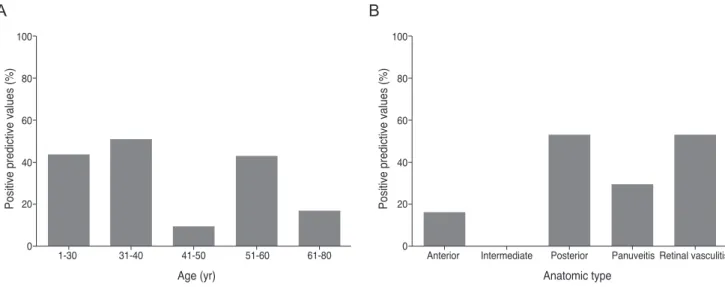
The positive predictive values for presumed TRU among patients with intraocular inflammation are shown in Fig. 4.
Values were 42.9%, 50.0%, 9.1%, 42.1%, and 16.7% in age groups 1, 2, 3, 4, and 5, respectively. Values were 15.0%, 0.0%, 51.7%, and 28.6% in patients with anterior, interme- diate, posterior, and panuveitis, respectively. In patients with and without retinal vasculitis, the positive predictive value of the IGRA was 52.2% and 22.8%, respectively. The IGRA test had greater positive predictive values in patients with a younger age (≤40 years), posterior uveitis, and reti- nal vasculitis. The positive predictive value of the IGRA among patients with intraocular inflammation was 70%
when all of these factors were present.
Multiple logistic regressions were performed to identify factors associated with the IGRA results. Age was inde- pendently associated with a positive IGRA result (p < 0.001), but the anatomic type of inflammation was not (p = 0.176).
The odds ratio for age adjusted for presumed TRU and ana- tomic type was 1.06 (95% confidence interval [CI], 1.03 to 1.09), and that for age group adjusted for the same confound- ing variables was 1.90 (95% CI, 1.41 to 2.56; p < 0.001). Likeli- hood ratios were calculated for assessing the value of perform- ing the IGRA diagnostic test. The positive likelihood ratio was 3.57 and the negative likelihood ratio was 0.00 in our study.
Fig. 3. Interferon-gamma release assay (IGRA)-positive rates among the four anatomic types of uveitis and between patients with and without retinal vasculitis. (A) The IGRA-positive rates were significantly different among the four anatomic groups (p = 0.001). (B) Pa- tients with retinal vasculitis had higher positive IGRA rate (p < 0.001) than those without retinal vasculitis. p-values were obtained using chi-square tests.
Anterior 0
20 40 60 80 100
Intermediate Posterior Panuveitis
Percentage of positive IGRA results
p = 0.001
With vasculitis 0
20 40 60 80 100
Without vasculitis
Percentage of positive IGRA results
p < 0.001
Anterior 0
20 40 60 80 100
Intermediate Posterior Panuveitis
False positive rate (%) False positive rate (%)
p = 0.002
With vasculitis 0
20 40 60 80 100
Without vasculitis p = 0.004
A B
Fig. 2. Interferon-gamma release assay (IGRA)-positive rates (A) and false-positive rates (B) among the five age groups examined. Both rates were significantly different among age groups (all p-values <0.001). p-values were obtained by chi-square tests for trend.
0 1-30 (n = 37) 31-40 (n = 41) 41-50 (n = 37) 51-60 (n = 36) 61-80 (n = 30) 20
40 60 80 100
Percentage of positive IGRA results
Age (yr) p < 0.001
0 1-30 (n = 37) 31-40 (n = 41) 41-50 (n = 37) 51-60 (n = 36) 61-80 (n = 30) 20
40 60 80 100
False positive (%)
Age (yr) p < 0.001
A B
Discussion
Intraocular inflammation can have many different ori- gins and presents with a wide spectrum of clinical mani- festations. If the underlying inflammatory cause is treat- able, prompt and appropriate treatment can result in a favorable outcome. In the current study, we examined the IGRA as a diagnostic tool for presumed TRU in Korean patients with intraocular inflammation. Our results suggest that the IGRA is highly sensitive and moderately specific for TRU. A test used for TB screening should have good sensitivity and acceptable specificity [14], so we believe that IGRA can be used as a screening test for presumed TRU in Korea. Unfortunately, the TST (Mantoux test) is not useful as a screening tool because of the high false-positive rate in the Korean population, which has a very high BCG vaccination rate. Therefore, an alternative test is needed for the Korean population, and in this study, we evaluated the IGRA as a screening method for TRU.
However, the false-positive rate (1-specificity) of IGRA was not low and cannot be neglected. Therefore, clinicians should consider which patients are more likely to have true TRU. Our analyses showed that younger age (≤ 40 years), a positive IGRA, and the presence of posterior uveitis and retinal vasculitis were all predictive of TRU.
Broad-based posterior synechiae, retinal vasculitis (with or without choroiditis), and serpiginous choroiditis in pa- tients with latent or manifest TB are clinically suggestive of TRU in TB-endemic areas [15-17]. This may explain
why the positive predictive value of IGRA was greater in patients with posterior uveitis and retinal vasculitis. Sever- al studies [15,16,18,19] have reported that vasculitis can oc- cur from a TB infection, and it may be associated with hy- persensitivity to MTB [20].
We examined how a positive IGRA result should be in- terpreted in patients with suspected TB uveitis. Because our older patients tended to have a higher IGRA positivity rate, a positive result is likely insufficient to make a defini- tive TRU diagnosis, especially in elderly patients with in- traocular inflammation. This result is comparable to those of Kang et al. [5], who reported that positive IGRA rates are positively and linearly correlated with patient age. In our study, the age of patients with and without TRU were not significantly different, so this relationship did not re- sult from inherent group differences in age.
We also examined which patients should have the IGRA performed for diagnosing presumed TRU. Patients with in- traocular inflammation usually undergo extensive diagnos- tic work-ups to identify the underlying inflammatory cause.
Performing the IGRA on all patients with intraocular in- flammation would not be cost-effective. Therefore, we rec- ommend having the IGRA done for patients with a clinical presentation suggestive of TRU, including retinal vasculitis and posterior uveitis. In addition, because a positive IGRA result in a young patient with posterior uveitis or retinal vasculitis likely indicates TRU, the test should be heavily considered if a concurrent TB infection is suspected.
Several limitations of our study require consideration.
Fig. 4. Positive predictive values of the interferon-gamma release assay for presumed tuberculosis-related uveitis in patients with intraoc- ular inflammation (A) in the age groups and (B) in the anatomic type subgroups.
0 1-30 31-40 41-50 51-60 61-80
20 40 60 80 100
Positive predictive values (%)
Age (yr)
0 Anterior Intermediate Posterior Panuveitis Retinal vasculitis 20
40 60 80 100
Positive predictive values (%)
Anatomic type
A B
Our calculations for positive and negative predictive values are limited by the absence of normal control data. Ang et al. [21] recommend that TRU be diagnosed by considering clinical presentation and the IGRA and TST results. Our internists did not perform the TST because prior BCG vac- cination is associated with high false positive rates and the Korean population has widely received the BCG vaccine (95.6% of children [22], 95.7% of adults [5]). Therefore, we could not compare or relate TST and IGRA results in this study, even though this comparison is crucial for evaluat- ing the relative diagnostic value of the IGRA. Additionally, further investigations are needed to evaluate the true rela- tionship between positive IGRA result rates and age among normal, healthy control subjects. Lastly, prospec- tive studies that include a larger number of patients may provide more concrete information on the usefulness of the IGRA in diagnosing TRU.
In conclusion, the IGRA may be a useful screening test for presumed TRU in Korea. Patients with positive results should be further evaluated with other reliable tests for TB (e.g., sputum culture, HRCT, anti-TB medication response) to rule-out or confirm TRU. Patient age and uveitis ana- tomic location may be helpful in interpreting the IGRA re- sults with respect to a TRU diagnosis. In particular, a posi- tive IGRA result is highly suggestive of TRU in Korea in a younger patient (≤ 40 years) with posterior uveitis and reti- nal vasculitis.
Conflict of Interest
No potential conflict of interest relevant to this article was reported.
Acknowledgements
This work was supported by a grant (2012R1A2A- 2A 02012821) funded by the National Research Foundation in Korea.
References
1. Centers for Disease Control and Prevention (CDC). Trends in tuberculosis: United States, 2004. MMWR Morb Mortal
Wkly Rep 2005;54:245-9.
2. Dye C, Scheele S, Dolin P, et al. Consensus statement.
Global burden of tuberculosis: estimated incidence, preva- lence, and mortality by country. WHO Global Surveillance and Monitoring Project. JAMA 1999;282:677-86.
3. Centers for Disease Control and Prevention (CDC). Ex- panded tuberculosis surveillance and tuberculosis morbidi- ty: United States, 1993. MMWR Morb Mortal Wkly Rep 1994;43:361-6.
4. Tabbara KF. Tuberculosis. Curr Opin Ophthalmol 2007;18:493-501.
5. Kang YA, Lee HW, Yoon HI, et al. Discrepancy between the tuberculin skin test and the whole-blood interferon gamma assay for the diagnosis of latent tuberculosis infec- tion in an intermediate tuberculosis-burden country. JAMA 2005;293:2756-61.
6. Itty S, Bakri SJ, Pulido JS, et al. Initial results of QuantiF- ERON-TB Gold testing in patients with uveitis. Eye (Lond) 2009;23:904-9.
7. Ang M, Htoon HM, Chee SP. Diagnosis of tuberculous uveitis: clinical application of an interferon-gamma release assay. Ophthalmology 2009;116:1391-6.
8. Feng Y, Diao N, Shao L, et al. Interferon-gamma release assay performance in pulmonary and extrapulmonary tu- berculosis. PLoS One 2012;7:e32652.
9. Critselis E, Amanatidou V, Syridou G, et al. The effect of age on whole blood interferon-gamma release assay re- sponse among children investigated for latent tuberculosis infection. J Pediatr 2012;161:632-8.
10. Diagnostic Standards and Classification of Tuberculosis in Adults and Children. This official statement of the Ameri- can Thoracic Society and the Centers for Disease Control and Prevention was adopted by the ATS Board of Direc- tors, July 1999. This statement was endorsed by the Council of the Infectious Disease Society of America, September 1999. Am J Respir Crit Care Med 2000;161(4 Pt 1):1376-95.
11. Mazurek GH, Jereb J, Lobue P, et al. Guidelines for using the QuantiFERON-TB Gold test for detecting Mycobacteri- um tuberculosis infection, United States. MMWR Recomm Rep 2005;54(RR-15):49-55.
12. Mazurek GH, Jereb J, Vernon A, et al. Updated guidelines for using interferon gamma release assays to detect Myco- bacterium tuberculosis infection: United States, 2010.
MMWR Recomm Rep 2010;59(RR-5):1-25.
13. Madariaga MG, Jalali Z, Swindells S. Clinical utility of in- terferon gamma assay in the diagnosis of tuberculosis. J
Am Board Fam Med 2007;20:540-7.
14. Grimes DA, Schulz KF. Uses and abuses of screening tests.
Lancet 2002;359:881-4.
15. Gupta A, Bansal R, Gupta V, et al. Ocular signs predictive of tubercular uveitis. Am J Ophthalmol 2010;149:562-70.
16. Gupta A, Gupta V, Arora S, et al. PCR-positive tubercular retinal vasculitis: clinical characteristics and management.
Retina 2001;21:435-44.
17. Gupta V, Gupta A, Rao NA. Intraocular tuberculosis: an update. Surv Ophthalmol 2007;52:561-87.
18. Hoh HB, Kong VY, Jaais F. Tuberculous retinal vasculitis.
Med J Malaysia 1998;53:288-9.
19. Reny JL, Challe G, Geisert P, et al. Tuberculosis-related retinal vasculitis in an immunocompetent patient. Clin In- fect Dis 1996;22:873-4.
20. Bodaghi B, LeHoang P. Ocular tuberculosis. Curr Opin Ophthalmol 2000;11:443-8.
21. Ang M, Wong W, Ngan CC, Chee SP. Interferon-gamma release assay as a diagnostic test for tuberculosis-associated uveitis. Eye (Lond) 2012;26:658-65.
22. Lee H, Dockrell HM, Kim DR, et al. The current status of BCG vaccination in young children in South Korea. Tuberc Respir Dis (Seoul) 2012;72:374-80.