2020 Keimyung University School of Medicine This is an open-access article distributed under the terms of the Creative Commons Attribution license (http://creativecommons.org/licenses/by-nc/4.0/), which permits unrestricted use, distribution, and reproduction in any medium, provided the original work is properly cited.
Metabolism-Related Genes in Human Kidney Clear Cell Carcinoma Using The Cancer Genome Atlas.
Woo-Jae Park1, Jee Young Park2, Taeg Kyu Kwon2,3, Jong-Wook Park2,3, Shin Kim2,3
1Department of Biochemistry, College of Medicine, Gachon University, Incheon, Korea
2Department of Immunology, Keimyung University School of Medicine, Daegu, Korea
3Institute of Medical Science, Keimyung University, Daegu, Korea
The sphingolipid rheostat concept states that the cellular fate is largely determined by various sphingolipid metabolites and the associated signaling pathways. Aberrant regulation of the sphingolipid metabolism-related components is closely associated with cancer survival and death, including aspects like cancer development, prolifer- ation, progression, and response to anticancer drugs. In the present study, we inves- tigated the expression and prognostic significance of the sphingolipid metabo- lism-related genes in clear cell renal cell carcinoma (ccRCC), the most common pathological subtype of kidney cancer, using an RNA-sequencing dataset of The Cancer Genome Atlas Kidney Clear Cell Carcinoma (TCGA KIRC) cohort. Expres- sion levels of various sphingolipid metabolism-related genes were significantly al- tered in ccRCC tissues compared with those of normal solid tissues. Notably, the ex- pression of B4GALNT1, BNIP3, DEGS1, GAL3ST1, S1PR4, SLC26A10, SMPDL3A, and SPHK1 was significantly upregulated, whereas the expression of B4GALT6, HPGD, LPAR1, SFTPB, ST6GALNAC5, and UGT8 was significantly downregulated in ccRCC tissues. Notably, among these significantly-altered sphingolipid metabo- lism-related genes, the Kaplan-Meier survival analyses showed that high expression levels of B4GALNT1, SLC26A10, and SPHK1 were associated with a poor prognosis of patients with ccRCC, whereas high expression levels of BNIP3, HPGD, and SMP- DL3A were associated with a better prognosis. Taken together, our study suggests that B4GALNT1, SLC26A10, SPHK1, BNIP3, HPGD, and SMPDL3A may be novel prognostic biomarkers and targets for a therapeutic strategy to improve the treat- ment of ccRCC.
Keywords: Sphingolipid metabolism, clear cell renal cell carcinoma, The cancer ge- nome atlas
Received: March 25, 2020 Revised: May 12, 2020 Accepted: May 15, 2020 Corresponding Author:
Shin Kim, M.D., Ph.D.
Department of Immunology, Keimyung University School of Medicine 1095 Dalgubeol-daero, Dalseo-gu, Daegu, 42601, Korea
Tel: +82-53-258-7359 Fax: +82-53-258-7355 E-mail: [email protected] pISSN 2092-8335 · eISSN 2733-5380 Keimyung Med J 2020 [Epub ahead of print]
https://doi.org/10.46308/kmj.2020.00101
Original Article
Keywords: Clear cell renal cell carcinoma, Sphingolipid metabolism, The Cancer Genome Atlas
Received: March 25, 2020 Revised: May 12, 2020 Accepted: May 15, 2020 Corresponding Author:
Shin Kim, M.D., Ph.D.
Department of Immunology, Keimyung University School of Medicine, 1095 Dalgubeol-daero, Dalseo-gu, Daegu 42601, Korea
Tel: +82-53-258-7359 Fax: +82-53-258-7355 E-mail: [email protected] pISSN 2092-8335 · eISSN 2733-5380 Keimyung Med J 2020 39(1):14-22 https://doi.org/10.46308/kmj.2020.00101
Original Article
Metabolism-related Genes in Human Kidney Clear Cell Carcinoma Using The Cancer Genome Atlas
Woo-Jae Park1, Jee Young Park2, Taeg Kyu Kwon2,3, Jong-Wook Park2,3, Shin Kim2,3
1Department of Biochemistry, College of Medicine, Gachon University, Incheon, Korea
2Department of Immunology, Keimyung University School of Medicine, Daegu, Korea
3Institute of Medical Science, Keimyung University, Daegu, Korea
Abbreviations: B4GALNT1, beta-1,4-N-acetyl-galactosaminyltransferase 1;
B4GALT6, beta-1,4-galactosyltransferase 6; BNIP3, BCL2 interacting protein 3;
ccRCC, clear cell renal cell carcinoma; CERS, Ceramide synthase; CRC, Col- orectal cancer; DEGS1, delta 4-desaturase, sphingolipid 1; GAL3ST1, galac- tose-3-O-sulforansferase 1; HPGD, 15-hydroxyprostaglandin dehydrogenase;
LPAR1, lysophosphatidic acid receptor 1; NST, normal solid tissue; PHK1, sphingosine kinase 1; S1PR4, sphingosine-1-phosphate receptor 4; SFTPB, sur- factant protein B; SLC26A10, solute carrier family 26 member 10; SMPDL3A, sphingomyelin phosphodiesterase acid like 3A; ST6GALNAC5, ST6 N-acetyl- galactosaminide alpha-2,6-sialyltransferase 5; TCGA KIRC, The Cancer Ge- nome Atlas Kidney Clear Cell Carcinoma; UGT8, UDP glycosyltransferase 8.
Introduction
Renal cell carcinoma (RCC), derived from the kidney paren- chyma accounts for almost 90% of all kidney tumors [1]. Accord- ing to histology of the RCC subtypes recommended by the World Health Organization, RCC is divided into three main sub- types, including clear cell renal cell carcinoma (ccRCC), papillary renal cell carcinoma, and chromophobe renal cell carcinoma, among which ccRCC is the most common subtype (80%-90%) [2]. Despite an adequate surgical resection, recurrence and me- tastasis occur in 25% of the patients due to the aggressiveness of ccRCC [3]. To improve this clinical limitation, there has been ex- tensive research to find potential biomarkers that can predict the prognosis for patients with ccRCC using genomic profiling ap- proaches such as microarray analysis, mutation analysis, and gene expression analysis [4-6]. Moreover, recent advances in bio- technology such as RNA sequencing (RNAseq) have revolution- ized various fields of research, providing new insight into com- plex diseases such as cancer with respect to identification of the causes, establishment of targets for therapeutic strategies, and ex- ploration of new prognostic biomarkers.
The sphingolipid rheostat is a biochemical concept that in- volves regulation of the interconversion of sphingolipid me- tabolites, including ceramide, sphingosine, and sphin- gosine-1-phosphate (S1P), and their related signaling path- ways, which play an important role in cell fate determination [7]. Accumulating evidences indicate that the dysregulated sphingolipid metabolism-related genes have clinicopathologi- cal relevance in various human cancers [8,9]. For example, a high expression of sphingosine kinase 1 (SPHK1) was signifi- cantly associated with a worse outcome in survival analysis of patients with breast cancer [10], and a high expression of cer- amide synthase (CERS) 5 was significantly associated with a poor prognosis in colorectal cancer (CRC) patients [11].
However, research is scarce on the expression and prognostic potential of various sphingolipid metabolism-related genes in ccRCC. Thus, in the present study, we investigated the mRNA expression levels of the dysregulated sphingolipid metabo- lism-related genes in ccRCC using gene expression RNAseq data from The Cancer Genome Atlas Kidney Clear Cell Car- cinoma (TCGA KIRC) cohort and their prognostic relevance.
Materials and Methods
Gene expression databases and cluster analysis
The gene expression RNAseq dataset (level 3, dataset ID:
TCGA.KIRC.sampleMap/HiSeqV2) and clinical characteris-
tic datasets (TCGA.KIRC.sampleMap/KIRC_clinicalMatrix and survival/KIRC_survival.txt) for the TCGA KIRC cohort were downloaded from the UCSC Xena database (https://
xena.ucsc.edu). The RNAseq dataset provides gene-level tran- script estimates with log2(x+1)-transformed counts normal- ized using the RNA-Seq by Expectation-Maximization (RSEM) normalization package. The RNAseq dataset of TCGA KIRC comprises 606 samples, including 533 primary tumor tissues, one additional new primary tumor tissue, and 72 normal solid tissues (NST). The mRNA expression levels of the sphingolipid metabolism-related genes [12] were sorted from the TCGA KIRC data. We calculated the |log2Fold- Change (FC)| to screen for genes with more than 2-fold dif- ferences in expression levels between groups. This study met the publication guidelines provided by TCGA (http://www.
cancer.gov/about-nci/organization/ccg/research/structru- al-genomics/tcga/ using-tcga/citing-tcga). Cluster analysis was performed using the Cluster 3.0 software [13] to classify the samples into statistically similar groups, and the resulting heatmaps were visualized in TreeView [14] (version 1.6). Ex- pression levels in the heatmaps have been quantile normal- ized [15], median-centered, and scaled for visualization.
Kaplan-Meier (KM) survival analysis with log rank test Prior to a survival analysis, we calculated the median gene expression values of the selected sphingolipid metabolism-re- lated genes from TCGA KIRC and determined the values as the cutoff values for survival analysis. Based on the median value of each gene, ccRCC samples of TCGA KIRC was divid- ed into 2 groups. The survival of the two groups was analyzed using the KM method, and statistical significance was as- sessed using the log-rank test.
Statistical analysis
Statistical analysis was performed with the SPSS software (version 25.0; IBM SPSS, Armonk, NY, USA). The Kolmogor- ov-Smirnov test was used for testing normality of the data.
Differences between groups were then statistically analyzed using the Student’s t-test. The association between inter-indi- vidual mRNA levels of the sphingolipid metabolism-related genes was assessed using the Pearson’s correlation coefficients for continuous variables. In general, p < 0.05 was considered to denote significance in all statistical analyses performed in the study.
Results
Altered expression levels of the sphingolipid metabo- lism-related genes in TCGA KIRC cohort.
The heatmap revealed that various sphingolipid metabo- lism-related genes were dysregulated in ccRCC compared with NST of patients with ccRCC (Fig. 1). The Student’s t-test
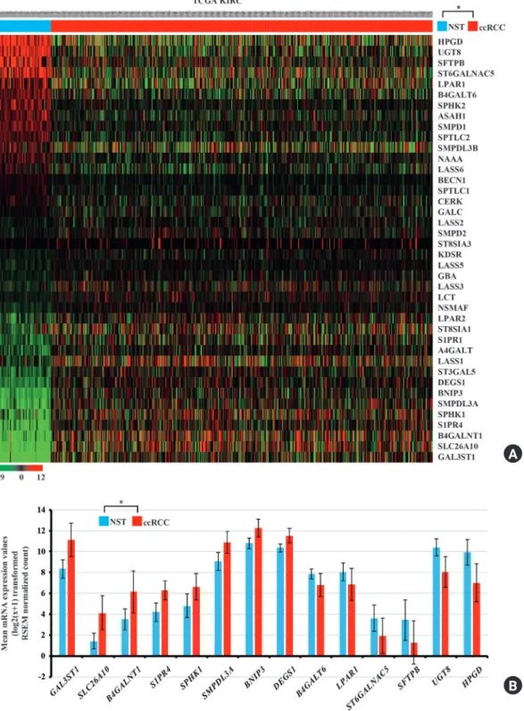
confirmed that various sphingolipid metabolism-related genes were significantly dysregulated in ccRCC tissues com- pared with NST (p < 0.05; Fig. 2A). Specifically, be- ta-1,4-N-acetyl-galactosaminyltransferase 1 (B4GALNT1), the BCL2 interacting protein 3 (BNIP3), delta 4-desaturase, sphingolipid 1 (DEGS1), galactose-3-O-sulforansferase 1 (GAL3ST1), the sphingosine-1-phosphate receptor 4 (S1PR4),
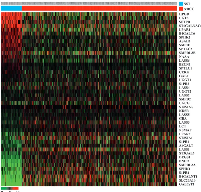
Fig. 1. Heatmap presenting the relative mRNA expression levels of the sphingolipid metabolism-related genes in the TCGA KIRC cohort. The data are presented in matrix format in which each row represents an individual gene and each column represents a single tissue. Each cell in the matrix represents the expression level of a gene feature in an individual tissue. The red and green colors in cells reflect relative high and low expression levels, respectively, as indicated in the scale bar. The samples were ordered from NSTs to ccRCC samples according to the standardized expression level of each gene as indicated. TCGA KIRC, The Cancer Genome Atlas kidney clear cell carcinoma; ccRCC, clear cell renal cell carcinoma; NST, normal solid tissue.
A
B
Fig. 2. (A) Heatmap showing significantly dysregulated mRNA expression of the sphingolipid metabolism-related genes in ccRCC tissues compared with the NST of TCGA KIRC cohort. The data are presented in matrix format in which each row represents an individual gene and each column represents a single tissue. Each cell in the matrix represents the expression level of a gene feature in an individual tissue. The red and green colors in cells reflect relative high and low expression levels, respectively, as indicated in the scale bar. The samples were ordered from NSTs to ccRCC samples according to the standardized expression level of each gene as indicated. *p<0.05 (NST versus ccRCC). (B) Relative mRNA levels of various sphingolipid metabolism-related genes in TCGA KIRC samples. *Student t-test, p<0.05,
|log2FoldChange| ≥ 1.0. TCGA KIRC, The Cancer Genome Atlas kidney clear cell carcinoma; ccRCC, clear cell renal cell carcinoma; NST, normal solid tissue.
the solute carrier family 26 member 10 (SLC26A10), sphingo- myelin phosphodiesterase acid like 3A (SMPDL3A), and SPHK1 were significantly upregulated in ccRCC, whereas be- ta-1,4-galactosyltransferase 6 (B4GALT6), 15-hydroxyprosta- glandin dehydrogenase (HPGD), the lysophosphatidic acid receptor 1 (LPAR1), the surfactant protein B (SFTPB), ST6 N-acetylgalactosaminide alpha-2,6-sialyltransferase 5 (ST- 6GALNAC5), and UDP glycosyltransferase 8 (UGT8) were significantly downregulated in ccRCC compared to their re- spective expression in NST (the Student’s t-test, p < 0.05,
|log2FC| ≥ 1.0; Fig. 2B). These 14 dysregulated sphingolipid metabolism-related genes may therefore play an important role in the ccRCC pathophysiology.
Inter-individual correlations between sphingolipid metabo- lism-related genes showing altered mRNA expression levels in ccRCC.
Based on the Pearson’s correlation coefficient, we found 69 significant correlations between the mRNA expression levels
of the 14 significantly dysregulated sphingolipid metabo- lism-related genes in ccRCC sorted from TCGA KIRC cohort (Table 1).
Survival analysis of the significantly dysregulated sphingo- lipid metabolism-related genes in ccRCC.
Survival data were available for a total of 605 ccRCC patients for the KM survival analysis. The KM plot and log-rank test in- dicated that higher mRNA expression levels of B4GALNT1, SLC26A10, and SPHK1 were associated with an unfavorable overall survival in ccRCC patients (Fig. 3A-3C; p = 0.013, p <
0.001, and p < 0.001, respectively). However, higher mRNA ex- pression levels of BNIP3, HPGD, and SMPDL3A were associated with a favorable overall survival in ccRCC patients (Fig. 3D-3F, p
= 0.005, p < 0.001, and p = 0.009, respectively).
Discussion
Since the sphingolipid rheostat was proposed as an import- Table 1. Pearson’s correlation analysis between inter-individual components of sphingolipid metabolism-related genes
Samples Correlations between components Pearson’s correlation coefficient value p-Value*
TCGA KIRC tissues from TCGA KIRC cohort (n = 533) B4GALNT1 and B4GALT6 -0.271 < 0.001
B4GALNT1 and BNIP3 -0.129 0.003
B4GALNT1 and GAL3ST1 -0.267 < 0.001
B4GALNT1 and SLC26A10 0.768 < 0.001
B4GALNT1 and SMPDL3A -0.244 < 0.001
B4GALNT1 and SPHK1 0.181 < 0.001
B4GALNT1 and ST6GALNAC5 0.087 0.045
B4GALNT1 and UGT8 -0.110 0.011
B4GALT6 and DEGS1 -0.141 0.001
B4GALT6 and GAL3ST1 -0.127 0.003
B4GALT6 and HPGD 0.158 < 0.001
B4GALT6 and S1PR4 -0.232 < 0.001
B4GALT6 and SFTPB 0.352 < 0.001
B4GALT6 and SLC26A10 -0.346 < 0.001
B4GALT6 and SMPDL3A 0.120 0.006
B4GALT6 and SPHK1 -0.255 < 0.001
B4GALT6 and ST6GALNAC5 -0.183 < 0.001
B4GALT6 and UGT8 0.347 < 0.001
BNIP3 and DEGS1 0.123 0.004
BNIP3 and GAL3ST1 0.410 < 0.001
BNIP3 and HPGD -0.106 0.014
BNIP3 and LPAR1 -0.201 < 0.001
BNIP3 and SFTPB -0.353 < 0.001
BNIP3 and SLC26A10 -0.104 0.016
BNIP3 and SMPDL3A 0.419 < 0.001
BNIP3 and SPHK1 -0.320 < 0.001
*Pearson’s correlation coefficient analysis.
ant concept in determining the cell fate, many studies have focused on the contribution of sphingolipid metabolism to the mechanism of cancer development, as well as for the iden- tification of new targets for a cancer therapeutic strategy and prognostic factors [16]. However, the expression and potential prognostic markers of the sphingolipid metabolism-related genes remain to be investigated for many cancer types, in- cluding ccRCC. In this study, using the RNAseq data of TCGA KIRC cohort, we evaluated the dysregulation of sphin- golipid metabolism-related genes in ccRCC and identified potential prognostic markers.
We identified eight sphingolipid metabolism-related genes (B4GALNT1, BNIP3, DEGS1, GAL3ST1, S1PR4, SLC26A10, SMPDL3A, and SPHK1) that were significantly upregulated in ccRCC in TCGA-KIRC cohort, and the KM survival analysis showed that higher expression levels of B4GALNT1, SLC26A10, and SPHK1 were significantly correlated with a worse prognosis. These results were found to be consistent with recent studies showing that worse overall survival was
Fig. 3. Survival analyses of the sphingolipid metabolism-related genes in ccRCC samples of TCGA KIRC cohort. Kaplan-Meier plot of overall survival in subjects with high versus low (A) B4GALNT1, (B) SLC26A10, (C) SPHK1, (D) BNIP3, (E) HPGD, and (F) SMPDL3A mRNA expression. TCGA KIRC, The Cancer Genome Atlas kidney clear cell carcinoma; ccRCC, clear cell renal cell carcinoma.
significantly associated with upregulated SPHK1 expression in non-small cell lung cancer [17] and that B4GALNT1 ex- pression was upregulated in ccRCC [18]. Although SLC26A10 expression was found to be significantly upregulated in col- orectal carcinoma compared to adenoma, the prognostic sig- nificance of SLC26A10 was not evident [19]. SPHK1 is a ma- jor enzyme of the S1P metabolism pathway, and plays an im- portant role in tumorigenesis [20], cancer angiogenesis [21], intercellular communication of the tumor environment [22], and formation of the prometastatic environment [23].
B4GALNT1 is a key enzyme in generation of the gangliosides GM2/GD2, and has also been shown to be involved in tumor- igenesis [24]. Thus, our results suggest that SPHK1 and B4GALNT1 may play crucial roles as oncogenes in ccRCC.
Similarly, we hypothesized that BNIP3 and SMPDL3A also functioned as oncogenes in ccRCC, and that their higher ex- pression levels may contribute to poor prognoses. Notewor- thy, BNIP3 and SMPDL3A were significantly upregulated in ccRCC, but higher expression levels of these genes were sig- A
D
B
E
B
F B4GALNT1
Time (days)
Time (days)
- High expression - Low exexpression
Time (days)
Time (days)
Time (days)
Time (days) BNIP3
SLC26A10
HPGD
SPHK1
SMPDL3A
Overall survivalOverall survival Overall survivalOverall survival Overall survivalOverall survival
1.0 0.8 0.6 0.4 0.2 0.0
1.0 0.8 0.6 0.4 0.2 0.0
1.0 0.8 0.6 0.4 0.2 0.0
1.0 0.8 0.6 0.4 0.2 0.0
1.0 0.8 0.6 0.4 0.2 0.0
1.0 0.8 0.6 0.4 0.2 0.0
0 0 0
0 P = 0.013
P = 0.005 P = 0.009
P < 0.001
P < 0.001
P < 0.001
0 0
1000 1000 1000
1000 1000
1000
2000 2000 2000
2000 2000
2000
3000 3000 3000
3000 3000
3000
4000 4000 4000
4000 4000
4000
5000 5000 5000
5000 5000
5000
nificantly correlated with better prognoses in TCGA-KIRC cohort. BNIP3 is a well-known tumor suppressor gene that regulates cell death and mitophagy [25], and its higher ex- pression level has previously been correlated with a good prognosis [26], providing further support that BNIP3 plays an important role as a tumor suppressor gene in the ccRCC pathophysiology. However, further investigation is needed to clarify the reason for the increase in BNIP3 expression levels in ccRCC tissues compared with the levels in NST. SMPDL3A functions as a liver X receptor target gene; however, little is known about its potential roles in cancer. Conversely, the oth- er six dysregulated sphingolipid metabolism-related genes (B4GALT6, HPGD, LPAR1, SFTPB, ST6GALNAC5, and UGT8) were significantly downregulated in ccRCC compared with those in NST. Notably, higher expression levels of HPGD were significantly correlated with a better prognosis in TC- GA-KIRC cohort. HPGD is responsible for regulation of pros- taglandin metabolism [27] and also acts as a tumor suppres- sor gene [28]. A decreased level of HPGD was correlated with a poor prognosis in patients with triple-negative breast cancer [29], which was found to be in line with our results, suggest- ing an important role in the pathophysiology of ccRCC.
Moreover, in the present study we provide the first evidence for the significantly-altered regulation of SLC26A10, B4GALT6, and SMPDL3A and their prognostic significance in ccRCC. However, previous studies have demonstrated con- flicting results in finding a poor prognosis associated with higher expression of BNIP3 in patients with non-small cell lung cancer [30] and uveal melanoma [31], and higher ex- pression of HPGD in patients with breast cancer [32]. There- fore, more detailed analysis will be needed to determine the regulatory mechanisms of sphingolipid metabolism-related genes and their association with the pathophysiology of ccRCC.
Significant correlations between the sphingolipid metabo- lism-related genes have been reported in various CRC co- horts, including CERS2 and CERS4, CERS4 and CERS5, and CERS5 and CERS6 [33]. Inter-individual correlations between the significantly dysregulated sphingolipid metabolism-relat- ed genes of ccRCC revealed 69 significant correlations; how- ever, no genes were correlated with these six CERS genes as none of these genes showed significantly-altered expression levels in ccRCC compared with those in NST. Therefore, can- cer signaling pathways altered in ccRCC may be related to the correlation between the sphingolipid metabolism-related genes other than the CERS genes.
In conclusion, we identified the significantly-altered sphin-
golipid metabolism-related genes and their prognostic signifi- cance in patients with ccRCC using TCGA-KIRC cohort. No- tably, our study suggests that B4GALNT1, SLC26A10, SPHK1, BNIP3, HPGD, and SMPDL3A may be potential prognostic biomarkers and promising therapeutic candidates for ccRCC.
Acknowledgements
We would like to thank all the members of our research group for their enthusiastic participation in this study.
Funding
The present study was supported by the National Research Foundation of Korea (grant funded by the Korea Government Ministry of Science,
ICT and Future Planning; grant nos. 2014R1A5A2010008, NRF-2016R1D1A1B04930619, and NRF-2019R1I1A3A0 1063114).
Availability of data and materials
The datasets analyzed during the present study are available from The Cancer Genome Atlas (https://www.cancer.gov/
tcga) and the UCSC Xena (https://xena.ucsc.edu). The data- sets generated in the present study are available from the cor- responding author upon reasonable request.
Authors contributions
WJP and SK contributed to the conception and design of the study, analysis of the data, interpretation of results, and the writing of the manuscript. JYP and SK contributed to the acquisition of data and the writing of the manuscript. TKK, JWP, and SK reviewed the manuscript.
SK edited the manuscript. All authors read and approved the manuscript and agree to be accountable for all aspects of the research in ensuring that the accuracy and integrity of any part of the work are appropriately investigated and resolved.
Ethics approval
Not applicable.
Patient consent
Not applicable.
Conflict of interest
All authors declare no conflicts-of-interest related to this article.
References
1. Bhatt JR, Finelli A. Landmarks in the diagnosis and treatment of renal cell carcinoma. Nat Rev Urol. 2014;11:517-25.
2. Kovacs G, Akhtar M, Beckwith BJ, Bugert P, Cooper CS, Dela- hunt B, et al. The Heidelberg classification of renal cell tumours.
J Pathol. 1997;183:131-3.
3. MacLennan S, Imamura M, Lapitan MC, Omar MI, Lam TB, Hilvano-Cabungcal AM, et al. Systematic review of oncological outcomes following surgical management of localised renal can- cer. Eur Urol. 2012;61:972-93.
4. Li Z, Liu J, Zhang X, Fang L, Zhang C, Zhang Z, et al. Prognostic significance of cyclin D1 expression in renal cell carcinoma: a systematic review and meta-analysis. Pathol Oncol Res. 2019.
DOI: 10.1007/s12253-019-00776-0.
5. Kojima T, Shimazui T, Horie R, Hinotsu S, Oikawa T, Kawai K, et al. FOXO1 and TCF7L2 genes involved in metastasis and poor prognosis in clear cell renal cell carcinoma. Genes Chro- mosomes Cancer. 2010;49:379-89.
6. Li X, Tan X, Yu Y, Chen H, Chang W, Hou J, et al. D9S168 mi- crosatellite alteration predicts a poor prognosis in patients with clear cell renal cell carcinoma and correlates with the down-reg- ulation of protein tyrosine phosphatase receptor delta. Cancer.
2011;117:4201-11.
7. Cuvillier O, Pirianov G, Kleuser B, Vanek PG, Coso OA, Gut- kind S, et al. Suppression of ceramide-mediated programmed cell death by sphingosine-1-phosphate. Nature. 1996;381:800-3.
8. Ryland LK, Fox TE, Liu X, Loughran TP, Kester M. Dysregula- tion of sphingolipid metabolism in cancer. Cancer Biol Ther.
2011;11:138-49.
9. Sedic M, Grbcic P, Pavelic SK. Bioactive sphingolipids as bio- markers predictive of disease severity and treatment response in cancer: current status and translational challenges. Anticancer Res. 2019;39:41–56.
10. Geffken K, Spiegel S. Sphingosine kinase 1 in breast cancer. Adv Biol Regul. 2018;67:59-65.
11. Fitzgerald S, Sheehan KM, Espina V, O'Grady A, Cummins R, Kenny D, et al. High CerS5 expression levels associate with re- duced patient survival and transition from apoptotic to autopha- gy signalling pathways in colorectal cancer. J Pathol Clin Res.
2015;1:54-65.
12. Ruckhaberle E, Rody A, Engels K, Gaetje R, von Minckwitz G,
Schiffmann S, et al. Microarray analysis of altered sphingolipid metabolism reveals prognostic significance of sphingosine ki- nase 1 in breast cancer. Breast Cancer Res Treat. 2008;112:41-52.
13. de Hoon MJ, Imoto S, Nolan J, Miyano S. Open source clustering software. Bioinformatics. 2004;20:1453-4.
14. Saldanha AJ. Java Treeview--extensible visualization of microar- ray data. Bioinformatics. 2004;20:3246-8.
15. Evans C, Hardin J, Stoebel DM. Selecting between-sample RNA- Seq normalization methods from the perspective of their as- sumptions. Brief Bioinform. 2018;19:776-92.
16. Newton J, Lima S, Maceyka M, Spiegel S. Revisiting the sphingo- lipid rheostat: evolving concepts in cancer therapy. Exp Cell Res.
2015;333:195-200.
17. Wang Y, Shen Y, Sun X, Hong TL, Huang LS, Zhong M. Prog- nostic roles of the expression of sphingosine-1-phosphate me- tabolism enzymes in non-small cell lung cancer. Transl Lung Cancer Res. 2019;8:674-81.
18. Yang H, Li W, Lv Y, Fan Q, Mao X, Long T, et al. Exploring the mechanism of clear cell renal cell carcinoma metastasis and key genes based on multi-tool joint analysis. Gene. 2019;720. DOI:
10.1016/j.gene.2019.144103.
19. Carvalho B, Sillars-Hardebol AH, Postma C, Mongera S, Terhaar Sive Droste J, Obulkasim A, et al. Colorectal adenoma to carci- noma progression is accompanied by changes in gene expression associated with ageing, chromosomal instability, and fatty acid metabolism. Cell Oncol (Dordr). 2012;35:53-63.
20. Nava VE, Hobson JP, Murthy S, Milstien S, Spiegel S. Sphin- gosine kinase type 1 promotes estrogen-dependent tumorigene- sis of breast cancer MCF-7 cells. Exp Cell Res. 2002;281:115-27.
21. Dai L, Liu Y, Xie L, Wu X, Qiu L, Di W. Sphingosine kinase 1/
sphingosine-1-phosphate (S1P)/S1P receptor axis is involved in ovarian cancer angiogenesis. Oncotarget. 2017;8:74947-61.
22. Schneider G. S1P signaling in the tumor microenvironment. Adv Exp Med Biol. 2020;1223:129-53.
23. Schneider G, Bryndza E, Abdel-Latif A, Ratajczak J, Maj M, Tar- nowski M, et al. Bioactive lipids S1P and C1P are prometastatic factors in human rhabdomyosarcoma, and their tissue levels in- crease in response to radio/chemotherapy. Mol Cancer Res.
2013;11:793-807.
24. Yoshida H, Koodie L, Jacobsen K, Hanzawa K, Miyamoto Y, Ya- mamoto M. B4GALNT1 induces angiogenesis, anchorage inde- pendence growth and motility, and promotes tumorigenesis in melanoma by induction of ganglioside GM2/GD2. Sci Rep.
2020;10. DOI: 10.1038/s41598-019-57130-2.
25. Chourasia AH, Macleod KF. Tumor suppressor functions of BNIP3 and mitophagy. Autophagy. 2015;11:1937-8.
26. Chourasia AH, Tracy K, Frankenberger C, Boland ML, Sharifi
MN, Drake LE, et al. Mitophagy defects arising from BNip3 loss promote mammary tumor progression to metastasis. EMBO Rep. 2015;16:1145-63.
27. Zhang Y, Desai A, Yang SY, Bae KB, Antczak MI, Fink SP, et al.
Tissue regeneration. Inhibition of the prostaglandin-degrading enzyme 15-PGDH potentiates tissue regeneration. Science.
2015;348. DOI: 10.1126/science.aaa2340.
28. Wolf I, O'Kelly J, Rubinek T, Tong M, Nguyen A, Lin BT, et al.
15-Hydroxyprostaglandin dehydrogenase is a tumor suppressor of human breast cancer. Cancer Res. 2006;66:7818-23.
29. Kochel TJ, Goloubeva OG, Fulton AM. Upregulation of cycloox- ygenase-2/prostaglandin E2 (COX-2/PGE2) pathway member multiple drug resistance-associated protein 4 (MRP4) and downregulation of prostaglandin transporter (PGT) and 15-prostaglandin dehydrogenase (15-PGDH) in triple-negative breast cancer. Breast Cancer (Auckl). 2016;10:61-70.
30. Giatromanolaki A, Koukourakis MI, Sowter HM, Sivridis E, Gibson S, Gatter KC, et al. BNIP3 expression is linked with hy- poxia-regulated protein expression and with poor prognosis in non-small cell lung cancer. Clin Cancer Res. 2004;10:5566-71.
31. Jiang Z, Yu F, Li M. Upregulation of BCL2 19 kD protein-inter- acting protein 3 (BNIP3) is predictive of unfavorable prognosis in uveal melanoma. Med Sci Monit. 2018;24:4711-7.
32. Lehtinen L, Vainio P, Wikman H, Reemts J, Hilvo M, Issa R, et al.
15-Hydroxyprostaglandin dehydrogenase associates with poor prognosis in breast cancer, induces epithelial-mesenchymal transition, and promotes cell migration in cultured breast cancer cells. J Pathol. 2012;226:674-86.
33. Jang SW, Park WJ, Min H, Kwon TK, Baek SK, Hwang I, et al.
Altered mRNA expression levels of the major components of sphingolipid metabolism, ceramide synthases and their clinical implication in colorectal cancer. Oncol Rep. 2018;40:3489-500.