Journal of the American Heart Association
ORIGINAL RESEARCH
Five-Year Risk of Acute Myocardial Infarction After Acute Ischemic Stroke in Korea
Keon-Joo Lee, MD; Seong-Eun Kim, PhD; Jun Yup Kim , MD; Jihoon Kang , MD; Beom Joon Kim , MD, PhD;
Moon-Ku Han , MD, PhD; Kang-Ho Choi , MD, PhD; Joon-Tae Kim, MD, PhD; Dong-Ick Shin, MD, PhD;
Jae-Kwan Cha, MD, PhD; Dae-Hyun Kim, MD, PhD; Dong-Eog Kim, MD, PhD; Wi-Sun Ryu, MD, PhD;
Jong-Moo Park, MD, PhD; Kyusik Kang , MD, PhD; Jae Guk Kim, MD; Soo Joo Lee, MD, PhD; Mi-Sun Oh , MD;
Kyung-Ho Yu, MD, PhD; Byung-Chul Lee, MD, PhD; Hong-Kyun Park , MD; Keun-Sik Hong, MD, PhD;
Yong-Jin Cho, MD, PhD; Jay Chol Choi , MD, PhD; Sung Il Sohn , MD, PhD; Jeong-Ho Hong, MD, PhD;
Moo-Seok Park, MD; Tai Hwan Park, MD, PhD; Sang-Soon Park, MD, PhD; Kyung Bok Lee, MD, PhD;
Jee-Hyun Kwon, MD, PhD; Wook-Joo Kim, MD; Jun Lee, MD, PhD; Ji Sung Lee , PhD; Juneyoung Lee, PhD;
Philip B. Gorelick , MD, MPH; Hee-Joon Bae , MD, PhD; on behalf of the CRCS-K Registry Investigators*
BACKGROUND: The long-term incidence of acute myocardial infarction (AMI) in patients with acute ischemic stroke (AIS) has not been well defined in large cohort studies of various race-ethnic groups.
METHODS AND RESULTS: A prospective cohort of patients with AIS who were registered in a multicenter nationwide stroke regis- try (CRCS-K [Clinical Research Collaboration for Stroke in Korea] registry) was followed up for the occurrence of AMI through a linkage with the National Health Insurance Service claims database. The 5-year cumulative incidence and annual risk were estimated according to predefined demographic subgroups, stroke subtypes, a history of coronary heart disease (CHD), and known risk factors of CHD. A total of 11 720 patients with AIS were studied. The 5-year cumulative incidence of AMI was 2.0%.
The annual risk was highest in the first year after the index event (1.1%), followed by a much lower annual risk in the second to fifth years (between 0.16% and 0.27%). Among subgroups, annual risk in the first year was highest in those with a history of CHD (4.1%) compared with those without a history of CHD (0.8%). The small-vessel occlusion subtype had a much lower in- cidence (0.8%) compared with large-vessel occlusion (2.2%) or cardioembolism (2.4%) subtypes. In the multivariable analysis, history of CHD (hazard ratio, 2.84; 95% CI, 2.01–3.93) was the strongest independent predictor of AMI after AIS.
CONCLUSIONS: The incidence of AMI after AIS in South Korea was relatively low and unexpectedly highest during the first year after stroke. CHD was the most substantial risk factor for AMI after stroke and conferred an approximate 5-fold greater risk.
Key Words: acute ischemic stroke ■ acute myocardial infarction ■ coronary heart disease ■ prospective cohort study ■ risk factors
Ischemic stroke and coronary heart disease (CHD) are the major causes of death worldwide.1 They share risk factors and strategies for secondary prevention, such as use of antithrombotics and sta- tin therapy.2,3 The coprevalence of CHD has been re- ported in one fifth of stroke patients.4
Although the incidence of acute myocardial infarc- tion (AMI) after acute ischemic stroke (AIS) has been reported in several studies,5,6 changes in risk factor profiles and recent advances in secondary prevention strategies may have led to differences in the occur- rence of AMI after stroke over time.7–9 For example,
Correspondence to: Hee-Joon Bae, MD, PhD, Department of Neurology, Seoul National University College of Medicine, Cerebrovascular Center, Seoul National University Bundang Hospital, 82, Gumi-ro 173 beon-gil, Bundang-gu, Seongnam-si, Gyeonggi-do 13620, South Korea. E-mail: [email protected] Supplementary material for this article is available at https://www.ahajo urnals.org/doi/suppl/10.1161/JAHA.120.018807
*A complete list of the CRCS-K Registry Investigators can be found in the Supplemental Material.
For Sources of Funding and Disclosures, see page 9.
© 2020 The Authors. Published on behalf of the American Heart Association, Inc., by Wiley. This is an open access article under the terms of the Creative Commons Attribution-NonCommercial-NoDerivs License, which permits use and distribution in any medium, provided the original work is properly cited, the use is non-commercial and no modifications or adaptations are made.
JAHA is available at: www.ahajournals.org/journal/jaha
Downloaded from http://ahajournals.org by on April 26, 2021
there was no study focusing on the incidence of poststroke AMI after high-intensity statin was intro- duced to the clinical practice guideline in year 2008,10 whereas much lower incidence of AMI could be ex- pected compared with previous reports, according to the use of high-intensity statin in patients with AIS.
Furthermore, as ischemic stroke is a heterogeneous disorder with diverse causes,11 patient characteristics, cardiovascular risk profiles, and poststroke progno- ses may vary by ischemic stroke subtype.12,13 Thus, one might expect that the risk of AMI in patients with stroke may differ by the aforementioned characteris- tics. However, there were only few studies with small number of subjects that explored the incidence of AMI according to stroke subtypes.5,14
Determination of the risk and predictors of AMI after AIS is important for the establishment of prevention strategies for both AMI and stroke recurrence. In this context, using a large AIS cohort from a nationwide multicenter stroke registry in South Korea, we estimated
the incidence of AMI after stroke, and explored factors that might heighten risk of poststroke AMI.
METHODS Study Population
This study was based on a multicenter prospec- tive stroke registry, the CRCS-K (Clinical Research Collaboration for Stroke in Korea) registry.15,16 Patients with AIS who were admitted to 14 tertiary or academic hospitals between January 2011 and November 2013 and gave consent for a linkage to secondary administrative data were included. The CRCS-K registry database was linked to a national claims database of the National Health Insurance Service (NHIS).17 The NHIS has provided all citizens of South Korea with a universal health insurance pro- gram since 1989, and 97.1% of the Korean popula- tion is covered by this program.17 The NHIS claims database contains information on beneficiaries’ de- mographic characteristics, diagnostic codes, pro- cedures, and prescription records for hospitalization and outpatient care. The claims database was linked to the CRCS-K registry database using the claim serial number that was generated for each claim at each hospital for the purpose of reimbursement.18
After the linkage process, patients were selected according to the following inclusion criteria: (1) aged
≥18 years, (2) admission within 7 days after symptom onset, and (3) corresponding stroke lesions docu- mented on brain imaging (magnetic resonance imaging or computed tomography). We excluded those whose stroke subtype was not specified.
Variables
Clinical information on demographics, vascular risk factors, stroke characteristics, and other potential risk factors of CHD was directly obtained from the CRCS-K registry database. The following vascular risk factors were chosen for study: hypertension, diabetes mel- litus, hyperlipidemia, history of CHD, atrial fibrillation, history of stroke or transient ischemia attack, and smoking status. Symptomatic carotid artery disease was defined as stenosis of >50% or occlusion of the proximal internal carotid artery or the common carotid artery ipsilateral to the ischemic lesion documented on magnetic resonance imaging, computed tomography, or conventional cerebral angiography.19 Initial stroke severity was measured according to the National Institutes of Health Stroke Scale score, and functional status at discharge was assessed with the modified Rankin Scale. Stroke subtypes were determined by vascular neurologists in charge of patients’ treatment, according to the Trial of Org 10172 in Acute Stroke Treatment classification, with some modifications20;
CLINICAL PERSPECTIVE
What Is New?
• In a multicenter prospective registry of Korean patients with acute ischemic stroke, 5-year cu- mulative incidence of acute myocardial infarc- tion was 2%, and was highest during the first year after stroke onset.
• Notably, patients with history of coronary heart disease showed a 5-fold risk of acute myocar- dial infarction after stroke onset, and those with cardioembolism subtype had a higher risk than other subtypes.
What Are the Clinical Implications?
• In general, the risk of acute myocardial infarc- tion after ischemic stroke was low.
• However, patients with certain risk factors, such as history of coronary heart disease, need at- tention during the first year after stroke event.
Nonstandard Abbreviations and Acronyms
AIS acute ischemic stroke
CRCS-K Clinical Research Collaboration for Stroke in Korea
NHIS National Health Insurance Service
NOMAS Northern Manhattan Study OxVasc Oxford Vascular Study
SPARCL Stroke Prevention by Aggressive Reduction in Cholesterol Levels
Downloaded from http://ahajournals.org by on April 26, 2021
the modifications were developed for the purpose of considering magnetic resonance imaging findings and the results of the reperfusion therapy for deciding the cause of ischemic stroke.20 In addition, the following variables were collected: antiplatelet and statin therapy administration before the index stroke event and at discharge, baseline systolic blood pressure, and labo- ratory data, such as low-density lipoprotein (LDL) cho- lesterol level, high-density lipoprotein (HDL) cholesterol level, and glomerular filtration rate (GFR).21
AMI was defined by the admission event claimed under the disease code (International Classification of Diseases, Tenth Revision [ICD-10]) I21* after index stroke through linkage with the NHIS claims database. The AMI events were further specified as ST-segment–elevation myocardial infarction (MI) (codes I21.0–I21.3), non–ST- segment–elevation MI (code I21.4), and fatal MI, if a patient died within 28 days after an AMI event.22 Data on mortal- ity were obtained from national vital statistics reports.
Ethical Approval
The collection of clinical information and linkage of the information with secondary databases for the purpose of stroke research with informed consent were approved by the local ethic committees at all participating centers. The use of the CRCS-K registry database and its linkage with the NHIS claims database for this study were approved further by the Institutional Review Board of Seoul National University Bundang Hospital (No. B-1511/322–106).
The data will not be available to other researchers for the purpose of reproducing the results because of local legal regulations regarding access to patient-level data.
Statistical Analysis
The baseline characteristics of study subjects were summarized as frequencies and percentages for cat- egorical variables and as means with SDs for continu- ous variables. If a variable was recorded in form of a scale (eg, National Institutes of Health Stroke Scale or modified Rankin Scale) or skewed, it was summarized as the median and interquartile range.
The cumulative incidence of AMI up to 5 year after the index stroke was estimated using the cumulative in- cidence function, with mortality treated as a competing risk for AMI, and is displayed on a yearly basis. The cu- mulative incidence of AMI was determined for all study subjects according to predetermined subgroups: aged
≤70 versus >70 years, sex, history of hypertension, di- abetes mellitus, CHD, and atrial fibrillation, presence of symptomatic carotid artery disease, and stroke sub- type. The differences between subgroups were evalu- ated using the Grey test.
To determine the time trend of poststroke AMI inci- dence and to identify the point of the highest risk of AMI,
the annual risk of AMI after index stroke was calculated on the basis of the survival estimates obtained using the Grey method for all study subjects and via a post hoc analysis in subgroups according to history of CHD, stroke subtype, history of atrial fibrillation, and presence of symptomatic carotid artery disease.23 For the first year, the monthly risk was calculated using the same method.
A Cox proportional hazards regression analysis was performed to explore risk factors of AMI after AIS. The following predetermined covariates were entered into the models: age, sex, vascular risk fac- tors listed above, antiplatelet and statin use before index stroke, systolic blood pressure, and laboratory data, including LDL cholesterol, HDL cholesterol, and GFR. To include a competing risk analysis into the Cox models, a subdistribution hazard model using the Fine and Grey method was adopted; a cause-specific hazard model treating the competing events as censored observations is also presented as supporting data.24 Age, LDL cholesterol, HDL cholesterol, GFR, and systolic blood pressure were dichotomized as follows: aged ≤70 versus >70 years, LDL cholesterol <100 versus ≥100 mg/dL, HDL cho- lesterol <40 versus ≥40 mg/dL, and GFR <60 versus
≥60 mL/min per 1.73 m2. Considering multicollinear- ity, stroke subtype and symptomatic carotid artery disease/atrial fibrillation were alternatively introduced into the multivariable models.
All tests were performed using SAS version 9.4 (SAS Institute Inc, Cary, NC), and statistical significance was declared with a 2-sided P value of <0.05.
RESULTS
Of 15,114 patients with AIS who were registered in the database during the study period and provided con- sent, 11 720 were analyzed for the study (Figure S1).
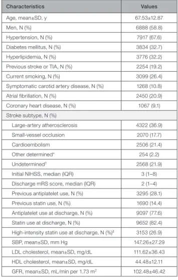
The mean age was 67 years, and 59% were men. Two thirds of the study subjects had hypertension, and one third had diabetes mellitus or hyperlipidemia. Of the subjects, 9% had a history of CHD and 11% had symptomatic carotid artery disease. As for stroke sub- types, 37% had large-artery atherosclerosis, 18% had small-vessel occlusion, and 21% had cardioembolism.
About 14% of the patients were using statin before index stroke event, and 82% were prescribed statin at discharge (Table 1).25
The median follow-up duration was 4.0 years (in- terquartile range, 3.2–4.9 years), and AMI occurred in 218 patients (1.9%), while 3270 patients (27.9%) died during this period. The cumulative incidence of AMI was 1.1% at 1 year and 2.0% at 5 years (Table 2 and Figure S2). Among the AMI events, there were 69 (31.7%) ST-segment–elevation MIs and 52 (23.9%) non–ST-segment–elevation MIs, whereas other 97 (44.5%) were not specified as either
Downloaded from http://ahajournals.org by on April 26, 2021
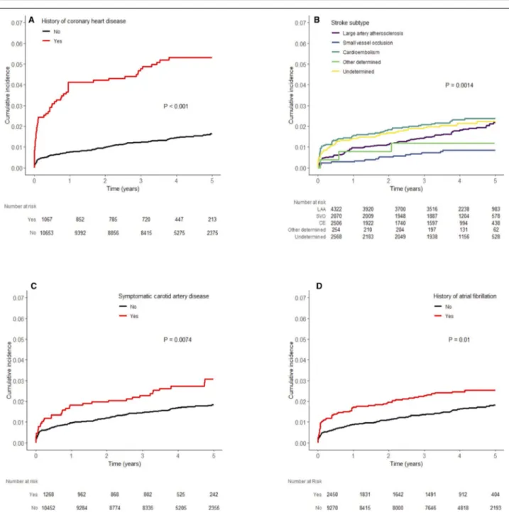
ST-segment–elevation MI or non–ST-segment–ele- vation MI. AMI events were fatal in 33 patients (15.1%) (Table S1). Ninety-six (44.0%) patients were admitted to the same hospital, where they were treated for index stroke. The subgroup analysis showed the greatest difference in 5-year cumulative incidence between those with and without a history of CHD (5.3% ver- sus 1.6%; P<0.001), followed by those with and with- out symptomatic carotid artery disease (3.1% versus 1.8%; P=0.007). There was no significant difference
between men and women. The patients with cardi- oembolism (2.38%), undetermined cause (2.23%), and large-artery atherosclerosis (2.18%) had a sim- ilar 5-year cumulative incidence, whereas those with small-vessel occlusion had a much lower cumulative incidence (0.84%; P=0.001) (Table 2 and Figure 1).
The 5-year cumulative incidence of AMI was higher in ex-smokers (2.91%) than in current smokers (1.92%) and nonsmokers (1.79%; P=0.012) (Figure S3).
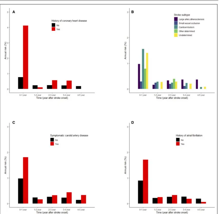
The annual risk was highest in the first year after the index stroke: 1.1% in the first year, followed by 0.2%, 0.3%, 0.3%, and 0.2% in the second, third, fourth, and fifth years, respectively. When we focused on the first year, the monthly risk was highest in the first month (0.51%), followed by the second month (0.13%), and then was <0.1% in the third month and afterwards (Figure S4). The annual risk in patients with history of CHD in the first year was 4.1%, which was much higher than the risk in the following years and in those without a history of CHD. The annual risk in the first year was lowest in patients with small-vessel occlusion (0.3%) compared with those with cardioembolism (1.6%), undetermined stroke cause (1.4%), and large-artery occlusion (1.0%). Also, those with symptomatic carotid artery disease (1.8%) and atrial fibrillation (1.7%) had higher annual risk in the first year than those without the conditions (Figure 2).
In the multivariable analysis, a history of CHD was the strongest predictor of poststroke AMI (Table 3).
Also, among stroke subtypes, cardioembolism, fol- lowed by undetermined cause and large-artery ath- erosclerosis, was associated with a higher risk of poststroke AMI compared with small-vessel occlu- sion. Other cardiovascular risk factors, such as hy- pertension, diabetes mellitus, and history of stroke or transient ischemia attack, also increased the risk of poststroke AMI. Age >70 years increased the risk in the cause-specific hazard model, but not in the subdistribution hazard model (Table S2). In addition, when atrial fibrillation and symptomatic carotid artery disease were entered into the models as an alter- native to stroke subtype, they increased the risk in the cause-specific hazard models, but did not in the subdistribution hazard models (Table S3). The results of when age, systolic blood pressure, LDL choles- terol, HDL cholesterol, and GFR systolic are treated as continuous variables are provided in Tables S4 and S5.
DISCUSSION
Using a recent large cohort of patients with AIS from a nationwide multicenter stroke registry in South Korea, we report on the 5-year incidence of poststroke AMI.
Among various subgroups, those of older age and
Table 1. Baseline Characteristics of the Study Subjects (N=11 720)
Characteristics Values
Age, mean±SD, y 67.53±12.87
Men, N (%) 6888 (58.8)
Hypertension, N (%) 7917 (67.6)
Diabetes mellitus, N (%) 3834 (32.7)
Hyperlipidemia, N (%) 3776 (32.2)
Previous stroke or TIA, N (%) 2254 (19.2)
Current smoking, N (%) 3099 (26.4)
Symptomatic carotid artery disease, N (%) 1268 (10.8)
Atrial fibrillation, N (%) 2450 (20.9)
Coronary heart disease, N (%) 1067 (9.1)
Stroke subtype, N (%)
Large-artery atherosclerosis 4322 (36.9)
Small-vessel occlusion 2070 (17.7)
Cardioembolism 2506 (21.4)
Other determined* 254 (2.2)
Undetermined† 2568 (21.9)
Initial NIHSS, median (IQR) 3 (1–8)
Discharge mRS score, median (IQR) 2 (1–4) Previous antiplatelet use, N (%) 3295 (28.1)
Previous statin use, N (%) 1690 (14.4)
Antiplatelet use at discharge, N (%) 9097 (77.6) Statin use at discharge, N (%) 9652 (82.4) High-intensity statin use at discharge, N (%)‡ 3153 (26.9)
SBP, mean±SD, mm Hg 147.26±27.29
LDL cholesterol, mean±SD, mg/dL 111.62±36.43 HDL cholesterol, mean±SD, mg/dL 44.48±12.11 GFR, mean±SD, mL/min per 1.73 m2 102.48±46.42 GFR indicates glomerular filtration rate; HDL, high-density lipoprotein;
IQR, interquartile range; LDL, low-density lipoprotein; mRS, modified Rankin Scale; NIHSS, National Institutes of Health Stroke Scale; SBP, systolic blood pressure; and TIA, transient ischemic attack.
*Other determined cause was defined as rare causes of ischemic stroke with diverse entities (details in study by Ko et al20).
†Undetermined cause was defined as follows: (1) not being classified as large-artery atherosclerosis, small-vessel disease, or cardioembolism and no cardioembolic source despite comprehensive workups; (2) no workup for stroke cause; or (3) coexistence of >1 stroke cause.
‡According to the American College of Cardiology/American Heart Association Guideline on Treatment of Blood Cholesterol (2013) (≥50%
reduction in LDL cholesterol).
Downloaded from http://ahajournals.org by on April 26, 2021
those with vascular risk factors, especially a history of CHD, had a higher cumulative incidence up to 5 years after AIS. The incidence also differed according to stroke subtype; those with small-vessel occlusion had lower incidence compared with those with cardioem- bolism, undetermined cause, and large-artery athero- sclerosis. The risk of AMI was highest in the first year after AIS.
Several previous studies have reported incidence of AMI after stroke.5,6 However, these studies were limited by small sample size or not being confined to patients with AIS.14,26–28 A recent meta-analysis of >130 000 patients showed that the annual risk of poststroke AMI is 1.67%.5 However, this meta-analysis reported on patient data from observational studies and clinical trials of diverse populations, and follow-up durations and definitions of AMI across those studies differed.
In addition, the study could not examine whether the incidence of AMI varied by stroke subtype because of a lack of such data. In contradistinction, our study ana- lyzed the incidence of poststroke AMI using real-world data starting at stroke onset, and the analysis was stratified by various subgroups, including the follow- ing factors: demographics, vascular risks, and stroke subtype.
We found a generally low risk of AMI, with the an- nual risk <1%, except in study subjects with known CHD during the first year after AIS. This is lower than what was reported in previous studies.6,14,26,29 The re- cent meta-analysis, however, showed a continuous decrease in the incidence of poststroke AMI,5 which might be attributed to improvement in poststroke management, including administration of statin. It is noteworthy that our study was conducted after the
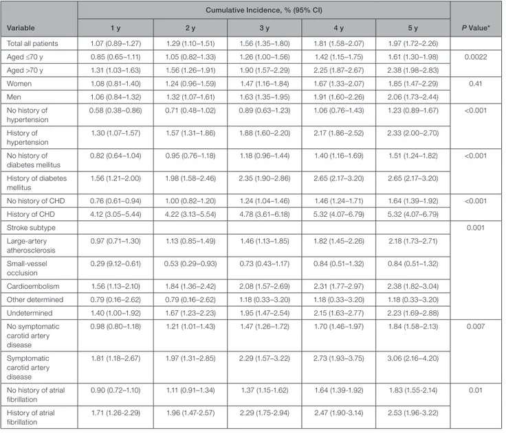
Table 2. Cumulative Incidences of AMI After AIS for All Subgroups
Variable
Cumulative Incidence, % (95% CI)
P Value*
1 y 2 y 3 y 4 y 5 y
Total all patients 1.07 (0.89–1.27) 1.29 (1.10–1.51) 1.56 (1.35–1.80) 1.81 (1.58–2.07) 1.97 (1.72–2.26)
Aged ≤70 y 0.85 (0.65–1.11) 1.05 (0.82–1.33) 1.26 (1.00–1.56) 1.42 (1.15–1.75) 1.61 (1.30–1.98) 0.0022 Aged >70 y 1.31 (1.03–1.63) 1.56 (1.26–1.91) 1.90 (1.57–2.29) 2.25 (1.87–2.67) 2.38 (1.98–2.83)
Women 1.08 (0.81–1.40) 1.24 (0.96–1.59) 1.47 (1.16–1.84) 1.67 (1.33–2.07) 1.85 (1.47–2.29) 0.41 Men 1.06 (0.84–1.32) 1.32 (1.07–1.61) 1.63 (1.35–1.95) 1.91 (1.60–2.26) 2.06 (1.73–2.44)
No history of hypertension
0.58 (0.38–0.86) 0.71 (0.48–1.02) 0.89 (0.63–1.23) 1.06 (0.76–1.43) 1.23 (0.89–1.67) <0.001
History of hypertension
1.30 (1.07–1.57) 1.57 (1.31–1.86) 1.88 (1.60–2.20) 2.17 (1.86–2.52) 2.33 (2.00–2.70)
No history of diabetes mellitus
0.82 (0.64–1.04) 0.95 (0.76–1.18) 1.18 (0.96–1.44) 1.40 (1.16–1.69) 1.51 (1.24–1.82) <0.001
History of diabetes mellitus
1.56 (1.21–2.00) 1.98 (1.58–2.46) 2.35 (1.90–2.86) 2.65 (2.17–3.20) 2.65 (2.17–3.20)
No history of CHD 0.76 (0.61–0.94) 1.00 (0.82–1.20) 1.24 (1.04–1.46) 1.46 (1.24–1.71) 1.64 (1.39–1.92) <0.001 History of CHD 4.12 (3.05–5.44) 4.22 (3.13–5.54) 4.78 (3.61–6.18) 5.32 (4.07–6.79) 5.32 (4.07–6.79)
Stroke subtype 0.001
Large-artery atherosclerosis
0.97 (0.71–1.30) 1.13 (0.85–1.49) 1.46 (1.13–1.85) 1.82 (1.45–2.26) 2.18 (1.73–2.71)
Small-vessel occlusion
0.29 (9.12–0.61) 0.53 (0.29–0.93) 0.73 (0.43–1.17) 0.84 (0.51–1.32) 0.84 (0.51–1.32)
Cardioembolism 1.56 (1.13–2.10) 1.84 (1.36–2.42) 2.08 (1.57–2.69) 2.31 (1.77–2.97) 2.38 (1.82–3.04) Other determined 0.79 (0.16–2.62) 0.79 (0.16–2.62) 1.18 (0.33–3.20) 1.18 (0.33–3.20) 1.18 (0.33–3.20) Undetermined 1.40 (1.00–1.92) 1.67 (1.23–2.23) 1.95 (1.47–2.54) 2.15 (1.63–2.77) 2.23 (1.69–2.88) No symptomatic
carotid artery disease
0.98 (0.80–1.18) 1.21 (1.01–1.43) 1.47 (1.26–1.72) 1.70 (1.46–1.97) 1.84 (1.58–2.13) 0.007
Symptomatic carotid artery disease
1.81 (1.18–2.67) 1.97 (1.31–2.85) 2.29 (1.57–3.22) 2.73 (1.93–3.75) 3.06 (2.16–4.20)
No history of atrial fibrillation
0.90 (0.72–1.10) 1.11 (0.91–1.34) 1.37 (1.15-1.62) 1.64 (1.39-1.92) 1.83 (1.55-2.14) 0.01
History of atrial fibrillation
1.71 (1.26-2.29) 1.96 (1.47-2.57) 2.29 (1.75-2.94) 2.47 (1.90-3.14) 2.53 (1.96-3.22)
AIS indicates acute ischemic stroke; AMI, acute myocardial infarction; and CHD, coronary heart disease.
*P values determined with the Grey test.
Downloaded from http://ahajournals.org by on April 26, 2021
SPARCL (Stroke Prevention by Aggressive Reduction in Cholesterol Levels) trial, which demonstrated the beneficial effect of high-intensity statin in a predomi- nantly AIS population.30 More than 80% of our study subjects were on statin therapy at hospital discharge, even though only 30% were diagnosed as having hy- perlipidemia. There was a 68% increase in statin use after stroke; only 14% were on statin before index stroke (Table 1). A secondary analysis of the SPARCL trial showed that patients randomized to high-inten- sity statin experienced coronary events less frequently
compared with those randomized to placebo, regard- less of stroke subtype, and the reduction in coronary events was greater than that for stroke events.31
Thus, it may not be surprising that our study showed a much lower incidence of AMI than previous studies. For example, the aforementioned meta-anal- ysis included only 5 of 58 studies that enrolled study subjects during the period after the publication of the SPARCL trial in 2006.5 Despite the overall low inci- dence of poststroke AMI in our study, the high risk of AMI in the first year after AIS and in those with a
Figure 1. Cumulative incidence of acute myocardial infarction after ischemic stroke among subgroups.
A, History of coronary artery disease. B, Stroke subtype. C, Symptomatic carotid artery disease. D, Atrial fibrillation. CE, cardioembolism; LAA, large artery atherosclerosis; SVO, small vessel occlusion.
Downloaded from http://ahajournals.org by on April 26, 2021
history of CHD suggests that it may be prudent to focus surveillance and preventative cardiovascular therapy for AMI on those with a history of CHD and certain vascular risk factors or specific stroke sub- types, as noted in our study.
The incidence of AMI was highest in the first year and especially during the first 2 months after AIS (Figure S4). Higher recurrence rate during the early period after index event is shown in patients with AMI or stroke.12,32,33 Not sufficiently controlled vascular risk factors or comorbidities during the early period after index event and ongoing disease process, such as
atherosclerosis or inflammation, provoked by index event could be explainable mechanisms.32,34,35 Also, poststroke cardiac arrhythmias could be another pos- sible cause of AMI after AIS.36
The incidence of AMI in East Asia, for example, may be lower than in traditional Western countries.37 The estimated age-standardized incidence rate of hospital- ized MI was 39 cases per 100 000 person-years in 2011 in Korea,38 which was much lower than that of 677 per 100 000 person-years in the same year in the United States.39 Differences in dietary and lifestyle fac- tors may explain, at least in part, the lower incidence
Figure 2. Annual risk of acute myocardial infarction after index stroke event.
A, History of coronary artery disease. B, Stroke subtype. C, Symptomatic carotid artery disease. D, Atrial fibrillation.
Downloaded from http://ahajournals.org by on April 26, 2021
of AMI in East Asian countries.37 Our results provide valuable race-ethnic observations on East Asian pa- tients with stroke.
Current smokers seemed not to have higher in- cidence of AMI after AIS in our study (Table 3 and Figure S3). This could be explained in line with the well-known “smokers’ paradox” of better prognosis in smokers than nonsmokers.40 This phenomenon is usually explained as current smokers are much younger and healthier than nonsmokers at the time of index event, and we were also able to find in our study that current smokers were much younger compared with ex-smokers or nonsmokers (age, 60.6±12.4 versus 69.2±11.2 versus 70.2±12.3 years in current smokers, ex-smokers, and nonsmokers, respectively).
Our study results are similar to those of earlier work. First, the 10-year risk of AMI after ischemic stroke or transient ischemia attack in the OxVasc (Oxford Vascular Study) cohort, with a median fol- low-up duration of 5.6 years, was much higher in those with CHD.29 However, the 10-year risk in those without CHD was about 8%, which may not be con- cordant with our results. This might be explained by the older age of the non-CHD population (mean
age, 73 years) and the earlier enrollment epoch (from 2002) in the OxVasc. Second, patients with embolic stroke had a significantly higher risk of poststroke AMI in the NOMAS (Northern Manhattan Study).14,26 Atrial fibrillation is known to be a risk factor of AMI, as atrial fibrillation may cause thromboembolism, and high heart rates evoked by atrial fibrillation might impair coronary perfusion and lead to demand (type 2) MI.41 The comparable risk in those with stroke of undetermined cause could be explained by the co- existence of large-artery atherosclerosis and cardio- embolic sources and the potential for the existence of undetected (ie, subclinical) AF in this stroke sub- type.11 The recent widespread use of non–vitamin K oral anticoagulants seemed to have a little effect on the AMI incidence in this study as they were not al- lowed for preventing atrial fibrillation–related stroke in most cases until the middle of 2015 in Korea.42 Third, the risk of poststroke AMI was highest in the first year after index stroke in a study performed by the Virtual International Stroke Trials Archive collaboration,34 al- though the aforementioned recent meta-analysis did not show any time trend.5 This discrepancy may be explained by the fact that our study and the study by the Virtual International Stroke Trials Archive collabo- ration included only patients with AIS and started to capture AMI from the early period after stroke onset, whereas, among 58 studies included in the recent meta-analysis, 15 studies confined study subjects to AIS and 9 to those enrolled within 7 days from onset.
Our study has limitations. First, as AMI events were captured using the diagnosis code of the claims data, the incidence of AMI could be underestimated or over- estimated. Previous validation studies in South Korea, however, reported that the accuracy of the diagnosis code for AMI was >70%.43,44 Second, the study sub- jects were South Koreans who were admitted to ac- ademic or tertiary stroke centers, which may limit the generalizability of the study results. Third, AMI events occurring during hospitalization and associated with the index stroke were not included. Fourth, because of the Personal Information Protection Act, only the pa- tients who gave their informed consent for a linkage between the registry and the secondary administrative databases were included in this study. Although the consent rate was as high as 92.4%,45 there could be a possible selection bias, such as the fact that those who were more disabled or died during hospitalization were not included in our study.
To date, our study is the single largest one reporting on the incidence of AMI in a single race-ethnic group over a relatively long period of time after AIS. Although the general incidence of poststroke AMI seems to be low, individuals with a history of CHD have a higher risk and thus may require more surveillance and preventa- tive attention, especially during the first year after stroke.
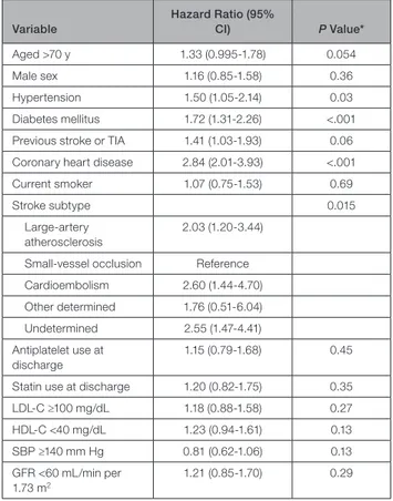
Table 3. Predictors of AMI Up to 5 Years After AIS Variable
Hazard Ratio (95%
CI) P Value*
Aged >70 y 1.33 (0.995-1.78) 0.054
Male sex 1.16 (0.85-1.58) 0.36
Hypertension 1.50 (1.05-2.14) 0.03
Diabetes mellitus 1.72 (1.31-2.26) <.001 Previous stroke or TIA 1.41 (1.03-1.93) 0.06 Coronary heart disease 2.84 (2.01-3.93) <.001
Current smoker 1.07 (0.75-1.53) 0.69
Stroke subtype 0.015
Large-artery atherosclerosis
2.03 (1.20-3.44)
Small-vessel occlusion Reference Cardioembolism 2.60 (1.44-4.70) Other determined 1.76 (0.51-6.04) Undetermined 2.55 (1.47-4.41) Antiplatelet use at
discharge
1.15 (0.79-1.68) 0.45
Statin use at discharge 1.20 (0.82-1.75) 0.35
LDL-C ≥100 mg/dL 1.18 (0.88-1.58) 0.27
HDL-C <40 mg/dL 1.23 (0.94-1.61) 0.13
SBP ≥140 mm Hg 0.81 (0.62-1.06) 0.13
GFR <60 mL/min per 1.73 m2
1.21 (0.85-1.70) 0.29
AIS indicates acute ischemic stroke; AMI, acute myocardial infarction;
GFR, glomerular filtration rate; HDL-C, high-density lipoprotein cholesterol;
LDL-C, low-density lipoprotein cholesterol; SBP, systolic blood pressure; and TIA, transient ischemic attach.
*Subdistribution hazard model by Fine and Grey.
Downloaded from http://ahajournals.org by on April 26, 2021
ARTICLE INFORMATION
Received August 4, 2020; accepted November 12, 2020.
Affiliations
From the Department of Neurology and Cerebrovascular Center, Seoul National University Bundang Hospital, Seoul National University College of Medicine, Seongnam, Republic of Korea (K.-J.L., S.-E.K., J.Y.K., J.K., B.J.K., M.-K.H., H.-J.B.); Department of Neurology, Chonnam National University Hospital, Gwangju, Republic of Korea (K.-H.C., J.-T.K.); Department of Neurology, Chungbuk National University Hospital, Cheongju, Republic of Korea (D.-I.S.); Department of Neurology, Dong-A University Hospital, Dong-A University College of Medicine, Busan, Republic of Korea (J.-K.C., D.- H.K.); Department of Neurology, Dongguk University Ilsan Hospital, Goyang, Republic of Korea (D.-E.K., W.-S.R.); Department of Neurology, Eulji General Hospital, Seoul, Republic of Korea (J.-M.P., K.K.); Department of Neurology, Eulji University Hospital, Eulji University, Daejeon, Republic of Korea (J.G.K., S.J.L.); Department of Neurology, Hallym University Sacred Heart Hospital, Anyang, Republic of Korea (M.-S.O., K.-H.Y., B.-C.L.); Department of Neurology, Ilsan Paik Hospital, Inje University, Goyang, Republic of Korea (H.-K.P., K.-S.H., Y.-J.C.); Department of Neurology, Jeju National University Hospital, Jeju National University School of Medicine, Jeju, Republic of Korea (J.C.C.); Department of Neurology, Keimyung University Dongsan Medical Center, Daegu, Republic of Korea (S.I.S., J.-H.H.); Department of Neurology, Seoul Medical Center, Seoul, Republic of Korea (M.-S.P., T.H.P., S.-S.P.);
Department of Neurology, College of Medicine, Soonchunhyang University Hospital, Seoul, Republic of Korea (K.B.L.); Department of Neurology, Ulsan University Hospital, Ulsan University College of Medicine, Ulsan, Republic of Korea (J.-H.K., W.-J.K.); Department of Neurology, Yeungnam University Hospital, Daegu, Republic of Korea (J.L.); Clinical Research Center, Asan Medical Center, Seoul, Republic of Korea (J.S.L.); Department of Biostatistics, Korea University College of Medicine, Seoul, Republic of Korea (J.L.); Davee Department of Neurology, Feinberg School of Medicine, Chicago, IL (P.B.G.);
and Department of Translational Neuroscience, College of Human Medicine, Michigan State University, Grand Rapids, MI (P.B.G.).
Acknowledgments
We would like to thank the National Health Insurance Service (NHIS) for giv- ing us the opportunity to analyze the claims data (national health information data; NHIS-2015-4-022-1) for this study.
Sources of Funding
This report was supported partially by funding from the research of Korea Centers for Disease Control and Prevention (code 2017ER620102).
Disclosures
None.
Supplementary Material
CRCS-K Registry Investigators Tables S1–S5
Figures S1–S4
REFERENCES
1. Roth GA, Abate D, Abate KH, Abay SM, Abbafati C, Abbasi N, Abbastabar H, Abd-Allah F, Abdela J, Abdelalim A, et al. Global, re- gional, and national age-sex-specific mortality for 282 causes of death in 195 countries and territories, 1980–2017: a systematic analysis for the Global Burden of Disease Study 2017. Lancet. 2018;392:1736–1788.
DOI: 10.1016/S0140 -6736(18)32203 -7.
2. Kernan WN, Ovbiagele B, Black HR, Bravata DM, Chimowitz MI, Ezekowitz MD, Fang MC, Fisher M, Furie KL, Heck DV, et al. Guidelines for the prevention of stroke in patients with stroke and transient isch- emic attack: a guideline for healthcare professionals from the American Heart Association/American Stroke Association. Stroke. 2014;45:2160–
2236. DOI: 10.1161/STR.00000 00000 000024.
3. Smith SC, Benjamin EJ, Bonow RO, Braun LT, Creager MA, Franklin BA, Gibbons RJ, Grundy SM, Hiratzka LF, Jones DW, et al. AHA/ACCF secondary prevention and risk reduction therapy for patients with coronary and other atherosclerotic vascular disease: 2011 update: a guideline from the American Heart Association and American College of
Cardiology Foundation. Circulation. 2011;124:2458–2473. DOI: 10.1161/
CIR.0b013 e3182 35eb4d.
4. Calvet D, Touzé E, Varenne O, Sablayrolles JL, Weber S, Mas JL.
Prevalence of asymptomatic coronary artery disease in ischemic stroke patients: the Precoris Study. Circulation. 2010;121:1623–1629. DOI:
10.1161/CIRCU LATIO NAHA.109.906958.
5. Boulanger M, Béjot Y, Rothwell PM, Touzé E. Long-term risk of myo- cardial infarction compared to recurrent stroke after transient ischemic attack and ischemic stroke: systematic review and meta-analysis. J Am Heart Assoc. 2018;7:e007267. DOI: 10.1161/JAHA.117.007267.
6. Touzé E, Varenne O, Chatellier G, Peyrard S, Rothwell PM, Mas JL. Risk of myocardial infarction and vascular death after transient ischemic attack and ischemic stroke: a systematic review and me- ta-analysis. Stroke. 2005;36:2748–2755. DOI: 10.1161/01.STR.00001 90118.02275.33.
7. Rosenson RS, Farkouh ME, Mefford M, Bittner V, Brown TM, Taylor B, Monda KL, Zhao H, Dai Y, Muntner P. Trends in use of high-intensity sta- tin therapy after myocardial infarction, 2011 to 2014. J Am Coll Cardiol.
2017;69:2696–2706.
8. Otite FO, Liaw N, Khandelwal P, Malik AM, Romano JG, Rundek T, Sacco RL, Chaturvedi S. Increasing prevalence of vascular risk factors in patients with stroke: a call to action. Neurology. 2017;89:1985–1994.
DOI: 10.1212/WNL.00000 00000 004617.
9. Bogiatzi C, Hackam DG, McLeod AI, Spence JD. Secular trends in isch- emic stroke subtypes and stroke risk factors. Stroke. 2014;45:3208–
3213. DOI: 10.1161/STROK EAHA.114.006536.
10. Adams RJ, Albers G, Alberts MJ, Benavente O, Furie K, Goldstein LB, Gorelick P, Halperin J, Harbaugh R, Johnston SC, et al. Update to the AHA/ASA recommendations for the prevention of stroke in patients with stroke and transient ischemic attack. Stroke. 2008;39:1647–1652. DOI:
10.1161/STROK EAHA.107.189063.
11. Adams HP, Bendixen BH, Kappelle LJ, Biller J, Love BB, Gordon DL, Marsh EE. Classification of subtype of acute ischemic stroke: defini- tions for use in a multicenter clinical trial: TOAST: Trial of Org 10172 in Acute Stroke Treatment. Stroke. 1993;24:35–41. DOI: 10.1161/01.
STR.24.1.35.
12. Kang K, Park TH, Kim N, Jang MU, Park SS, Park JM, Ko Y, Lee SJ, Lee KB, Lee J, et al. Recurrent stroke, myocardial infarction, and major vascular events during the first year after acute ischemic stroke: the multicenter prospective observational study about recurrence and its determinants after acute ischemic stroke I. J Stroke Cerebrovasc Dis.
2016;25:656–664.
13. Grau AJ, Weimar C, Buggle F, Heinrich A, Goertler M, Neumaier S, Glahn J, Brandt T, Hacke W, Diener HC. Risk factors, outcome, and treatment in subtypes of ischemic stroke: the German stroke data bank.
Stroke. 2001;32:2559–2566. DOI: 10.1161/hs1101.098524.
14. Dhamoon MS, Tai W, Boden-Albala B, Rundek T, Paik MC, Sacco RL, Elkind MSV. Risk of myocardial infarction or vascular death after first ischemic stroke: the northern Manhattan study. Stroke. 2007;38:1752–
1758. DOI: 10.1161/STROK EAHA.106.480988.
15. Kim BJ, Park JM, Kang K, Lee SJ, Ko Y, Kim JG, Cha JK, Kim DH, Nah HW, Han MK, et al. Case characteristics, hyperacute treatment, and outcome information from the clinical research center for stroke-fifth division registry in South Korea. J Stroke. 2015;17:38–53. DOI: 10.5853/
jos.2015.17.1.38.
16. Kim BJ, Han MK, Park TH, Park SS, Lee KB, Lee BC, Yu KH, Cha JK, Kim DH, Lee J, et al. Current status of acute stroke management in Korea: a report on a multicenter, comprehensive acute stroke registry.
Int J Stroke. 2014;9:514–518. DOI: 10.1111/ijs.12199.
17. Seong SC, Kim YY, Khang YH, Park JH, Kang HJ, Lee H, Do CH, Song JS, Bang JH, Ha S, et al. Data resource profile: the national health in- formation database of the national health insurance service in South Korea. Int J Epidemiol. 2017;46:799–800.
18. Lee KB, Lee JG, Kim BJ, Kim JY, Lee KJ, Han MK, Park JM, Kang K, Cho YJ, Park HK, et al. The epidemiology of fracture in patients with acute ischemic stroke in Korea. J Korean Med Sci. 2019;34:e164.
19. Anderson GB, Ashforth R, Steinke DE, Ferdinandy R, Findlay JM.
CT angiography for the detection and characterization of carotid ar- tery bifurcation disease. Stroke. 2000;31:2168–2174. DOI: 10.1161/01.
STR.31.9.2168.
20. Ko Y, Lee SJ, Chung JW, Han MK, Park JM, Kang K, Park TH, Park SS, Cho YJ, Kong KS, et al. MRI-based algorithm for acute ischemic stroke subtype classification. J Stroke. 2014;16:161. DOI: 10.5853/
jos.2014.16.3.161.
Downloaded from http://ahajournals.org by on April 26, 2021
21. Levey AS, Bosch JP, Lewis JB, Greene T, Rogers N, Roth D; Modification of Diet in Renal Disease Study Group. A more accurate method to es- timate glomerular filtration rate from serum creatinine: a new prediction equation. Ann Intern Med. 1999;130:461–470.
22. Asaria P, Elliott P, Douglass M, Obermeyer Z, Soljak M, Majeed A, Ezzati M. Acute myocardial infarction hospital admissions and deaths in England: a national follow-back and follow-forward record-linkage study. Lancet Public Health. 2017;2:e191–e201. DOI: 10.1016/S2468 -2667(17)30032 -4.
23. Rutten-Jacobs LCA, Maaijwee NAM, Arntz RM, Schoonderwaldt HC, Dorresteijn LD, Van der Vlugt MJ, Van Dijk EJ, De Leeuw FE. Long-term risk of recurrent vascular events after young stroke: the FUTURE study.
Ann Neurol. 2013;74:592–601.
24. Austin PC, Lee DS, Fine JP. Introduction to the analysis of survival data in the presence of competing risks. Circulation. 2016;133:601–609.
DOI: 10.1161/CIRCU LATIO NAHA.115.017719.
25. Stone NJ, Robinson JG, Lichtenstein AH, Bairey Merz CN, Blum CB, Eckel RH, Goldberg AC, Gordon D, Levy D, Lloyd-Jones DM, et al. 2013 ACC/AHA guideline on the treatment of blood cholesterol to reduce atherosclerotic cardiovascular risk in adults: a report of the American College of Cardiology/American Heart Association Task Force on Practice Guidelines. Circulation. 2014;129:1–45. DOI: 10.1161/01.
cir.00004 37738.63853.7a.
26. Dhamoon MS, Sciacca RR, Rundek T, Sacco RL, Elkind MSV. Recurrent stroke and cardiac risks after first ischemic stroke: the Northern Manhattan Study. Neurology. 2006;66:641–646. DOI: 10.1212/01.
wnl.00002 01253.93811.f6.
27. Amarenco P, Lavallée PC, Labreuche J, Ducrocq G, Juliard JM, Feldman L, Cabrejo L, Meseguer E, Guidoux C, Adraï V, et al.
Coronary artery disease and risk of major vascular events after ce- rebral infarction. Stroke. 2013;44:1505–1511. DOI: 10.1161/STROK EAHA.111.000142.
28. Venketasubramanian N, Röther J, Bhatt DL, Pasquet B, Mas JL, Alberts MJ, Hill MD, Aichner F, Steg PG. Two-year vascular event rates in pa- tients with symptomatic cerebrovascular disease: the reach registry.
Cerebrovasc Dis. 2011;32:254–260.
29. Boulanger M, Li L, Lyons S, Lovett NG, Kubiak MM, Silver L, Touzé E, Rothwell PM. Essen risk score in prediction of myocardial infarction after transient ischemic attack or ischemic stroke without prior coro- nary artery disease. Stroke. 2019;50:3393–3399. DOI: 10.1161/STROK EAHA.119.025831.
30. Amarenco P, Bogousslavsky J, Callahan A, Goldstein LB, Hennerici M, Rudolph AE, Sillesen H, Simunovic L, Szarek M, Welch KMA, et al.
High-dose atorvastatin after stroke or transient ischemic attack. N Engl J Med. 2006;355:549–559.
31. Amarenco P, Goldstein LB, Sillesen H, Benavente O, Zweifler RM, Callahan A, Hennerici MG, Zivin JA, Welch KMA. Coronary heart dis- ease risk in patients with stroke or transient ischemic attack and no known coronary heart disease: findings from the stroke prevention by aggressive reduction in cholesterol levels (SPARCL) trial. Stroke.
2010;41:426–430. DOI: 10.1161/STROK EAHA.109.564781.
32. Smolina K, Wright FL, Rayner M, Goldacre MJ. Long-term survival and recurrence after acute myocardial infarction in England, 2004 to 2010.
Circ Cardiovasc Qual Outcomes. 2012;5:532–540.
33. Chen Y, Wright N, Guo Y, Turnbull I, Kartsonaki C, Yang L, Bian Z, Pei P, Pan D, Zhang Y, et al. Mortality and recurrent vascular events after first incident stroke: a 9-year community-based study of 0·5 million Chinese adults. Lancet Glob Health. 2020;8:e580–e590.
34. Prosser J, MacGregor L, Lees KR, Diener HC, Hacke W, Davis S. Predictors of early cardiac morbidity and mortality after isch- emic stroke. Stroke. 2007;38:2295–2302. DOI: 10.1161/STROK EAHA.106.471813.
35. Wang H, Eitzman DT. Acute myocardial infarction leads to acceleration of atherosclerosis. Atherosclerosis. 2013;229:18–22. DOI: 10.1016/j.
ather oscle rosis.2013.04.004.
36. Kallmünzer B, Breuer L, Kahl N, Bobinger T, Raaz-Schrauder D, Huttner HB, Schwab S, Köhrmann M. Serious cardiac arrhythmias after stroke: incidence, time course, and predictors-a systematic, pro- spective analysis. Stroke. 2012;43:2892–2897. DOI: 10.1161/STROK EAHA.112.664318.
37. Ueshima H, Sekikawa A, Miura K, Turin TC, Takashima N, Kita Y, Watanabe M, Kadota A, Okuda N, Kadowaki T, et al. Cardiovascular disease and risk factors in Asia: a selected review. Circulation.
2008;118:2702–2709. DOI: 10.1161/CIRCU LATIO NAHA.108.790048.
38. Kim RB, Kim HS, Kang DR, Choi JY, Choi NC, Hwang S, Hwang JY.
The trend in incidence and case-fatality of hospitalized acute myo- cardial infarction patients in Korea, 2007 to 2016. J Korean Med Sci.
2019;34:1–12.
39. Sacks NC, Ash AS, Ghosh K, Rosen AK, Wong JB, Rosen AB.
Trends in acute myocardial infarction hospitalizations: are we seeing the whole picture? Am Heart J. 2015;170:1211–1219. DOI: 10.1016/j.
ahj.2015.09.009.
40. Aune E, Røislien J, Mathisen M, Thelle DS, Otterstad JE. The “smoker’s paradox” in patients with acute coronary syndrome: a systematic re- view. BMC Med. 2011;9:97. DOI: 10.1186/1741-7015-9-97.
41. Soliman EZ, Safford MM, Muntner P, Khodneva Y, Dawood FZ, Zakai NA, Thacker EL, Judd S, Howard VJ, Howard G, et al. Atrial fibrillation and the risk of myocardial infarction. JAMA Intern Med. 2014;174:107–114.
42. Ko YJ, Kim S, Park K, Kim M, Yang BR, Kim MS, Lee J, Park BJ. Impact of the health insurance coverage policy on oral anticoagulant prescrip- tion among patients with atrial fibrillation in Korea from 2014 to 2016. J Korean Med Sci. 2018;33:1–12.
43. Kim H, Yun JE, Lee SH, Jang Y, Jee SH. Validity of the diagnosis of acute myocardial infarction in Korean National Medical Health Insurance claims data: the Korean Heart Study. Korean Circ J. 2012;42:10–15.
44. Ryu SY, Park JK, Suh I, Jee SH, Park J, Kim CB, Kim KS. The accuracy of myocardial infarction diagnosis in medical insurance claims. Yonsei Med J. 2000;41:570–576.
45. Kim JY, Lee KJ, Kang J, Kim BJ, Han MK, Kim SE, Lee H, Park JM, Kang K, Lee SJ, et al. Development of stroke identification algorithm for claims data using the multicenter stroke registry database. PLoS One.
2020;15:1–15. DOI: 10.1371/journ al.pone.0228997.
Downloaded from http://ahajournals.org by on April 26, 2021
SUPPLEMENTAL MATERIAL
Downloaded from http://ahajournals.org by on April 26, 2021
Beom Joon Kim, MD, PhD (Seoul National University Bundang Hospital, site
investigator); Hee-Joon Bae, MD, PhD (Seoul National University Bundang Hospital, site investigator); Moon-Ku Han, MD, PhD (Seoul National University Bundang Hospital; site investigator); Jihoon Kang, MD (Seoul National University Bundang Hospital; site investigator); Jun Yup Kim, MD (Seoul National University Bundang Hospital; site investigator); Joon-Tae Kim, MD, PhD (Chonnam National University Hospital, site investigator); Ki-Hyun Cho, MD, PhD (Chonnam National University Hospital, site investigator); Man-Seok Park, MD, PhD (Chonnam National University Hospital, site investigator); Kang-Ho Choi, MD (Chonnam National University
Hospital, site investigator); Dong-Ick Shin, MD, PhD (Chungbuk National University Hospital, site investigator); Jae-Kwan Cha, MD, PhD (Dong-A University Hospital, site investigator); Dae-Hyun Kim, MD, PhD (Dong-A University Hospital, site investigator); Dong-Eog Kim, MD, PhD (Dongguk University Ilsan Hospital, site investigator); Wi-Sun Ryu, MD, PhD (Dongguk University Ilsan Hospital, site investigator); Jong-Moo Park, MD, PhD (Eulji General Hospital, site investigator);
Kyusik Kang, MD, PhD (Eulji General Hospital, site investigator); Soo Joo Lee, MD, PhD (Eulji University Hospital, site investigator); Jae Guk Kim, MD (Eulji University Hospital, site investigator); Mi-Sun Oh, MD, PhD (Hallym University Sacred Heart Hospital, site investigator); Kyung-Ho Yu, MD, PhD (Hallym University Sacred Heart Hospital, site investigator); Byung-Chul Lee, MD, PhD (Hallym University Sacred Heart Hospital, site investigator); Keun-Sik Hong, MD, PhD (Inje University Ilsan Paik Hospital, site investigator); Yong-Jin Cho, MD, PhD (Inje University Ilsan Paik Hospital, site investigator); Jay Chol Choi, MD, PhD (Jeju National University
Downloaded from http://ahajournals.org by on April 26, 2021
Medical Center, site investigator); Jeong-Ho Hong, MD, PhD (Keimyung University Dongsan Medical Center, site investigator); Tai Hwan Park, MD, PhD (Seoul Medical Center, site investigator); Sang-Soon Park, MD (Seoul Medical Center, site
investigator); Jee-Hyun Kwon, MD, PhD (Ulsan University Hospital, site
investigator), Wook-Joo Kim, MD (Ulsan University Hospital, site investigator);
Kyung Bok Lee, MD, PhD (Soon Chun Hyang University Hospital, site investigator);
Jun Lee, MD, PhD (Yeungnam University Medical Center, site investigator);
Ji Sung Lee, PhD (Asan Medical Center, statistical analysis), Juneyoung Lee, PhD (Korea University, statistical analysis and data management)
Downloaded from http://ahajournals.org by on April 26, 2021
CI, cumulative incidence; MI, myocardial infarction; STEMI, ST-elevation myocardial infarction;
NSTEMI, non-ST-elevation myocardial infarction
Cumulative incidence, % (95% CI)
1-year 2-year 3-year 4-year 5-year
Fatal MI 0.2 (0.1-0.3) 0.2 (0.1-0.3) 0.3 (0.2-0.4) 0.3 (0.2-0.4) 0.3 (0.2-0.4)
N of patients 23 25 31 33 33
Non-fatal MI 0.9 (0.7-1.1) 1.1 (0.9-1.3) 1.3 (1.1-1.5) 1.5 (1.3-1.8) 1.7 (1.5-2.0)
N of patients 102 126 152 174 183
STEMI 0.4 (0.3-0.5) 0.4 (0.3-0.6) 0.5 (0.4-0.7) 0.6 (0.5-0.7) 0.6 (0.5-0.8)
N of patients 41 50 61 67 69
NSTEMI 0.2 (0.1-0.3) 0.3 (0.2-0.4) 0.3 (0.2-0.5) 0.4 (0.3-0.6) 0.5 (0.4-0.6)
N of patients 22 30 39 47 50
Unspecified 0.5 (0.4-0.7) 0.6 (0.5-0.8) 0.7 (0.6-0.9) 0.8 (0.7-1.0) 0.9 (0.7-1.1)
N of patients 62 71 83 93 97
Downloaded from http://ahajournals.org by on April 26, 2021
stroke (Cause-specific hazard model) .
CI, confidence interval; TIA, transient ischemic attack; SBP, systolic blood pressure; LDL, low-density lipoprotein; HDL, high-density lipoprotein; GFR, glomerular filtration rate
Hazard ratio (95% CI)
Acute myocardial infarction Mortality
Age>70 1.54 (1.15-2.06) 3.78 (3.47-4.11)
Male sex 1.15 (0.84-1.57) 1.04 (0.97-1.12)
Hypertension 1.50 (1.05-2.15) 1.06 (0.97-1.15)
Diabetes 1.78 (1.36-2.35) 1.32 (1.23-1.42)
Previous stroke or TIA 1.37 (1.01-1.85) 1.23 (1.14-1.33)
Coronary heart disease 2.87 (2.07-3.97) 1.14 (1.03-1.26)
Current smoker 1.08 (0.76-1.54) 1.02 (0.93-1.12)
Stroke subtype
Large artery atherosclerosis 2.12 (1.25-3.59) 1.89 (1.65-2.17)
Small vessel occlusion [Reference] [Reference]
Cardioembolism 2.93 (1.61-5.32) 2.74 (2.36-3.17)
Other determined 1.99 (0.58-6.85) 2.95 (2.24-3.88)
Undetermined 2.77 (1.60-4.78) 2.34 (2.03-2.70)
Antiplatelet use at discharge 1.05 (0.71-1.53) 0.73 (0.67-0.79)
Statin use at discharge 1.03 (0.71-1.50) 0.59 (0.54-0.64)
LDL-C≥100mg/dL 1.20 (0.89-1.61) 0.99 (0.92-1.07)
HDL-C<40mg/dL 1.21 (0.93-1.58) 0.96 (0.90-1.04)
SBP≥140mmHg 0.81 (0.62-1.07) 1.04 (0.97-1.11)
GFR<60mL/min/1.73m2 1.39 (0.98-1.97) 1.95 (1.79-2.12)
Downloaded from http://ahajournals.org by on April 26, 2021