• 검색 결과가 없습니다.

A Case of Pleural Paragonimiasis Confused with Tuberculous Pleurisy

N/A
N/A
Protected

Academic year: 2021

Share "A Case of Pleural Paragonimiasis Confused with Tuberculous Pleurisy"

Copied!
4
0
0

로드 중.... (전체 텍스트 보기)

전체 글

(1)

A Case of Pleural Paragonimiasis Confused with Tuberculous Pleurisy

Junwhi Song, M.D.

1,

*, Goohyeon Hong, M.D.

1,

*, Jae-Uk Song, M.D.

1

, Wooyoul Kim, M.D.

1

, Seo Goo Han, M.D.

1

, Yousang Ko, M.D.

1

, Boksoon Chang, M.D.

1

, Byeong-Ho Jeong, M.D.

1

, Jung Seop Eom, M.D.

1

, Ji Hyun Lee, M.D.

1

, Byung Woo Jhun, M.D.

1

, Kyeongman Jeon, M.D.

1

, Hong Kwan Kim, M.D.

2

and Won-Jung Koh, M.D.

1

1

Division of Pulmonary and Critical Care Medicine, Department of Medicine,

2

Department of Thoracic Surgery, Samsung Medical Center, Sungkyunkwan University School of Medicine, Seoul, Korea

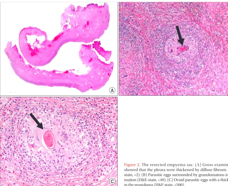
Here, we report a case of pleural paragonimiasis that was confused with tuberculous pleurisy. A 38-year-old man complained of a mild febrile sensation and pleuritic chest pain. Radiologic findings showed right pleural effusion with pleural thickening and subpleural consolidation. Adenosine deaminase (ADA) activity in the pleural effusion was elevated (85.3 IU/L), whereas other examinations for tuberculosis were negative. At this time, the patient started empirical anti-tuberculous treatment. Despite 2 months of treatment, the pleural effusion persisted, and video-assisted thoracoscopic surgery was performed. Finally, the patient was diagnosed with pleural paragonimiasis based on the pathologic findings of chronic granulomatous inflammation containing Paragonimus eggs. This case suggested that pleural paragonimiasis should be considered when pleural effusion and elevated ADA levels are observed.

Keywords: Adenosine Deaminase; Paragonimiasis; Pleural Effusion; Tuberculosis

neoplasm

2,3

. Routine laboratory test results with pleural fluid examination and radiologic findings are non-specific for pleu- ropulmonary paragonimiasis, often delaying diagnosis

4

.

Lymphocytic pleural effusions, often caused by tuberculosis, are commonly encountered in clinical practice. The measure- ment of adenosine deaminase (ADA) levels can facilitate the diagnosis of tuberculous effusions, but false-positive findings from lymphocytic effusions have been reported

5-7

. Here, we report clinical, radiologic, and laboratory features of a case that mimicked tuberculous pleurisy, but was ultimately diag- nosed as pleural paragonimiasis after surgical biopsy.

Case Report

A 38-year-old man developed right chest pain and dyspnea.

He had been healthy until 12 months previously, when he developed right spontaneous pneumothorax. After drainage with a chest tube at a local hospital, the pneumothorax im- proved. He had no history of tuberculosis and the ingestion history of raw crab or river fish was uncertain.

One month before his visit to our hospital, the patient re- Copyright © 2014

The Korean Academy of Tuberculosis and Respiratory Diseases.

All rights reserved.

Introduction

Pleuropulmonary paragonimiasis is a food-borne zoonosis commonly caused by the trematode Paragonimus wester- mani

1

. This parasitic infection has diverse symptoms and can mimic other conditions, such as mycobacterial infection or

CASE REPORT

http://dx.doi.org/10.4046/trd.2014.76.4.175

ISSN: 1738-3536(Print)/2005-6184(Online) • Tuberc Respir Dis 2014;76:175-178

175

Address for correspondence: Won-Jung Koh, M.D.

Division of Pulmonary and Critical Care Medicine, Department of Medicine, Samsung Medical Center, Sungkyunkwan University School of Medicine, 81 Irwon-ro, Gangnam-gu, Seoul 135-710, Korea

Phone: 82-2-3410-3429, Fax: 82-2-3410-3849 E-mail: [email protected]

*Junwhi Song and Goohyeon Hong contributed equally to this work.

Received: Dec. 7, 2012 Revised: Dec. 31, 2012 Accepted: Oct. 28, 2013

cc

It is identical to the Creative Commons Attribution Non-Commercial

License (http://creativecommons.org/licenses/by-nc/3.0/).

(2)

J Song et al.

176 Tuberc Respir Dis 2014;76:175-178 www.e-trd.org

ported right chest pain and mild dyspnea. Three weeks later, a chest radiograph taken a local hospital revealed right pleural effusion. Serous fluid (400 mL) was removed and pleural fluid analysis showed that the white blood cell (WBC) count was 700/mL (67% lymphocytes) and the ADA level was 85.3 IU/L.

The patient then started empirical anti-tuberculous treatment including isoniazid, rifampin, ethambutol, and pyrazinamide.

The patient was referred to our hospital because of persistent pleural effusion after 10 days of anti-tuberculous treatment.

Upon admission to our hospital, clinical laboratory testing showed that the blood leukocyte count was 8,600/mL, with a differential of 67.9% neutrophils and 5.2% eosinophils (nor- mal range, 0−9.3%). The erythrocyte sedimentation rate was 120 mm/hr and the C-reactive protein level had increased to 8.27 mg/dL. A human immunodeficiency virus antibody test was negative. Chest radiography and computed tomography revealed right loculated pleural effusion with diffuse pleural thickening (Figure 1).

A percutaneous catheter was inserted and analysis of as- pirated pleural fluid showed the following values: pH, 7.3;

WBC count, 100/mL (69% polymorphonuclear neutrophils, 28% lymphocytes, 0% eosinophils); protein, 6.9 g/dL; lactate dehydrogenase (LDH), 2,226 U/L (serum LDH, 246 U/L);

glucose, 12 mg/dL; and ADA, 84 IU/L. The patient continued anti-tuberculous treatment. In addition, percutaneous cath- eter drainage of pleural fluid was performed with intrapleural urokinase instillation for 5 days. Despite these efforts, the locu- lated pleural effusion did not improve.

Therefore, video-assisted thoracoscopic surgery was performed for decortication. Diffuse, dense fibrosis of the pleura and empyema sacs was observed. Adhesiolysis and decortication were the performed. Pathologic examination

showed chronic granulomatous inflammation with parasitic eggs, many eosinophils, and necrosis, which were consistent with paragonimiasis (Figure 2). Parasite-specific enzyme- linked immunosorbent assay after surgery was positive for Paragonimus. The patient was finally diagnosed with pleural paragonimiasis. Anti-tuberculous treatment was discontinued and praziquantel (25 mg/kg) was prescribed for 2 days. His symptoms and chest radiography were recovered 20 days after discharge. After 2 years of follow-up, the patient was free of symptoms and showed no pleural effusion on chest radiog- raphy.

Discussion

Paragonimiasis is an infection caused by lung flukes, in- cluding Paragonimus westermani and related subspecies.

Paragonimus is endemic in the Far East, including Korea, and in central South America and western Africa

8

. In these areas, reservoir hosts such as freshwater crab or crayfish harboring metacercariae of the parasite are an important source of hu- man infection. When humans ingest pickled or inadequately cooked food, metacercariae reach the small intestine and excyst. They then penetrate the intestinal wall and enter the peritoneal cavity, migrate into the diaphragm, pleura, and reach the lung. Most patients are asymptomatic, but can pres- ent hemorrhage or inflammation causing necrosis and cysts

9

. Microscopic demonstration of eggs in stool or sputum and detection of antibodies to Paragonimus are important for di- agnosis

10,11

. Surgical evaluation is also an important diagnostic option in complicated cases

4

.

Because pleural involvement of Paragonimus is a main

Figure 1. A 38-year-old man with pleural

effusion who was diagnosed with pleural

paragonimiasis. (A) Chest radiography

showed pleural effusion. (B) Chest

computed tomography showed pleural

thickening with pleural effusion. (C) Sub-

pleural consolidation.

(3)

Pleural paragonimiasis

http://dx.doi.org/10.4046/trd.2014.76.4.175 177

www.e-trd.org

presentation of pleuropulmonary paragonimiasis, the dif- ferentiation of this infection from other causes of pleural effu- sion is important

12

. Differentiation is especially important in tuberculosis-endemic areas because pleuropulmonary para- gonimiasis is often difficult to distinguish from tuberculosis, leading to misdiagnosis. Although peripheral blood or pleural fluid eosinophilia is reported with a high incidence in patients with paragonimiasis, a small proportion of patients do not have these characteristic findings

2,13

. Our patient did not pres- ent with eosinophilia in the peripheral blood or eosinophils in pleural fluid and bronchoalveolar lavage. Ova or parasite bodies were not found in the bronchoalveolar lavage or stool.

Furthermore, elevated ADA activity was suggestive of tuber- culous pleurisy. Percutaneous drainage was ineffective, and the final diagnosis was delayed until surgical pleural biopsy, although the history of previous pneumothorax could suggest the possibility of paragnonimiasis.

ADA activity in pleural fluid is a useful biomarker of tuber- culous pleurisy and provides a reliable basis for treatment decisions. However, high ADA levels can also be found in patients with other causes of pleural effusion

5-7,14

. Here, we report clinical, radiologic, and laboratory features of a case that mimicked tuberculous pleurisy, but was diagnosed with pleural paragonimiasis after surgical biopsy. In conclusion, the possibility of pleural paragonimiasis should be considered in the differential diagnosis of pleural effusion with elevated ADA levels in areas where tuberculosis and paragonimiasis could be prevalent.

Conflicts of Interest

No potential conflict of interest relevant to this article was reported.

Figure 2. The resected empyema sac. (A) Gross examination

showed that the pleura were thickened by diffuse fibrosis (H&E

stain, ×2). (B) Parasitic eggs surrounded by granulomatous inflam-

mation (H&E stain, ×40). (C) Ovoid parasitic eggs with a thick shell

in the granuloma (H&E stain, ×200).

(4)

J Song et al.

178 Tuberc Respir Dis 2014;76:175-178 www.e-trd.org

References

1. Nakamura-Uchiyama F, Mukae H, Nawa Y. Paragonimiasis: a Japanese perspective. Clin Chest Med 2002;23:409-20.

2. Jeon K, Koh WJ, Kim H, Kwon OJ, Kim TS, Lee KS, et al. Clini- cal features of recently diagnosed pulmonary paragonimiasis in Korea. Chest 2005;128:1423-30.

3. Song JU, Um SW, Koh WJ, Suh GY, Chung MP, Kim H, et al.

Pulmonary paragonimiasis mimicking lung cancer in a tertia- ry referral centre in Korea. Int J Tuberc Lung Dis 2011;15:674- 9.

4. Boland JM, Vaszar LT, Jones JL, Mathison BA, Rovzar MA, Colby TV, et al. Pleuropulmonary infection by Paragonimus westermani in the United States: a rare cause of eosinophilic pneumonia after ingestion of live crabs. Am J Surg Pathol 2011;35:707-13.

5. Lee YC, Rogers JT, Rodriguez RM, Miller KD, Light RW. Ad- enosine deaminase levels in nontuberculous lymphocytic pleural effusions. Chest 2001;120:356-61.

6. Jimenez Castro D, Diaz Nuevo G, Perez-Rodriguez E, Light RW. Diagnostic value of adenosine deaminase in nontubercu- lous lymphocytic pleural effusions. Eur Respir J 2003;21:220- 4.

7. Krenke R, Korczynski P. Use of pleural fluid levels of adenos- ine deaminase and interferon gamma in the diagnosis of

tuberculous pleuritis. Curr Opin Pulm Med 2010;16:367-75.

8. Kagawa FT. Pulmonary paragonimiasis. Semin Respir Infect 1997;12:149-58.

9. Im JG, Whang HY, Kim WS, Han MC, Shim YS, Cho SY. Pleu- ropulmonary paragonimiasis: radiologic findings in 71 pa- tients. AJR Am J Roentgenol 1992;159:39-43.

10. Slemenda SB, Maddison SE, Jong EC, Moore DD. Diagnosis of paragonimiasis by immunoblot. Am J Trop Med Hyg 1988;39:

469-71.

11. Xu ZB. Studies on clinical manifestations, diagnosis and con- trol of paragonimiasis in China. Southeast Asian J Trop Med Public Health 1991;22 Suppl:345-8.

12. Vidamaly S, Choumlivong K, Keolouangkhot V, Vannavong N, Kanpittaya J, Strobel M. Paragonimiasis: a common cause of persistent pleural effusion in Lao PDR. Trans R Soc Trop Med Hyg 2009;103:1019-23.

13. Taniguchi H, Mukae H, Matsumoto N, Tokojima M, Katoh S, Matsukura S, et al. Elevated IL-5 levels in pleural fluid of patients with paragonimiasis westermani. Clin Exp Immunol 2001;123:94-8.

14. Porcel JM, Esquerda A, Bielsa S. Diagnostic performance of

adenosine deaminase activity in pleural fluid: a single-center

experience with over 2100 consecutive patients. Eur J Intern

Med 2010;21:419-23.

수치

Figure 2. The resected empyema sac. (A) Gross examination  showed that the pleura were thickened by diffuse fibrosis (H&E  stain, ×2)

참조

관련 문서

2재화 2요소 헥셔-올린 모형에서는 어느 한 경제에서 어느 한 요소의 양이 증가하면, 그 요소를 집약적으로 사용하는 산업의 생산량은 증가하고 다른

Should a module be used with any of the absolute maximum ratings exceeded, the characteristics of the module may not be recovered, or in an extreme case, the module

All patients with fever, thrombocytopenia and leukocytopenia in SFTS endemic areas should be suspected of SFTS, even they don’t have his- tory of tick bite... Major exposure

The index is calculated with the latest 5-year auction data of 400 selected Classic, Modern, and Contemporary Chinese painting artists from major auction houses..

The cleanest animals in the facility should be easily serviced from the clean side of the cage- wash or clean cage storage areas, while rooms holding the dirtiest animals

After first field tests, we expect electric passenger drones or eVTOL aircraft (short for electric vertical take-off and landing) to start providing commercial mobility

1 John Owen, Justification by Faith Alone, in The Works of John Owen, ed. John Bolt, trans. Scott Clark, "Do This and Live: Christ's Active Obedience as the

Histogram shows the difference of pleural effusion volume depending on the experimental conditions in freshwater group... Youn Shin