• 검색 결과가 없습니다.

Diagnostic Tools of Pleural Effusion

N/A
N/A
Protected

Academic year: 2021

Share "Diagnostic Tools of Pleural Effusion"

Copied!
12
0
0

로드 중.... (전체 텍스트 보기)

전체 글

(1)

Diagnostic Tools of Pleural Effusion

Moon Jun Na, M.D., Ph.D.

Respiratory Division, Department of Internal Medicine, Konyang University College of Medicine, Daejeon, Korea

Pleural effusion is not a rare disease in Korea. The diagnosis of pleural effusion is very difficult, even though the patients often complain of typical symptoms indicating of pleural diseases. Pleural effusion is characterized by the pleural cavity filled with transudative or exudative pleural fluids, and it is developed by various etiologies. The presence of pleural effusion can be confirmed by radiological studies including simple chest radiography, ultrasonography, or computed tomography. Identifying the causes of pleural effusions by pleural fluid analysis is essential for proper treatments. This review article provides information on the diagnostic approaches of pleural effusions and further suggested ways to confirm their various etiologies, by using the most recent journals for references.

Keywords: Pleural Effusion; Pleurisy; Diagnosis

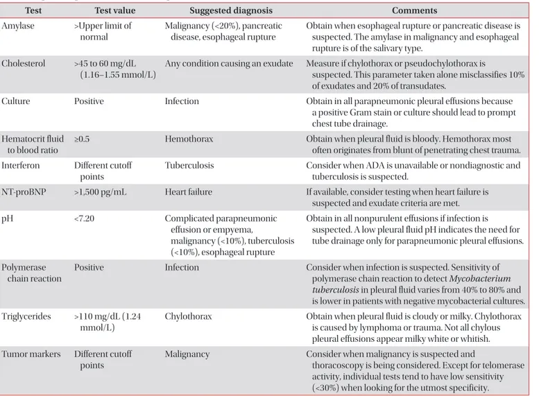
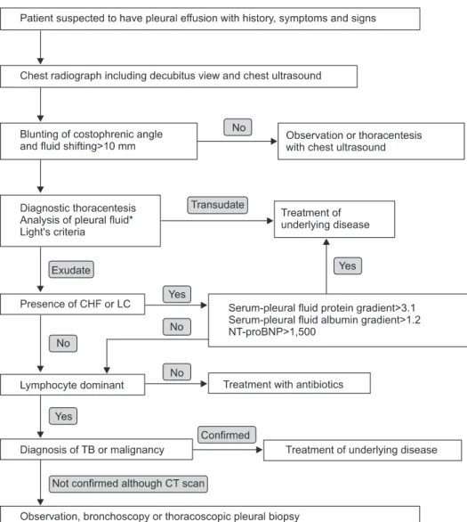
The first step of differential diagnosis or determination of pathogenesis for pleural fluid is to determine whether the patient has a transudative or exudative pleural effusion
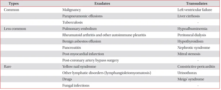
2

. Transudates are caused by increased hydrostatic pressures (e.g., heart failure), decreased oncotic forces (e.g., hypopro- teinemia), increased negative intrapleural pressure (e.g., atel- ectasis), or movement of ascitic fluid through the diaphragm (e.g., hepatic hydrothorax). In contrast, exudates are due to the increased capillary permeability and/or impaired lymphatic drainage which results from the proliferative (e.g., malignancy) or inflammatory (e.g., parapneumonic effusions) processes

3

.

This article aims to review the history taking and the physi- cal examinations of patients who were suspected to have symptoms and signs of pleural effusions, to provide precise ra- diological approaches including thoracic ultrasonography for diagnosing the presence of pleural effusions, and to diagnose the different causes of effusions by using the thoracentesis and pleural biopsy.

History, Symptoms and Signs

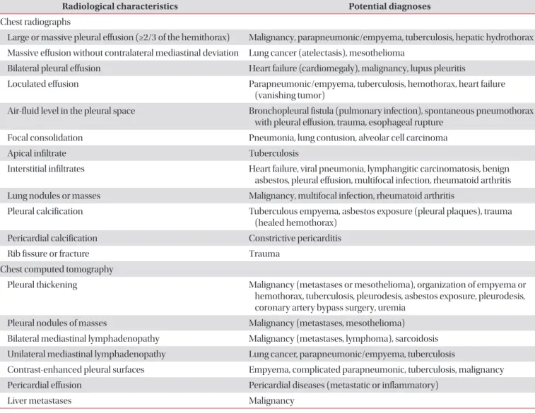
The clinical history, symptoms and signs may be very help- ful for evaluating many causes of the pleural effusions (Table 2)

3

. Especially, because the effusion by drugs is misdiagnosed, the clinical history which includes the medication history is important. If the causes of pleural effusions are suspected to by drugs, such drug list can be easily found in http://www.

pneumothox.com.

Copyright © 2014

The Korean Academy of Tuberculosis and Respiratory Diseases.

All rights reserved.

Introduction

The mean amount of pleural fluid in the normal is as small as 8.4±4.3 mL. Fluid that enters the pleural space can originate in the pleural capillaries, the interstitial spaces of the lung, the intrathoracic lymphatics, the intrathoracic blood vessels, or the peritoneal cavity. Pleural fluid is usually absorbed through the lymphatic vessels in the parietal pleura by means of stomas in the parietal pleura, or through the alternative tran- scytosis

1

. But various pathogenic mechanisms increase the amount of pleural fluids by increasing the rates of pleural fluid formation exceeding the rate of pleural fluid absorption (Table 1)

1

.

Address for correspondence: Moon Jun Na, M.D., Ph.D.

Division of Pulmonology, Department of Internal Medicine, Konyang University College of Medicine, 158 Gwanjeodong-ro, Seo-gu, Daejeon 302-718, Korea

Phone: 82-42-600-8821, Fax: 82-42-600-9090 E-mail: mjnamd@hanmail.net

Received: Mar. 28, 2014 Revised: Apr. 4, 2014 Accepted: Apr. 11, 2014

cc It is identical to the Creative Commons Attribution Non-Commercial License (http://creativecommons.org/licenses/by-nc/3.0/).

(2)

While small amounts of effusions are usually asymptomatic, the increasing effusion produces dyspnea, trepopnea, chest pain or cough. Dyspnea is the consequence of a combination of restrictive lung defects, a ventilation-perfusion mismatch, and a decrease in cardiac output

4

. Although large volumes of pleural effusions usually correlate with the degree of dys- pnea, dyspnea is frequently not proportional to the size of the pleural effusions. Patients with underlying lung diseases (chronic obstructive pulmonary disease [COPD], carcino- matous lymphangitis, pulmonary emboli) may experience intense dyspnea with just small-to-moderate sized pleural effusions. Trepopnea is a form of positional dyspnea in which the patients experience less dyspnea when lying on the side of the pleural effusion

3

. Chest pain accompanied with a pari- etal pleural inflammation clinically is a sharp localized pain, worsening on deep inspiration or coughing, and occasionally twisting or bending movements. A malignancy involving the parietal pleura frequently produces a chronic dull ache local- ized to the relevant anatomic region

5

. Pleuritic chest pain may be referred to the abdomen, and to the ipsilateral shoulder when the diaphragmatic pleura is involved. Cough is dry and nonproductive, a consequence of pleural inflammation or compression of the bronchial trees, but they are rarely helpful for diagnosing pleural effusions. If hemoptysis is combined, it may suggest endobronchial cancer or pulmonary thrombo- embolism

1

.

Upon physical examination, tactile fremitus in palpation is either absent or attenuated, and the percussion note over a pleural effusion is dull or flat. In auscultation, the breathing sounds are decreased or absent, and auscultatory percussion (Guarino’s second method) is abnormal. Pleural rubs often appear as pleural effusions which diminish in size, and will disappear once the effusion develops

5

.

Radiologic Diagnosis

1. Chest radiograph

If symptoms and signs are suspected to have pleural effu- sions, the chest radiograph is usually the diagnosing method.

1) Chest posteroanterior and lateral view: The chest posteroanterior (PA) view is the abnormal blunting of sharp lateral costophrenic angle when pleural fluid is over 200 mL.

In addition, the lateral radiography may show blunting of the sharp posterior costophrenic angle when the fluid exceeds 50 mL

6

. Increasing amounts of the effusion form a meniscus, opacify the lung, and obscure the diaphragmatic margin.

Radiological characteristics of subpulmonic effusions are el- evations of either one or both diaphragms, the displacement of the apex of the apparent diaphragm more laterally, and separation between the lower border of the lung and gastric bubble greater than 2 cm in the left-sided effusion

1

.

2) Anteroposterior radiograph: The anteroposterior (AP) chest radiography is abnormal when pleural fluid is over 300 mL. The earliest sign is blunting of the costophrenic angle.

Subsequently, it also causes increased density of the hemitho- rax, loss of the hemidiaphragm, and decreased visibility of the lower lobe vasculature

1

.

3) Lateral decubitus view: In the lateral decubitus view, pleural effusion is easily detected by free pleural fluids shifting between the dependent chest wall and the lower border of the lung. Diagnostic thoracentesis is safe when the distance of shifting is more than 10 mm. In general, bilateral decubitus chest radiographs should be ordered to assess the underlying lungs for infiltrates or atelectasis

1

.

4) Chest radiographic finding (Table 3): Useful radiologi- Table 1. General causes of pleural effusions

Mechanisms Examples

Increased pleural fluid formation

Increased interstitial fluid in the lung Left ventricular failure, pneumonia, and pulmonary embolus Increased intravascular pressure in pleura Right or left ventricular failure, superior vena caval syndrome

Increased permeability of the capillaries in the pleura Pleural inflammation, increased levels of vascular endothelial growth factor Increased pleural fluid protein level Pulmonary edema, hemothorax

Decreased pleural pressure Lung atelectasis or increased elastic recoil of the lung, trapped lung Increased fluid in peritoneal cavity Ascites or peritoneal dialysis

Disruption of the thoracic duct Chylothorax

Disruption of blood vessels in the thorax Hemothorax Decreased pleural fluid absorption

Obstruction of the lymphatics draining the parietal pleura Cancer, lymphoma

Elevation of systemic vascular pressures Superior vena caval syndrome or right ventricular failure

Disruption of the aquaporin system in the pleura

(3)

cal findings of pleural effusions are summarized in Table 3

3

. (1) Bilateral effusion: Bilateral pleural effusion is com- monly seen in heart failure. For bilateral effusion with a nor- mal heart size, the differential diagnosis should include ma- lignancy and, less commonly, lupus pleuritis and constrictive pericarditis

3

.

(2) Massive effusion more than half of hemithorax:

The most frequent cause of massive pleural effusions is ma- lignancy (55%), followed by complicated parapneumonic or empyema (22%), and tuberculosis (TB) (12%). If massive ef- fusions are without contralateral displacement of mediastinal structures, the endobronchial obstructions by lung cancer or Table 2. Medical history and physical examinations of pleural effusions

Findings Potential causes of the pleural effusion

History

Abdominal surgery Postabdominal surgery, subphrenic abscess, pulmonary embolism

Asbestos exposure Mesothelioma, benign asbestos pleural effusion

Cancer Malignancy, paramalignant effusions (pulmonary embolism, atelectasis,

postobstructive pneumonitis, postradiation therapy)

Cardiac procedures or myocardial infarction Pleural effusion secondary to coronary artery bypass surgery, post-cardiac injury syndrome

Cirrhosis Hepatic hydrothorax, spontaneous bacterial pleuritis

Collapse therapy for pulmonary TB Tuberculous or pyogenic empyema, pyothorax-associated lymphoma, trapped lung

Dialysis Heart failure, uremic pleuritis, pleural effusion secondary to peritoneal dialysis

Drug use Drug-related effusions (e.g., dasatinib, gonadotrophins, amiodarone)

Esophageal surgery, dilation or endoscopy Chylothorax, esophageal perforation

Heart failure Heart failure-related effusion

HIV Pneumonia, TB, primary effusion lymphoma, Kaposi sarcoma

Neurosurgery Intrathoracic migration of ventriculoperitoneal shunt, ventriculopleural shunt, duropleural fistula

Pancreatic disease Pancreatic effusion (pancreatic-pleural fistula) Radiofrequency ablation of lung or liver tumors Pleuritis secondary to radiofrequency ablation Recurrent episodes of pleuritic pain (plus fever,

abdominal pain or arthritis)

Familial Mediterranean fever

Rheumatic autoimmune diseases Rheumatoid pleurisy, lupus pleuritis, parapneumonic effusion, pulmonary arterial hypertension

Trauma Hemothorax, chylothorax

Symptoms

Fever Parapneumonic effusion/empyema, TB, viral pleuritis, lupus pleuritis

Hemoptysis Lung cancer, TB, pulmonary embolism, parapneumonic effusion

Weight loss Cancer, empyema, TB

Signs

Ascites Hepatic hydrothorax, ovarian cancer, Meig’s syndrome, constrictive pericarditis Distended abdominal veins, encephalopathy, spider nevi Cirrhosis

Pericardial rub Acute pericarditis

S3 gallop, elevated neck veins, positive abdomino-jugular test, displaced apical impulse

Heart failure

Unilateral calf pain or swelling Pulmonary embolism

Yellow dystrophic nails, lymphedema Yellow nail syndrome

TB: tuberculosis; HIV: human immunodeficiency virus.

(4)

mediastinum fixation by mesothelioma should be consid- ered

7

.

(3) Loculated effusion: The loculation of pleural space is caused by adhesions between contiguous pleural surfaces. It occurs most frequently in conditions that cause intense pleu- ral inflammations, such as empyema, hemothorax, or TB pleu- risy. In patients with congestive heart failure after treatment, the loculated effusion in fissure may simulate a mass, termed as the vanishing tumor or pseudotumor in chest PA view

1

.

(4) Combined pneumonia in lower lobe: The AP, PA, and lateral chest radiographs are not sensitive methods to identify parapneumonic effusions in patients with pneumonia, be- cause all views missed more than 10% of significant effusions.

The existence of a lower lobe parenchymal consolidation con- cealed the identification of some pleural effusions. Therefore, such considerations should be used for obtaining additional imaging, such as thoracic ultrasonography in patients with lower lobe parenchymal consolidations on plain film radio-

graphs

8

.

2. Thoracic ultrasonography

Thoracic ultrasonography (TUS) will detect the presence of as little as 5−50 mL of pleural fluids and is 100% sensitive for effuions

9

. Ultrasonography can be used under several different situations, including the following: 1) determining the pres- ence of pleural fluid; 2) identification of the appropriate loca- tions for an attempted thoracentesis, pleural biopsy, or chest tube placement; 3) identification of pleural fluid loculations; 4) distinction of pleural fluids from thickening; 5) semiquantita- tion on the amount of pleural fluids; 6) differentiation of a pyo- pneumothorax from a lung abscess; 7) assessment of whether a pleurodesis is present; and 8) evaluation of the trauma patient for the presence of a hemothorax or a pneumothorax

1

. TUS is also an useful instrument for the diagnosis and treat- ment of pleural diseases, especially in the intensive care units.

Table 3. Useful radiological signs in pleural effusions

Radiological characteristics Potential diagnoses

Chest radiographs

Large or massive pleural effusion (≥2/3 of the hemithorax) Malignancy, parapneumonic/empyema, tuberculosis, hepatic hydrothorax Massive effusion without contralateral mediastinal deviation Lung cancer (atelectasis), mesothelioma

Bilateral pleural effusion Heart failure (cardiomegaly), malignancy, lupus pleuritis

Loculated effusion Parapneumonic/empyema, tuberculosis, hemothorax, heart failure (vanishing tumor)

Air-fluid level in the pleural space Bronchopleural fistula (pulmonary infection), spontaneous pneumothorax with pleural effusion, trauma, esophageal rupture

Focal consolidation Pneumonia, lung contusion, alveolar cell carcinoma

Apical infiltrate Tuberculosis

Interstitial infiltrates Heart failure, viral pneumonia, lymphangitic carcinomatosis, benign asbestos, pleural effusion, multifocal infection, rheumatoid arthritis Lung nodules or masses Malignancy, multifocal infection, rheumatoid arthritis

Pleural calcification Tuberculous empyema, asbestos exposure (pleural plaques), trauma (healed hemothorax)

Pericardial calcification Constrictive pericarditis

Rib fissure or fracture Trauma

Chest computed tomography

Pleural thickening Malignancy (metastases or mesothelioma), organization of empyema or hemothorax, tuberculosis, pleurodesis, asbestos exposure, pleurodesis, coronary artery bypass surgery, uremia

Pleural nodules of masses Malignancy (metastases, mesothelioma) Bilateral mediastinal lymphadenopathy Malignancy (metastases, lymphoma), sarcoidosis Unilateral mediastinal lymphadenopathy Lung cancer, parapneumonic/empyema, tuberculosis

Contrast-enhanced pleural surfaces Empyema, complicated parapneumonic, tuberculosis, malignancy Pericardial effusion Pericardial diseases (metastatic or inflammatory)

Liver metastases Malignancy

(5)

Pleural fluids on ultrasonograph can be characterized ei- ther as echo free (anechoic), complex septated (fibrin strands or septa), complex nonseptated (heterogeneous echogenic material), or homogenrously echogenic

1

. If echogenecity is extremely high, immediate thoracentesis is required for dif- ferentiation of empyema or hemothorax. The TUS findings which suggest malignancy include parietal pleural thicken- ing of more than 1 cm, pleural nodularity and diaphragmatic thickness >7 mm (sensitivity 42% and specificity 95% for each criteria)

10

.

3. Computed tomography

Chest computed tomography (CT) in pleural effusion is available for differentiation of pleural collections or masses, detection of loculated fluid collections, demonstration of abnormalities in lung parenchyme, distinguishing empyema with air-fluid levels from lung abscess, identification of pleural thickening, evaluation of major and minor fissures, and distin- guishing benign and malignant effusions

1

. CT findings which are suggestive of malignancy are as follows: pleural nodular- ity, pleural rind, mediastinal pleural involvement, and pleural thickening greater than 1 cm

11

.

4. Magnetic resonance imaging

Magnetic resonance imaging of chest is currently less satis- factory and higher cost than CT or TUS in pleural disease due to poor spatial resolutions and motion artifacts

1

.

5. Positron emission tomography scan

Because the 18-fluorodeoxyglucose (

18

FDG) concentrated in malignant cells are more avid than normal tissues, the FDG- positron emission tomography is useful for differentiating be- nign and malignant pleural effusions including mesothelioma.

But, false positive scans may be due to infections (parapneu- monics, empyema, TB) and talc pleurodesis

3

.

Thoracentesis

If the thickness of the pleural fluid on decubitus radiograph, TUS or the CT scan is greater than 10 mm, diagnostic thora- centesis should be performed. Although the effusion is due to obvious congestive heart failure, the thoracentesis should be concerned under the following conditions: 1) not bilateral and comparable sized effusions; 2) pleuritic chest pain; 3) febrile;

and 4) no responses to diuretics. Using TUS is safe for small or loculated effusions

1

.

The main contraindication of thoracentesis is a hemorrhag- ic diathesis. Some guidelines recommend correcting an inter-

national normalized ratio to less than 2, transfusing platelets to more than 50,000/μL, or withholding certain medications (oral anticoagulants, heparin or clopidogrel) before perform- ing minimally invasive procedures such as thoracentesis

12

. Thoracentesis should not be attempted in local cutaneous conditions such as pyoderma or herpes zoster infection

1

.

The most common complication of thoracentesis is pneu- mothorax. The incidence of pneumothorax after thoracentesis with conventional techniques was 18%, whereas it was only 3% with the TUS

13

. Other common complications of thora- centesis are cough, chest pain, vasovagal reflux characterized by bradycardia and a decreased blood pressure, infection of the pleural space, hemothorax due to laceration of intercostal artery, splenic or hepatic laceration, soft tissue infection and seeding of the needle tract with tumor cells. Postprocedure chest radiograph is only recommended in aspiration of air, developing of symptoms, and the loss of tactile fremitus

1

.

To relieve dyspnea in massive pleural effusion, therapeutic thoracentesis is helpful. The removal as little as 300−500 mL at once is generally sufficient to relieve dyspnea in patients with undiagnosed effusions

3

. Although most physicians tend to avoid performing thoracentesis with more than 1,500 mL at one time, the re-expansion pulmonary edema is uncommon (0.5%), even when >1,000 mL of pleural fluids are removed

14

. To prevent re-expansion pulmonary edema, therapeutic tho- racentesis should be stopped in symptoms such as chest tight- ness, chest pain, dyspnea or more than minimal coughing

1

. If the dyspnea does not improve after therapeutic thoracentesis in malignant effusions, underlying diseases such as pulmo- nary carcinomatous lymphangitis, atelectasis, pulmonary or tumor embolism, or COPD should be considered

3

.

Analysis of Pleural Fluid

1. Preparation of pleural fluid sample for test

Approximately 20−40 mL of aspirated fluid should be im- mediately placed into appropriate anticoagulant (EDTA or heparin) coated tubes for biochemistry (5 mL), microbiology (5−10 mL), cytology (10−25 mL), and heparin coated syringe for the pH measurement. Pleural fluids should be analyzed within 4 hours of extraction. If pleural fluid contacts with air for a long time, the CO

2

will escape and the pH level will in- crease

3

. Measurements of pH level should be accomplished more accurately with a blood gas machine either than the pH indicator paper or pH meter

1

. For aerobic and anaerobic bac- terial cultures, the pleural fluids should be inoculated directly into the blood culture media by the bedside to improve posi- tively cultured results

15

.

Routine and optional tests of pleural fluids are summarized

in Tables 4 and 5

3

.

(6)

2. Differentiation of transudate and exudate

The first step in differential diagnosis of pleural effusion is to determine whether the effusion is transudative or exuda- tive. Light’s criteria (Table 6)

1

should be used to differentiate transudates and exudates by analyzing the levels of protein and lactate dehydrogenase (LDH) in the pleural fluid and serum. Transudate is mostly due to systemic diseases, such as congestive heart failure, liver cirrhosis, or nephrosis that easily treated with diuretics. Exudate resulted from local diseases, where the fluid originated, and further investigations should be directed at the genesis of the local disease

1

. Common causes of exudative pleural effusion is malignancy, parapneu- monic effusion and TB (Table 7)

16

.

Light’s criteria is the gold standard to differentiate the tran-

sudate and exudate over the last 30 years. However, the con- centrations of the biochemical components (protein, LDH, and albumin) in pleural fluids increase progressively during the diuretic therapy of patients with congestive heart failure

17

. In patients with congestive heart failure receiving diuretic therapy, the misclassification of transudate to exudate (29%) in Light’s criteria can be more accurately corrected in the albu- min gradient (>1.2 g/dL) than the protein gradient (>3.1 g/dL) between the serum and the pleural fluid. In patients with liver cirrhosis misclassified as exudate (18%) in Light’s criteria, their diagnostic accuracy increased more in albumin ratios (<0.6) than albumin or protein gradient

18

. Although the accuracy of the albumin gradient is higher than the protein gradient, it is recommend to use protein gradient easily obtained in Light’s criteria. Increased levels of pleural fluid or serum N-terminal Table 4. Routine pleural fluid tests for pleural effusions

Test Test value Suggested diagnosis Comments

Adenosine deaminase (ADA)

>40 U/L (667 nakt/L)

Tuberculosis (>90%), empyema (60%), complicated parapneumonic effusion (30%), malignancy (5%), rheumatoid arthritis

In the United States, ADA is no routinely requested because of the low prevalence of tuberculosis pleurisy.

Cytology Present Malignancy Actively dividing mesothelial cells can mimic and

adenocarcinoma.

Glucose <60 mg/dL (3.3 mmol/L)

Complicated parapneumonic effusion or empyema, tuberculosis (20%), malignancy (<10%), rheumatoid arthritis

In general, pleural fluids with a low glucose level also have low pH and high LDH levels.

Lactate

dehydrogenase (LDH)

>Two thirds of upper limits of normal for serum LDH

Any condition causing an exudate Very high levels of pleural fluid LDH (>1,000 U/L) typically are found in patients with complicated parapneumonic pleural effusion and in about 40% of those with tuberculosis pleurisy.

LDH fluid to serum ratio

>0.6 Any condition causing an exudate Most patients who meet the criteria for an exudative effusion with LDH but no with protein levels have either parapnuemonic effusions or malignancy.

Protein fluid to serum ratio

>0.5 Any condition causing an exudate A pleural fluid protein level >3 mg/dL suggests an exudate, but when taken alone this parameter misclassifies more than 10% of exudates and 15% of transudates.

Red blood cell count

>100,000/mm

3

(100×10

6

/L)

Malignancy, trauma, parapneumonic effusion, pumonary embolism

A fluid hematocrit <1% is nonsignificant.

White blood cell count and differential

>10,000/mm

3

(10×10

9

/L)

Empyema, other exudates (uncommon) In purulent fluids, leukocyte count is commonly much lower than expected because dead cells or other debris account for much of the turbidity.

Eosinophils >10% Not diagnostic The presence of air or blood in the pleural space is

a common cause. No diagnosis is ever obtained in an many as third of patients with eosinophilic pleural effusion.

Lymphocytes >50% Malignancy, tuberculosis, pulmonary embolism, coronary artery bypass surgery

Pleural fluid lymphocytosis >90% suggests tuberculosis or lymphoma.

Neutrophils >50% Parapneumonic effusion, pulmonary embolism, abdominal diseases

In about 7% of acute tuberculosis pleurisy

and 20% of malignant pleural effusions, a

neutrophilic fluid predominance can be seen.

(7)

pro-brain natriuretic peptide (NT-proBNP) is more diagnostic the heart failure-associated transudative effusion which are misclassified as exudate by Light’s criteria

19

.

3. Appearance of pleural fluid

The gross appearance of pleural fluid frequently serves as

useful diagnostic information. A reddish pleural fluid indi- cates that the blood is present (malignant disease, trauma, or pulmonary embolization), and a brownish tinge indicates that the blood has been present for a prolonged period. Because the gross bloody pleural fluid indicates hemothorax, a hema- tocrit (>1/2 of blood hematocrit) should be obtained. Black pleural fluids are pleural infections with Aspergillus niger or Rhizopus oryzae, or following massive bleeding due to meta- static carcinoma and melanoma. Turbid pleural fluids can occur from either increased cellular contents (pyothorax) or increased lipid contents (chylothorax or pseudochylothorax).

If the fluid is centrifuged, the supernatant is clear in pyothorax.

The odor of pleural fluids is important for finding the etiol- ogy. A feculent odor indicates anaerobic bacterial infection of pleural space, and the urine-like smell is related to urinotho- rax

1

. Although a watery appearance is may suggest transudate, only 13% of transudates are classified to be watery

20

.

Table 5. Optional pleural fluid tests for pleural effusions

Test Test value Suggested diagnosis Comments

Amylase >Upper limit of normal

Malignancy (<20%), pancreatic disease, esophageal rupture

Obtain when esophageal rupture or pancreatic disease is suspected. The amylase in malignancy and esophageal rupture is of the salivary type.

Cholesterol >45 to 60 mg/dL (1.16–1.55 mmol/L)

Any condition causing an exudate Measure if chylothorax or pseudochylothorax is

suspected. This parameter taken alone misclassifies 10%

of exudates and 20% of transudates.

Culture Positive Infection Obtain in all parapneumonic pleural effusions because

a positive Gram stain or culture should lead to prompt chest tube drainage.

Hematocrit fluid to blood ratio

≥0.5 Hemothorax Obtain when pleural fluid is bloody. Hemothorax most

often originates from blunt of penetrating chest trauma.

Interferon Different cutoff points

Tuberculosis Consider when ADA is unavailable or nondiagnostic and tuberculosis is suspected.

NT-proBNP >1,500 pg/mL Heart failure If available, consider testing when heart failure is suspected and exudate criteria are met.

pH <7.20 Complicated parapneumonic

effusion or empyema,

malignancy (<10%), tuberculosis (<10%), esophageal rupture

Obtain in all nonpurulent effusions if infection is suspected. A low pleural fluid pH indicates the need for tube drainage only for parapneumonic pleural effusions.

Polymerase chain reaction

Positive Infection Consider when infection is suspected. Sensitivity of polymerase chain reaction to detect Mycobacterium tuberculosis in pleural fluid varies from 40% to 80% and is lower in patients with negative mycobacterial cultures.

Triglycerides >110 mg/dL (1.24 mmol/L)

Chylothorax Obtain when pleural fluid is cloudy or milky. Chylothorax is caused by lymphoma or trauma. Not all chylous pleural effusions appear milky white or whitish.

Tumor markers Different cutoff points

Malignancy Consider when malignancy is suspected and

thoracoscopy is being considered. Except for telomerase activity, individual tests tend to have low sensitivity (<30%) when looking for the utmost specificity.

ADA: adenosine deaminase; NT-proBNP: N-terminal pro-b-type natriuretic peptide.

Table 6. Light’s criteria for identifying transudates and exudates

An exudative pleural effusion meets one or more of the following criteria, while a transudate meets none

Pleural fluid protein divided by serum protein>0.5

Pleural fluid lactate dehydrogenase (LDH) divided by serum LDH>0.6

Pleural fluid LDH>2/3 (67%) the upper normal limit for serum

LDH

(8)

4. Biochemical analysis of pleural fluid

1) Pleural fluid white blood cell differential counts: In patients with exudative pleural effusions, the differential cell counts provide clues for the etiology of pleural effusions. Exu- dative pleural effusions with predominantly polymorphonu- clear leukocytes (>50%) mean acute process, and such causes are parapneumonic effusion, pulmonary embolus, viral infec- tion, gastrointestinal disease, asbestos pleural effusion, malig- nant pleural disease, or acute TB pleurisy. Exudative pleural effusions with predominantly mononuclear cells (>50%) indicate chronic processes, and the most common causes are malignant disease, pulmonary embolization, pleural effusion following coronary arterial bypass surgery and TB. Causes of eosinophilic pleural effusions (>10%) are air (most common) or blood in the pleural space, malignancy, parapneumonic, transudates, TB, pulmonary embolism, asbestos-related pleu- ral effusion, drug reaction, parasitic disease and Churg-Strauss syndrome

1,21

.

2) Pleural fluid pH level: The sample of pleural fluid which measures the pH levels should be collected anaerobi- cally in a heparinized syringe, placed on ice, with residual lido- caine removed, and then analyzed within 1 hour by the blood gas analyzer. The accurate method of measuring pleural fluid pH level is through the blood gas analyzer rather than the pH meter, pH indicator stick, and litmus paper. But, the gross pus should be avoided when using the blood gas analyzer

22

. Expo- sures to local anesthetic (lidocaine) or heparin in the syringe decreases pH levels of pleural fluid. Pleural fluid pH level is also increased for the exposure of the sample to air (escape of CO

2

) and the delay of test for over 4 hours

23

.

Pleural fluid pH level may decrease as a result of increased acid productions by pleural fluid cells and bacteria (e.g., com-

plicated parapneumonic effusion and empyema, esophageal rupture) or by an abnormal pleural membrane that blocks hydrogen ion efflux from the pleural space into the circulatory system (e.g., malignancy, TB, and chronic rheumatoid pleu- risy)

3

. Pleural pH levels of 7.2 or less in the parapneumonic ef- fusion is related with a poor outcome, as well as pleural locu- lation and the needs of invasive procedure with thoracotomy tube drainage

1

. A low pleural fluid pH level (≤7.30) in patients with malignant pleural effusions showed greater cytology positivity, potentially worse outcomes, and poor responses to chemical pleurodesis as compared with patients of normal pleural fluid pH

24

.

3) Pleural fluid glucose: In general, the pleural fluid with low glucose (<60 mg/dL) also have a low pH (<7.20) and high LDH levels. A low pleural fluid glucose (<60 mg/dL) is mainly caused by complicated parapneumonic effusion, malignancy, TB and rheumatoid pleuritis, and caused rarely hemothorax, paragonimiasis, Churg-Strauss syndrome, and occasionally, lupus pleuritis

1,2

.

4) Pleural fluid adenosine deaminase: Adenosine de- aminase (ADA) is the enzyme that catalyzes the conversions of adenosine to inosine. In general, elevated effusions higher than the cutoff level of 40−45 U/L means TB effusion, but it is also possible in rheumatoid pleuritis or empyema. The increase in ADA activity with TB pleurisy is mainly due to ADA2, but the additional ADA isoenzymes are not helpful to diagnose TB pleurisy

1

. Notably, when pleural fluid ADA activ- ity is extremely high (>250 U/L), the empyema or lymphoma, rather than TB, should be the first consideration

25

.

5) Amylase: Elevation of pleural fluid amylase, defined levels greater than the upper limits of normal serum level (100−130 U/L) and a pleural fluid to serum ratio >1.0, developed in pan- creatic disease, esophageal rupture or malignancy

26

.

Table 7. Causes of transudative and exudative pleural effusions

Types Exudates Transudates

Common Malignancy Left ventricular failure

Parapneumonic effusions Liver cirrhosis

Tuberculosis -

Less common Pulmonary embolism Hypoalbuminemia

Rheumatoid arthritis and other autoimmune pleuritis Peritoneal dialysis

Benign asbestos effusion Hypothyroidism

Pancreatitis Nephrotic syndrome

Post-myocardial infarction Mitral stenosis

Post-coronary artery bypass surgery -

Rare Yellow nail syndrome Constrictive pericarditis

Other lymphatic disorders (lymphangioleiomyomatosis) Urinothorax

Drugs Meigs’ syndrome

Fungal infections -

(9)

6) Triglycerides: Chylothorax is a milky exudate with high pleural fluid triglycerides (TG) values more than 110 mg/dL.

Pleural fluid TG less than 50 mg/dL can exclude chylothorax.

In cases of pleural fluid TG 50−110 mg/dL, lipoprotein analy- sis is required for the demonstration of chylomicrons. For fast- ing or malnourished patients, lipoprotein analysis is necessary even with TG less than 50 mg/dL

27

.

7) C-reactive protein: A neutrophilic exudate with pleural fluid C-reactive protein (CRP) levels >45 mg/L will most likely be parapneumonic, and if pleural fluid CRP >100 mg/dL, it will be complicated parapneumonic effusion

28

. But, it is not clear whether the high CRP is superior to low pH levels (<7.2) and low glucose (<60 mg/dL) which diagnose the complicated parapneumonic effusions.

8) Procalcitonin: Procalcitonin (PCT) is a biomarker used to diagnose the systemic bacterial infection. Both the serum and pleural fluid PCT is higher in parapneumonic effusions than TB pleurisy or malignant effusion

29

. However, pleural fluid PCT lacks the ability of separating complicated parap- neumonic effusions from uncomplicated ones

30

. Thus, the diagnostic value of PCT is limited.

9) NT-proBNP: NT-proBNP is neurohormones released by ventricular cardiomyocytes in response to increased pressure or volume. Pleural fluid NT-proBNP is a very useful biomarker with high diagnostic accuracy for distinguishing pleural effu- sions of cardiac origins with 94% of sensitivity and specific- ity

31

. Because of the high correlations between the pleural and serum level of NT-proBNP, diagnostic thoracentesis may not be necessary in high serum NT-proBNP levels in patients with pleural effusion

32

. The most widely used cut-off point is 1,500 pg/mL. Pleural NT-proBNP rather than BNP can discriminate between transudates of hepatic or cardiac origins, and can also correctly identify more than 80% of cardiac transudates mislabeled by Light’s criteria

33

.

10) Tumor marker: Several tumor markers, such as car- cinoembryonic antigen (CEA), cancer antigen (CA) 125, CA 15-3, CYFRA 21-1 have low sensitivity (<30%) at cutoff val- ues with 100% specificity. But, if combined, the sensitivity is similar to that of pleural fluid cytology (approximately 50%)

34

. The pleural fluid mesothelin and fibulin-3 have been recently reported with good results for diagnosis of mesothelioma. But, pleural tumor marker measurements cannot replace the de- finitive cytohistological study

3

.

5. Cytological analysis of pleural fluid

The accuracy of cytologic examinations of malignant pleu- ral effusion is around 60% (range of 40−87%). Several factors influence the diagnostic yield of cytology. 1) Presence of paramalignant pleural effusion: negative malignant cells in congestive heart failure, pulmonary emboli, pneumonia, lym- phatic blockade, hypoproteinemia, atelectasis, or drugs and radiation; 2) tumor types: more frequent positive in adeno-

carcinoma than sarcoma; 3) the types of specimen examined:

greater positivity in the combination of both cell blocks and smears; 4) repeated examinations up to 2 times: positivity of 65% in the first specimen, further 27% in second specimen, and further only 5% in third specimen; 5) skill of cytologist; 6) higher tumor burden in the pleural space

1,35

. Combination of immunocytochemical markers that use epithelial membrane antigen, CEA, calretinin, and thyroid transcription factor-1 is very helpful for distinguishing benign mesothelial cells, meso- thelioma, and adnocarcinoma

19

.

6. Microbiologic analysis of pleural fluid

In pleural effusions due to bacterial infection, the gram stain and culture (both aerobic and anaerobic) of the pleural fluid should be obtained. Although the result of culture would be lower in patients with previous antibiotics treatment, the direct inoculation of pleural fluid into blood culture media at the bedside increased the identification rate of pathogen from 37.7% to 58.5% in one study

15

. Non-groupable streptococci (S.

viridans, S. milleri) and pneumococcus are the most com- monly isolated pathogens in community-acquired empyema, whereas the staphylococcal species (particularly methicillin- resistant Staphylococcus aureus), Enterococcus and Entero- bacteriaceae lead in causing hospital-acquired infections

3

.

Pleural fluid cultures are positive for less than 40% of Myco- bacterium tuberculosis and smears are virtually always nega- tive. If TB pleurisy is suspected, the use of a BACTEC system with bedside inoculation provides higher yields and faster results than the conventional methods

1,36

.

Undiagnosed Pleural Effusions after Initial Thoracentesis

If pleural fluid analysis and chest CT (for pulmonary emboli and abnormality in the chest) are not helpful for identifying the cause of pleural effusions, other options should be fol- lowed.

1. Observation

Observation is the best action if the patient is improving, such as pleural effusion due to viral illness that is self-limited.

If pleural fluids re-accumulate after therapeutic thoracentesis,

the time of re-accumulation is important to differentiate the

causes. When effusion re-accumulates rapidly within 24−72

hours, the clinician should consider transudative causes, such

as trapped lung, peritoneal dialysis, hepatic hydrothorax and

extravascular migration of a central venous catheter, with sa-

line or glucose infusion. Exudates that recur rapidly following

the thoracentesis can be produced in angiosarcoma, chylo-

thorax, lung entrapment by malignancy and parapneumonic

(10)

effusions, malignant ascites, Meigs syndrome and blood by iatrogenic haemothorax

26

. Effusions that typically persist for more than 6 months are limited to unexpandable lung, post- coronary artery bypass graft surgery, benign asbestos pleural effusion, rheumatoid pleurisy, lymphangioleiomyomatosis (chylothorax), cholesterol effusions, and yellow nail syn- drome

3

.

2. Bronchoscopy

Bronchoscopy is useful in patients with pleural effusions for one or more of the following: 1) pulmonary infiltrates in chest radiograph or CT scan; 2) hemoptysis; 3) massive pleural effu- sion more than three fourths of the hemithorax; and 4) medi- astinum shifted toward the side of the effusion

1

.

3. Pleural biopsy

Blind needle pleural biopsies were frequently performed

primarily to establish the diagnosis of TB pleurisy or malig- nancy. However, it is rarely indicated because the TB pleurisy is easily diagnosed by pleural fluid ADA>40 U/L, and the blind biopsy of malignant effusion is diagnostic only about 20% of the patients with cytology negative malignant effusions

2,37

.

CT-guided cutting needle biopsy provides a significantly higher diagnostic yield than the blind needle biopsy in pleural mass or pleural thickening

38

.

Thoracoscopy allows direct visualization of the pleural surface, biopsy of areas which appear to be abnormal, and therapeutic maneuvers such as complete fluid drainage and talc pleurodesis during the same procedure

3

. Thoracoscopy should only be performed when less invasive procedures are non-diagnostic. Malignant pleural effusion is suspected in 1) a symptomatic period of more than a month; 2) absence of fever; 3) blood-tinged or bloody pleural fluid; and 4) CT find- ings suggestive of malignancy (pulmonary or pleural masses, pulmonary atelectasis, or lymphadenopathy)

39

. Despite the medical thoracoscopy in one series, 12% (5/142) of the pa-

Figure 1. Algorithm for diagnostic ap- proaches for patients suspected of pleu- ral effusions. *Analysis of pleural fluid includes protein and lactate dehydroge- nase of pleural fluids and serum, gross appearance, red blood cell, white blood cell with differential count, pH levels, glu- cose, amylase, cholesterol, triglyceride, cytology, acid-fast bacilli stain, TB culture, TB-polymerase chain reaction, Gram stain, routine culture, carcinoembryonic antigen and adenosine deaminase of pleural fluids. CHF: congestive heart fail- ure; LC: liver cirrhosis; NT-proBNP: N-ter- minal pro-b-type natriuretic peptide; TB:

tuberculosis; CT: computed tomography.

(11)

tients initially diagnosed as nonspecific pleuritis/fiborsis were subsequently diagnosed with malignant mesothelioma after a mean interval of 9.8 months

40

.

Summary

For patients with clinical history, symptoms and signs re- lated to pleural diseases, physicians have to use diagnostic ra- diographical studies to diagnose pleural effusions, and the use of TUS is essential. Thoracentesis is necessary to identify the causes of pleural effusion, and it is a safe procedure without any complications when using the TUS. Through the analysis of pleural fluid, it is possible to differentiate between transu- dative or exudative pleural effusions, and to prove the causes of pleural effusions. Transudative pleural effusion is easily treated with corrections of underlying diseases, such as diuret- ics. If the causes of exudative pleural effusions are not proved by conventional diagnostic methods including CT scans, the next necessary steps are observations, bronchoscopy and/or pleural biopsy by using thoracoscopy. Diagnostic algorithm is summarized in Figure 1.

Conflicts of Interest

No potential conflict of interest relevant to this article was reported.

References

1. Light RW. Pleural diseases. 6th ed. Philadelphia: Lippincott Williams & Wilkins; 2013.

2. Light RW. Pleural effusions. Med Clin North Am 2011;95:

1055-70.

3. Porcel JM, Light RW. Pleural effusions. Dis Mon 2013;59:29- 57.

4. Diaz-Guzman E, Budev MM. Accuracy of the physical ex- amination in evaluating pleural effusion. Cleve Clin J Med 2008;75:297-303.

5. Brims FJ, Davies HE, Lee YC. Respiratory chest pain: diagno- sis and treatment. Med Clin North Am 2010;94:217-32.

6. Froudarakis ME. Diagnostic work-up of pleural effusions. Res- piration 2008;75:4-13.

7. Porcel JM, Vives M. Etiology and pleural fluid characteristics of large and massive effusions. Chest 2003;124:978-83.

8. Brixey AG, Luo Y, Skouras V, Awdankiewicz A, Light RW. The efficacy of chest radiographs in detecting parapneumonic ef- fusions. Respirology 2011;16:1000-4.

9. Qureshi NR, Gleeson FV. Imaging of pleural disease. Clin Chest Med 2006;27:193-213.

10. Qureshi NR, Rahman NM, Gleeson FV. Thoracic ultrasound

in the diagnosis of malignant pleural effusion. Thorax 2009;

64:139-43.

11. Yilmaz U, Polat G, Sahin N, Soy O, Gulay U. CT in differential diagnosis of benign and malignant pleural disease. Monaldi Arch Chest Dis 2005;63:17-22.

12. Patel IJ, Davidson JC, Nikolic B, Salazar GM, Schwartzberg MS, Walker TG, et al. Consensus guidelines for periproce- dural management of coagulation status and hemostasis risk in percutaneous image-guided interventions. J Vasc Interv Radiol 2012;23:727-36.

13. Raptopoulos V, Davis LM, Lee G, Umali C, Lew R, Irwin RS.

Factors affecting the development of pneumothorax associ- ated with thoracentesis. AJR Am J Roentgenol 1991;156:917- 20.

14. Jones PW, Moyers JP, Rogers JT, Rodriguez RM, Lee YC, Light RW. Ultrasound-guided thoracentesis: is it a safer method?

Chest 2003;123:418-23.

15. Menzies SM, Rahman NM, Wrightson JM, Davies HE, Shorten R, Gillespie SH, et al. Blood culture bottle culture of pleural fluid in pleural infection. Thorax 2011;66:658-62.

16. Hooper C, Lee YC, Maskell N; BTS Pleural Guideline Group.

Investigation of a unilateral pleural effusion in adults: British Thoracic Society Pleural Disease Guideline 2010. Thorax 2010;65 Suppl 2:ii4-17.

17. Romero-Candeira S, Fernandez C, Martin C, Sanchez-Paya J, Hernandez L. Influence of diuretics on the concentration of proteins and other components of pleural transudates in patients with heart failure. Am J Med 2001;110:681-6.

18. Bielsa S, Porcel JM, Castellote J, Mas E, Esquerda A, Light RW.

Solving the Light’s criteria misclassification rate of cardiac and hepatic transudates. Respirology 2012;17:721-6.

19. Porcel JM. Pleural fluid biomarkers: beyond the Light criteria.

Clin Chest Med 2013;34:27-37.

20. Villena V, Lopez-Encuentra A, Garcia-Lujan R, Echave- Sustaeta J, Martinez CJ. Clinical implications of appearance of pleural fluid at thoracentesis. Chest 2004;125:156-9.

21. Kalomenidis I, Light RW. Eosinophilic pleural effusions. Curr Opin Pulm Med 2003;9:254-60.

22. Putnam B, Elahi A, Bowling MR. Do we measure pleural fluid pH correctly? Curr Opin Pulm Med 2013;19:357-61.

23. Mishra EK, Rahman NM. Factors influencing the measure- ment of pleural fluid pH. Curr Opin Pulm Med 2009;15:353-7.

24. Heffner JE, Heffner JN, Brown LK. Multilevel and continuous pleural fluid pH likelihood ratios for evaluating malignant pleural effusions. Chest 2003;123:1887-94.

25. Porcel JM, Esquerda A, Bielsa S. Diagnostic performance of adenosine deaminase activity in pleural fluid: a single-center experience with over 2100 consecutive patients. Eur J Intern Med 2010;21:419-23.

26. Sahn SA. Getting the most from pleural fluid analysis. Respi- rology 2012;17:270-7.

27. Skouras V, Kalomenidis I. Chylothorax: diagnostic approach.

Curr Opin Pulm Med 2010;16:387-93.

(12)

28. Porcel JM, Bielsa S, Esquerda A, Ruiz-Gonzalez A, Falguera M.

Pleural fluid C-reactive protein contributes to the diagnosis and assessment of severity of parapneumonic effusions. Eur J Intern Med 2012;23:447-50.

29. Lee SH, Lee EJ, Min KH, Hur GY, Lee SY, Kim JH, et al. Procal- citonin as a diagnostic marker in differentiating parapneu- monic effusion from tuberculous pleurisy or malignant effu- sion. Clin Biochem 2013;46:1484-8.

30. Porcel JM, Vives M, Cao G, Bielsa S, Ruiz-Gonzalez A, Marti- nez-Iribarren A, et al. Biomarkers of infection for the differen- tial diagnosis of pleural effusions. Eur Respir J 2009;34:1383-9.

31. Janda S, Swiston J. Diagnostic accuracy of pleural fluid NT- pro-BNP for pleural effusions of cardiac origin: a systematic review and meta-analysis. BMC Pulm Med 2010;10:58.

32. Han CH, Choi JE, Chung JH. Clinical utility of pleural fluid NT- pro brain natriuretic peptide (NT-proBNP) in patients with pleural effusions. Intern Med 2008;47:1669-74.

33. Porcel JM. Utilization of B-type natriuretic peptide and NT- proBNP in the diagnosis of pleural effusions due to heart fail- ure. Curr Opin Pulm Med 2011;17:215-9.

34. Porcel JM, Vives M, Esquerda A, Salud A, Perez B, Rodriguez- Panadero F. Use of a panel of tumor markers (carcinoem- bryonic antigen, cancer antigen 125, carbohydrate antigen

15-3, and cytokeratin 19 fragments) in pleural fluid for the dif- ferential diagnosis of benign and malignant effusions. Chest 2004;126:1757-63.

35. Garcia LW, Ducatman BS, Wang HH. The value of multiple fluid specimens in the cytological diagnosis of malignancy.

Mod Pathol 1994;7:665-8.

36. Light RW. Update on tuberculous pleural effusion. Respirol- ogy 2010;15:451-8.

37. Prakash UB, Reiman HM. Comparison of needle biopsy with cytologic analysis for the evaluation of pleural effusion: analy- sis of 414 cases. Mayo Clin Proc 1985;60:158-64.

38. Maskell NA, Gleeson FV, Davies RJ. Standard pleural biopsy versus CT-guided cutting-needle biopsy for diagnosis of ma- lignant disease in pleural effusions: a randomised controlled trial. Lancet 2003;361:1326-30.

39. Ferrer J, Roldan J, Teixidor J, Pallisa E, Gich I, Morell F. Predic- tors of pleural malignancy in patients with pleural effusion undergoing thoracoscopy. Chest 2005;127:1017-22.

40. Davies HE, Nicholson JE, Rahman NM, Wilkinson EM, Davies RJ, Lee YC. Outcome of patients with nonspecific pleuritis/

fibrosis on thoracoscopic pleural biopsies. Eur J Cardiothorac

Surg 2010;38:472-7.

수치

Table 3. Useful radiological signs in pleural effusions
Table 5. Optional pleural fluid tests for pleural effusions
Table 7. Causes of transudative and exudative pleural effusions
Figure 1. Algorithm for diagnostic ap- ap-proaches for patients suspected of  pleu-ral effusions

참조

관련 문서

GRIPHON Prostacyclin (PGI2) Receptor Agonist In Pulmonary Arterial Hypertension IPAH idiopathic pulmonary arterial hypertension MCTD mixed connective tissue

Histogram shows the difference of pleural effusion volume depending on the experimental conditions in freshwater group... Youn Shin

,cl i ni calstatus,exerci se capaci ty, hemodynami c vari abl es, echocardi ographi c parameters, several markersofneurohumoralacti vati on,andbi ochemi

소구경 배관의 각 연결부분은 주로 용접으로 되어 있으며,구조의 특성에 따라 Socket-wel ded joi nt,Butt-wel ded joi nt,Speci alsl i p-on joi nt등의

Ejectors is a fluid transportation device for which a principle is used that high-pressure fluid are spouted through driving pipe and the pressure

따라서 기술혁신 성과에 따른 핵심원천기술 의 개발이 시급한 실정인데 기존의 IT, BT, NT 등 인접 과학기술만의 융합 으로는 기술혁신에 한계가 따름. 23)..

Taylor Series

A jet pump is a fluid carrying device which spouts fluid of high pressure from a driving engine pipe and absorbs fluid of high pressure whose pressure