• 검색 결과가 없습니다.

A Case of Giant, Benign Schwannoma Associated with Total Lung Collapse by Bloody Effusion

N/A
N/A
Protected

Academic year: 2021

Share "A Case of Giant, Benign Schwannoma Associated with Total Lung Collapse by Bloody Effusion"

Copied!
4
0
0

로드 중.... (전체 텍스트 보기)

전체 글

(1)

A Case of Giant, Benign Schwannoma Associated with Total Lung Collapse by Bloody Effusion

Benign schwannoma is the most common neurogenic tumor in the mediastinum. Mediastinal benign schwannomas are most often asymptomatic and rarely accompanied by bloody pleural effusion. In the clinical analysis of 7 cases of pulmonary schwannomas, pleural effusion, and blood invasion were evident in 3 patients with malignant schwannoma.

Herein, we report a rare case of giant, benign schwannoma presented with total collapse of right lung by massive, bloody pleural effusion.

Keywords: Neurilemmoma; Pleural Effusion

authors experienced a case of giant, benign schwannoma which presented with total collapse of right lung by massive, bloody pleural effusion. The case is reported here along with a literature review.

Case Report

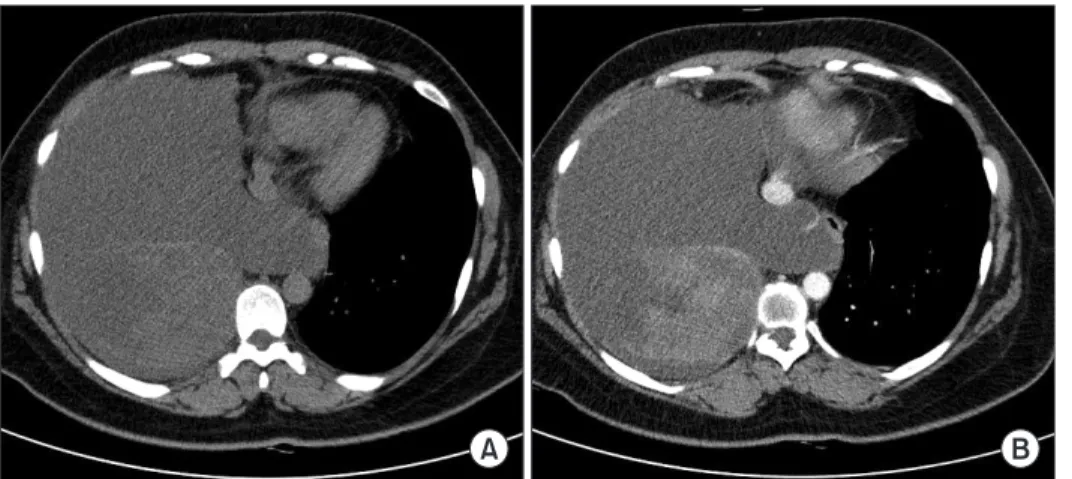
A 36-year-old female presented with dyspnea with onset one week prior. Chest X-ray showed total opacity in the right lung (Figure 1). Chest computed tomography revealed mas- sive pleural fluid collection with total passive atelectasis of right lung. In addition, an inhomogeneous mass was found at the posterior portion of fluid collection (Figure 2A). The mass was well-circumscribed and showed minimal enhancement by radio-contrast dye (Figure 2B). A closed thoracentesis was performed and the aspirated pleural fluid was grossly bloody.

Pleural fluid analysis was as follows: total protein 5.0 g/dL, lac- tate dehydrogenase 97 IU/L, glucose 96 mg/dL, pH 7.22, red blood cell 95,000/mm

3

, white blood cell 18/mm

3

(differential count was impossible due to the small number of leukocytes), adenosine deaminase 21.9 IU/L. Cytologic exam was negative for malignant cell and a culture of pleural fluid did not grow any significant respiratory pathogens. Video-assisted thoracic surgery (VATS) revealed that right pleural cavity was filled with bloody fluid and a dumbbell-shaped tumor was found Copyright © 2013

The Korean Academy of Tuberculosis and Respiratory Diseases.

All rights reserved.

Ju Young Jang, M.D.

1

, Jin Se Kim, M.D.

1

, Ju Won Choe, M.D.

2

, Mi Kyung Kim, M.D.

3

, Jae Woo Jung, M.D.

1

, Jae Chol Choi, M.D.

1

, Jong Wook Shin, M.D.

1

, In Won Park, M.D.

1

, Byoung Whui Choi, M.D.

1

and Jae Yeol Kim, M.D.

1

Departments of

1

Internal Medicine,

2

Chest Surgery, and

3

Pathology, Chung-Ang University College of Medicine, Seoul, Korea

Introduction

A variety of benign and malignant tumors of peripheral nerve origin can occur in the mediastinum. They are most frequently found in the posterior compartment of the medias- tinum and benign schwannoma is the most common tumor type. Mediastinal benign schwannomas are usually asymp- tomatic and if symptoms are present they usually develop by compression of nerve or blood vessel

1

. Benign schwannomas are rarely accompanied by pleural effusion and bloody effu- sion is usually associated with malignant schwannoma

2

. The

CASE REPORT

http://dx.doi.org/10.4046/trd.2013.75.2.71

ISSN: 1738-3536(Print)/2005-6184(Online) • Tuberc Respir Dis 2013;75:71-74

71

Address for correspondence: Jae Yeol Kim, M.D.

Department of Internal Medicine, Chung-Ang University Hospital, 102 Heukseok-ro, Dongjak-gu, Seoul 156-755, Korea

Phone: 82-2-6299-1396, Fax: 82-2-825-7571 E-mail: [email protected]

Received: Apr. 1, 2013 Revised: Apr. 30, 2013 Accepted: May 24, 2013

cc

It is identical to the Creative Commons Attribution Non-Commercial

License (http://creativecommons.org/licenses/by-nc/3.0/).

(2)

JY Jang et al.

72 Tuberc Respir Dis 2013;75:71-74 www.e-trd.org

in the posterior thorax wall (Figure 3). Because the mass was tightly fixated to the thoracic wall, VATS was converted into an open thoracotomy for the complete resection of the tumor.

Upon removal, the mass was revealed to be a whitish soft tumor measuring 10.0×12.0×3.0 cm with a yellow cut-surface (Figure 4A). Upon microscopic exam, the tumor was com- posed of spindle cells with elongated nuclei, forming interlac- ing bundle with focal nuclear palisading. Mitotic figures were rare (Figure 4B). Immunohistochemical studies showed a strongly positive reaction with S-100 protein (data not shown).

All of these findings are consistent with benign schwannoma.

In the afternoon of the operation day, the patient complained of dyspnea and chest X-ray showed total haziness in the right lung (Figure 5A), which must have developed by re-expansion pulmonary edema after removal of massive pleural effusion.

The patient was closely monitored in the intensive care unit with restriction of fluid administration. Several days later,

the pulmonary edema was resolved and the patient was dis- charged on the 10th day after operation. Chest X-ray taken a month after the surgery showed full expansion of right lung (Figure 5B).

Discussion

Neurogenic tumors comprise 19% to 39% of all mediastinal tumors. They develop from mediastinal peripheral nerve, sympathetic and parasympathetic ganglia, and embryonic remnants of neural tube

3

. Because posterior compartment of mediastinum includes spinal nerves, vagus nerve, and sympathetic chains, neurogenic tumors of the mediastinum are most commonly present in the posterior mediastinal compartment

4-6

. Among posterior mediastinal neurogenic tu- mors, schwannoma is the most common. Mediastinal benign schwannomas originate from Schwann cells. They affect both genders equally and develop predominantly in the third and

Figure 1. Chest posterior-anterior taken on the day of admission.

There is total opacity of the right lung.

Figure 2. Pre- and post-contrast chest computed tomography (CT). (A) Pre- contrast chest CT revealed massive pleu- ral fluid collection in the right lung with near total, passive atelectasis. (B) With contrast enhancement, relatively well- circumscribed mass was found in the posterior portion which showed minimal and inhomogeneous enhancement with radio-contrast dye.

Figure 3. Video-assisted thoracoscopic view. A dumbbell-shaped,

whitish tumor was found in the posterior thorax wall.

(3)

Benign schwannoma with bloody effusion

http://dx.doi.org/10.4046/trd.2013.75.2.71 73

www.e-trd.org

fourth decades of life

2

. Multiple tumors can be presented with neurofibromatosis. Mediastinal benign schwannomas are most often asymptomatic. However, sometimes mediastinal benign schwannomas can cause severe problems such as car- diac tamponade

7

or pleural effusion

8,9

, though bloody pleural effusion is usually associated with malignant schwannoma

2,10

. It is noteworthy that massive and bloody pleural effusion was associated with benign schwannoma in this case. Benign schwannomas are typically treated by surgical resection;

because VATS can decrease hospital stay and minimize post- operative complications, it has become the preferred method for resection for posterior neurogenic tumors

11

. In the present case, VATS was converted to an open thoracotomy because the mass was large and fixed to the posterior thoracic wall. It is important to note that postoperative course may be com- plicated in schwannoma cases that are accompanied by mas-

sive pleural effusion; our patient suffered from re-expansion pulmonary edema after removal of a large mass and massive pleural effusion.

In summary, benign schwannoma can be accompanied by massive, bloody pleural effusion and patient should be moni- tored carefully during the postoperative period for the devel- opment of re-expansion pulmonary edema.

References

1. Kim HG, Park SJ, Kwon SH, Hong SJ, Lee JS, Lee MS, et al. A case of schwannoma induced stenosis of superior mesenteric artery. Korean J Med 2004;66:86-90.

2. Shao J, Zhu XH, Shi JY, Ma J, Ge XJ, You ZQ. Primary pulmo- nary schwannoma: clinical analysis of 7 cases and review of Figure 4. Pathologic examination. (A) Upon gross evaluation, the mass was a whitish soft tumor measuring 10.0×12.0×3.0 cm with a yellow cut-surface. (B) Upon microscopic exam, the tumor was composed of spindle cells with elongated nuclei, forming interlacing bundle with fo- cal nuclear palisading. Mitotic figures were rare (H&E stain, ×100).

Figure 5. Early postoperative and post-

discharge chest posterior-anterior im-

ages (PAs). (A) A chest PA taken in the

afternoon of the operation showed total

haziness in the right lung by re-expansion

pulmonary edema. (B) A month after the

surgery the right lung was fully aerated

without infiltration.

(4)

JY Jang et al.

74 Tuberc Respir Dis 2013;75:71-74 www.e-trd.org

the literature. Zhonghua Jie He He Hu Xi Za Zhi 2003;26:3-6.

3. Marchevsky AM. Mediastinal tumors of peripheral nervous system origin. Semin Diagn Pathol 1999;16:65-78.

4. Fatimi SH, Bawany SA, Ashfaq A. Ganglioneuroblastoma of the posterior mediastinum: a case report. J Med Case Rep 2011;5:322.

5. Kambayashi T, Suzuki T. Neurogenic tumors originated from vagus nerve and intercostal nerve simultaneously; report of a case. Kyobu Geka 2011;64:516-9.

6. Lin MW, Chang YL, Lee YC, Huang PM. Non-functional para- ganglioma of the posterior mediastinum. Interact Cardiovasc Thorac Surg 2009;9:540-2.

7. Kato M, Shiota S, Shiga K, Takagi H, Mori H, Sekiya M, et al.

Benign giant mediastinal schwannoma presenting as car-

diac tamponade in a woman: a case report. J Med Case Rep 2011;5:61.

8. Ishibashi H, Akamatsu H, Sunamori M, Komori H, Shirasawa S, Nomoto S. A case of giant dumbbell shaped schwannoma with massive pleural effusion. Kyobu Geka 2001;54:742-6.

9. Cho JH, Kim KU, Park HK, Jeong YJ, Kim YD, Kim YS, et al.

Two cases of benign solitary schwannoma with pleural effu- sion. Tuberc Respir Dis 2007;63:78-82.

10. Miravitlles M, de Gracia J, Roca-Tey R, Monso E. Pleural effu- sion secondary to a malignant thoracic schwannoma. Med Clin (Barc) 1992;99:117.

11. Hazelrigg SR, Boley TM, Krasna MJ, Landreneau RJ, Yim AP.

Thoracoscopic resection of posterior neurogenic tumors. Am

Surg 1999;65:1129-33.

수치

Figure 3. Video-assisted thoracoscopic view. A dumbbell-shaped,  whitish tumor was found in the posterior thorax wall.
Figure 5. Early postoperative and post- post-discharge chest posterior-anterior  im-ages (PAs)

참조

관련 문서

As a result of this study, it showed that the majority of students in the department of aviation operation and aviation service selected their major with

2재화 2요소 헥셔-올린 모형에서는 어느 한 경제에서 어느 한 요소의 양이 증가하면, 그 요소를 집약적으로 사용하는 산업의 생산량은 증가하고 다른

웹 표준을 지원하는 플랫폼에서 큰 수정없이 실행 가능함 패키징을 통해 다양한 기기를 위한 앱을 작성할 수 있음 네이티브 앱과

_____ culture appears to be attractive (도시의) to the

The index is calculated with the latest 5-year auction data of 400 selected Classic, Modern, and Contemporary Chinese painting artists from major auction houses..

In the chapters of Journey of Renewal that describe the Mission Accountability Framework there will be flashbacks to the story of Richmond Corps, showing clearly how

1 John Owen, Justification by Faith Alone, in The Works of John Owen, ed. John Bolt, trans. Scott Clark, "Do This and Live: Christ's Active Obedience as the

Second, the analysis of the differences in sports participation time showed statistically significant differences in the operation of the competition and