INTRODUCTION
Cyanobacteria (blue-green algae) are common members of the phytoplankton in mesotrophic to hypereutrophic lakes, where it frequently domi- nates as a nuisance under stratified condition (Paerl, 1988). Massive accumulations of cyanobac- teria are worldwide in lakes and reservoirs during the summer. Especially, they abundantly inhibit- ing in freshwater ecosystems frequently form blooms in mesotrophic and eutrophic lakes and ponds. Anabaena spp. and Microcystis spp. are well known as one of the most common bloomforming cyanobacteria, which cause many problems for scenery, anxiety about toxicity (Carmichael, 1992;
Harada, 1996) and unpleasant odors (Kikuchi et al., 1973; Tsuchiya et al., 1992). Therefore, various microorganisms such as bacteria, actinomycetes, fungi, phages and amoebae have been treated in the termination and decomposition of cyanobac- teria blooms (Mitsutani et al., 1987; Fukami et al., 1992; Yamamoto et al., 1998; Jang et al., 2003;
Kim and Han, 2004).
In the present work, we isolated a bacterial strain AK-05 showing high lytic activity against a cyanobacterium, Anabaena cylindrica. Identifi- cation of the isolates based on 16S rDNA sequence information was conducted. Some specific charac- teristics of the isolate were investigated on a alga- lytic test. In addition, the effect of pH and temper- ature on the lytic activity was investigated to
─
─ 241 ──
Characterization of a Novel Alga-Lytic Bacterium, Acidovorax temperans AK- -05, Isolated from
an Eutrophic Lake for Degradation of Anabaena cylindrica
Kim, Jeong--Dong and Myung--Soo Han*
(Department of Life Science, Hanyang University, Seoul 133-791, Korea)
부영양 호수에서 분리한 Acidovorax temperans AK-05의 Anabaena cylindrica 분해 특성.
김정동∙한명수* (한양대학교 생명과학과)
부영양 호수로부터 살조 세균을 분리하고 동정한 결과 Anabaena cylindrica NIES-19를 유릴 탄 소원으로 이용하는 double layer 방법으로 15종의 살조세균을 분리하였으며 높은 살조 활성을 나 타내는4종의 살조세균AK-05, AK-07, AK-13 그리고AK-28을 선별하여 살조 능력을 비교하였 다. 이들 중에AK-05가 가장 높은 살조 활성을 나타내었으며 이를16S rDNA 염기서열을 분석한 결과 Acidovorax temperans와 99.5%의 유사성을 나타내어 Acidovorax temperans AK-05로 명명 하였다. A. temperans AK-05의 배양 여액을 A. cylindrica NIES-19에 뚜렷한 살조 활성을 나타냈 었으며, 이것의 살조 활성 능력을 분석한 결과 살조 활성에 관여하는 주요 물질은non-protein이 며 열에 안정적이었다. 이러한 살조 활성 능력은 알칼리 조건과 25~30�C 에서 가장 높게 나타냈 다. 따라서 이와 같은 특성은 일반적으로 알칼리 조건을 야기하는 Cyanobacteria에 의한 water blooms이 발생하는 호수에 적용하는데 매우 유리할 것으로 여겨진다.
Key words : alga-lytic bacteria, Acidovorax temperans, extracellular substances, algal blooms
* Corresponding author: Tel: ±82-2-2290-0956, Fax: ±82-2-2296-1741, E-mail: [email protected]
evaluate the applicability of the isolate as a biological control agent for cyanobacterial blooms.
MATERIALS AND METHODS
1. Host alga and culture conditions
A. cylindrica NIES-19 that used as host for alga-lytic bacteria was kindly supplied by Nation- al Institute for Environmental Studies, Japan, and a clonal axenic culture was cultivated and maintained in MDM medium including KNO2 10 g, MgSO4∙7H2O 2.5 g, K2HPO4 2.5 g, NaCl 1.0 g, A5 solution 1 mL, and Fe solution 1 mL per 1 liter under continuous illumination of cool white f luorescent lamps giving an incident light intensity of 35~80 µmE m-2s-1and at 25~28�C with agitation at 150 rpm on rotary shaker. A5 solution is composed with H3BO32.86 g, MnSO4∙ 7H2O 2.5 g, ZnSO4∙7H2O 0.222 g, Na2MoO4∙ 2H2O 0.021 g, CuSO4∙5H2O 0.079 g per 1 liter (Cattenholz, 1988). In addition, Fe solution is plus FeSO4∙7H2O 2.0 g and H2SO4 0.26 mL per 1 liter.
2. Isolation of alga-lytic bacteria
Various samples collected from Pal’tang reservoir and Sukchon Lake in Korea in which cyanobacteria are dominant phytoplankton dur- ing summer were used as the isolation source.
Initial screening was conducted using skim milk agar plates containing 10 g of skim milk (Difco), 1 g of yeast extract (Difco) and 15 g of agar in 1 liter of distilled water. Then, the double-layer method (Shilo, 1970) using A. cylindrica NIES- 19 as a sole nutrient was used for second screen- ing method (Yamamoto, 1981). Axgenic cultures of A. cylindrica NIES-19 were grown in MDM medium for 1 week, and 1 mL of A. cylindrica NIES-19 cultures was mixed with 1 mL of fil- tered (200µm filter) suspension of surface waters or sediment samples and molten MDM soft agar equilibrated to 50�C . The mixture was imme- diately poured onto a MDM soft agar plate. After the agar had solidified, the plates were incubated at 25~28�C with continuous illumination of cool white fluorescent lamps giving an incident light intensity of 35~80µE m-2s-1. Bacterial colonies that produced clear zones on lawns of A. cylindri- ca were picked, purified, and maintained on PY medium slants containing 1.0% (w/v) of peptone,
0.1% (w/v) of yeast extract and 1.5% (w/v) of agar (pH adjusted to 7.0). Isolated bacteria were stored at -80�C in PY medium supplemented with 20% (v/v) of glycerol.
3. Identification of isolates
The chromosomal DNA was isolated using a method described elsewhere (Yoon et al., 1997).
The amplif ication of the 16S rDNA was conducted using two primers according to Stackebrandt and Liesack (1993), 5’-GAGTTTGATCCTGGCTCAG- 3’ and 5’-AGAAAGGA-GGTGATCCAGCC-3’. A PCR was run for 35 cycles in a DNA thermal cycler, Genetic analyzer 377 (Perkin-Elmer, Boston, USA), employing the thermal profile according to Yoon et al. (1997). The 16S rDNA sequence of bacterial isolate AK-05 was aligned using CLUSTAL W software (Nigam et al., 2000).
The evolutionary distance matrices were calcu- lated with the DNADIST program within the PHYLIP package (Felsenstein, 1993). The sequence of representative species of the genus Acidovorax and related taxa were cited using the GenBank Database. The values of 16S rDNA similarity were calculated from the alignment, while the evolu- tionary distances were calculated using a Kimura two-parameter correction. A phylogenetic tree was constructed using the neighbor-joining method (Saitou and Nei, 1987) based on the calculated distance matrix.
4. Extraction and measurement of cyanobact- erial chlorophyll a
A solution of 90% methanol was used for extrac- tion of chlorophyll a (chl-a) from cyanobacteria.
Measurement of chlorophyll a was according to the method of Parson et al. (1984). Cyanobacteria filtrated by GF/C (Whatman, Uppsala, Sweden) were subjected to extraction with 90% acetone at 4�C for 24 h. After extraction, the solid suspension was removed by centrifugation. Then, the absor- bance of extracts at 750, 647, 644 and 630 nm was measured using a spectrophotometer (model HP8453, Hewlett Packard, MI, USA). The concen- tration of chlorophyll a was calculated using fol- lowing equation.
Chlorophyll a (µg L-1) = Ca× (VaVb-1
) Ca : Enumerated wavelength value Va : amount of 90% aceton (mL) Vb : amount of sample filtered (L)
5. Assay of alga-lytic activity
The alga-lytic activity of the isolates was tested as fallows. Supernatants of the bacterial isolates cultivated at 30�C and 120 rpm for 24 h in Erlen- meyer f lask including PY medium were separated from the cells used for the assays and were suspended in sterile lake water avoid any inf lu- ence of chemicals from the MDM medium during the assay. The lake water used in this study was collected from Pal’tang reservoir and sterilized by membrane f iltration (0.2-µm pore size, Whatman, Uppsala, Sweden). The concentration of host cyanobacterial cells, 1×10⁷ cells mL-1, was added in the sterilized lake water and the supernatant of the bacterial isolates was mixed with lake water containing host cyanobacterial cells at a ratio of 1 : 1 (v/v). Alga-lytic activities were evaluated by the change in chlorophyll a concentration or in cyanobacterial cells.
6. Alga-lytic activity of supernatant under heat and proteinase treatment conditions Several treatments were carried out on the bacterial supernatant to investigate the change of alga-lytic activity. For heat treatment, bacterial supernatant was deposed into a test tube and incubated 90�C in a water bath for 30 min. A 1 mL of bacterial supernatant was incubated at 55�C in a water bath for 3 after mixing with 5 µL of proteinase-K solution (Sigma).
7. Effect of various pHs and temperatures The Anabaena-lytic activity of bacterial super- natant was investigated under different pH con- ditions between 4.5~11.0 generated using NaOH and HCl. To determine the inf luence of NaOH or HCl additions on cyanobacterial cells, both dis- tilled water and fresh PY medium the pH of
which was adjusted by NaOH or HCl were used as a control. And the effect of temperature in the Anabaena-lytic activity of the bacterial super- natant toward A. cylindrica was investigated at various temperatures, 5, 10, 15, 20, 25 and 30�C .
RESULTS AND DISCUSSION 1. Isolation and identification of alga-lytic
bacteria
Fifteen isolates showing clear zones on the host A. cylindrica NIES-19 over-layer agar were productively isolated. Among the isolates, only four-isolates lyse A. cylindrica NIES-19 when co-culturing was carried out. The alga-lytic activities of the supernatants of four isolates were shown in Table 1. Cyanobacterial chloro- phyll a was reduced from 10 to 45% by the super- natants of four isolates after 48 h of incubation.
The isolate AK-05 exhibited the highest activity.
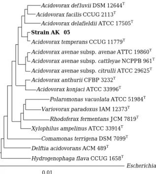
This strain was identified based on 16S rDNA sequence and showed 99.5% homology with Aci- dovorax temperans CCUG 11779T(Fig. 1). To illu- minate its lytic mechanism, the alga-lytic test by bacterial supernatant in the present or absence of A. temperans AK-05 cells was carried out. The cells of A. cylindrica NIES-19 were highly aggre- gated by the cells of AK-05, but no significant difference in the alga-lytic activity based on the chlorophyll a measurement was observed during 48 h of incubation. It indicates that the alga-lytic activity of A. temperans AK-05 was attributed to extracellular products and direct contact with bac- teria cells was not responsible for the lyses of cyanobacterial cells.
2. Alga-lytic activity
After examination of the alga-lytic activity against A. cylindrica, the chlorophyll a was de- creased to 42% of the initial chlorophyll; however, 99.8% of the cyanobacterial cells were complete destroyed (Table 2). This differences in the residual amounts of chlorophyll a and cyanobac- terial cells demonstrated that alga-lytic substance degraded the cell or cells wall but not chlorophyll a. In addition, relationship between bacterial growth phase and alga-lytic activity was inves- tigated. As shown in Fig. 1, the alga-lytic activity of the supernatant was minimal, although the growth of A. temperans AK-05 reached the sta-
Table 1. Reduction ratios of chlorophyll a of A. cylindrica NIES-19 upon incubation with the bacterial supernatants of four isolates cultivated at 30�C for 48 h in PY medium.
Bacterial isolate aReduction ratios of Chl-a (%)
AK-05 45
AK-07 23
AK-13 26
AK-26 10
aReduction ration = (decreased amount of Chl-a / the initial amount of Chl-a)×100
tionary. In exponential growth phase, the alga- lytic activity gradually increased. The super- natant obtained from AK-05 cultivated for 72 h exhibited the highest activity. This explained that the alga-lytic substances were produced after sta- tionary phase. The producing of antibiotics during the early stages is common in bacteria (Katz and Demain, 1977). These results suggested that alga- lytic substances produced from A. temperans AK- 05 are possibly antibiotic analogues.
3. Alga-lytic activity under several conditions The alga-lytic activity of the bacterial super- natant under several conditions was investigated.
PY medium was used as a control to evaluate any
inf luence of components in the culture medium.
In Fig. 3, cyanobacterial cells was not decreased even after 96 h incubation with PY medium and it is suggesting that PY medium did not inf luence on cyanobacterial cells. On the other, the bacterial supernatant lyses 90% of the cyanobacteria cells over a period of 72 h. Therefore, it was obvious that some substance produced by A. temperans AK-05 in supernatant were responsible for lyses. To test the heat stability of the alga-lytic substance in the supernatant, alga-lytic activity was investigated after heat treatment at 90�C . As shown in Fig. 4, 75% of cells were degraded after 60 h of incuba- tion. Although the alga-lytic activity was some- what decreased by heat treatment, the activity of bacterial supernatant were still reminded in high.
The result indicated that the major alga-lytic substances were heat stable. To determine whether the alga-lytic substances were proteins or not, proteinase-K was treated. 66% reduction of alga-lytic activity by bacterial supernatant pre- treated with proteinase-K was observed. This result demonstrated that the major alga-lytic substances were not degraded by proteinase-K.
Table 2. Reduction ratio of chlorophyll a and cell number of A. cylindrica NIES-19 after treatment of the supernatant of A. temperans AK-05.
Treatment
aChlorophyll a bCell number cReduction ratio of cReduction ratio of (mg L-1) (cells mL-1) chlorophyll a (%) cell number (%)
Supernatant 1.05 2.0×10⁵ 58 99.8
PY medium 2.43 3.1×10⁷ 2.8 3.9
aThe initial cells: 1.0×10⁸, bThe initial chlorophyll a: 2.5 mg L-1, cReduction ratio = (decreased amount/the initial amount)×100
Fig. 1. Phylogenetic tree based on 16S rDNA sequences showing the positions of strain AK-05, the type strains of Acidovorax species and the represen- tatives of some other related taxa. Scale bar repre- sents 0.01 substitutions per nucleotide position.
Escherichia coli 0.01
Hydrogenophaga flava CCUG 1658T Delftia acidovorans ACM 489T Xylophilus ampelinus ATCC 33914T
Acidovorax konjaci ATCC 33996T Acidovorax anthurii CFBP 3232T
Acidovorax avenae subsp. citrulli ATCC 29625T Acidovorax avenae subsp. cattleyae NCPPB 961T Acidovorax avenae subsp. avenae ATTC 19860T Acidovorax temperans CCUG 11779T
Strain AK--05
Comamonas terrigena DSM 7099T Variovorax paradoxus IAM 12373T
Rhodoferax fermentans JCM 7819T Polaromonas vacuolata ATCC 51984T Acidovorax facilis CCUG 2113T
Acidovorax delafieldii ATCC 17505T Acidovorax def luvii DSM 12644T
Fig. 2. Alga-lytic activity of the bacterial supernatant of A. temperans strain AK-05 related with bacterial growth phages.
Cell number(×106cells mL-1) Acivity(%)
Incubation time (h)
0 12
140 120 100 80 60 40 20 0
100
80
60
40
20
24 36 48 60 72 0
Nevertheless, A. temperans AK-05 made clear zone on skim milk medium, it produced any protease.
Thus, protease contributing alga-lytic activity is considered minor.
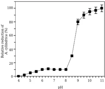
4. Effect of pH and temperature
Alga-lytic activities of the supernatant of bacterial culture investigated at different pH values were shown in Fig 4. Anabaena-cells were not ruptured in the acidic pHs, but, in the alkaline conditions, cyanobacterial cells were dramatically decreased. i.e., cyanobacterial cells were not degraded by the bacterial supernatant at pH 4, but 100% cell lyses were detected at alkaline condition, pH 11. Cyanobacterial blooms often alkalify the aquatic environments because some cyanobacteria including the genus Microcystis and Anabaena can use HCO3more efficiently than CO2(Matsuda et al., 1999). Accordingly, this alka- line pH-stable characteristic of alga-lytic sub- stances produced by A. temperans AK-05 is advan- tage for application to control water bloom in aquat- ic environments.
Temperature is very important factor in the con- trol of water blooms in eutrophic lakes (Tujimura et al., 2001). Therefore, the effect of temperature
of alga-lytic activity to degrade A. cylindrica NIES-19 was examined at various temperatures (Fig. 5). At 25 and 30�C , the cells of A. cylindrica were almost lysed up to 100%. This is telling that the cells were ruined more competently as the temperature increases. At 4�C , the alga-lytic activity was not detected since the cyanobacterial cell membrane was considered to motionless at
Fig. 3. Alga-lytic activity of the bacterial supernatant of A. temperans strain AK-05 against A. cylindrica NIES-19 under several conditions. Samples are (○), PY medium as a control; (■), bacterial super- natants without any treatment; (▲), bacterial supernatant pretreated with heat; (●), bacterial supernatant pretreated with proteinase-K.
Fig. 4. Influence of pHs on alga-lytic activity of A. tempe- rans strain AK-05 bacterial supernatant against A. cylindrica NIES-19.
Incubation time (h)
Relative cells(%)
0 0 20 40 60 80 100
12 24 36 48 60 72 84 96
pH Relative reduction of A. cylindrica(%)
4 0 20 40 60 80 100
5 6 7 8 9 10 11
Fig. 5. Inf luence of temperature on alga-lytic activity of A. temperans strain AK-05 bacterial supernatant against A. cylindrica NIES-19. Symbols are bac- terial supernatants treated at (○), 3�C; (●), 10�C;
(△), 15�C; (▲), 20�C; (□), 25�C; (■) 30�C.
Incubation time (h) Relative cells of A. cylindrica(%)
0 12
0 20 40 60 80 100
24 36 48 60 72 84 96
low temperature (Goldman and Carpenter, 1994;
Tujimura et al., 2001). In consequent, optimal culture conditions for growth of cyanobacteria such as high temperature and alkaline pH can make active transport of the alga-lytic sub- stances to cyanobacterial cells and successive rapid lyses.
ABSTRACT
Isolation and identification of alga-lytic bac- teria were carried out. Fifteen isolates of alga- lytic bacteria were screened by the double layer method using A. cylindrica NIES-19 as a sole nutrient and four isolates among them were compared with their alga-lytic activity. The isolate AK-05 exhibiting the highest alga-lytic activity was identified as Acidovorax temperans base on its 16S rDNA sequence. The culture supernatant of the isolate AK-05 was reliable for the alga-lytic. Alga-lytic activity assays of culture supernatant revealed that the major sub- stances for alga-lytic activity were non-proteins and heat stable. The highest alga-lytic activity was practical under alkaline conditions and at 25~30�C . It is indicating an advantage for the application of water blooms by cyanobacteria in eutrophic lakes where the pH is generally in alka- line region.
ACKNOWLEDGMENTS
This study was supported by the National Research Laboratory Program (2000-N-NL- 01-C-290) of the Korean Ministry of Science and Technology.
REFERENCES
Carmichael, W.W. 1992. A status report of planktonic cyanobacteria (blue-green algae and their toxins).
#EPA/600/R-92/079. United States Environ- mental Protection Agency, Washington, DC, USA.
Castenholz, R.W. 1988. Culturing methods for cyanobacteria. Methods Enzymol. 167: 8-92.
Felsenstein, J. 1993. PHYLIP: Phylogenetic Infe- rence Package. Version 3.5. Seattle, University of Washington, Washington, USA.
Fukami, K., A. Yuzawa, T. Nishijima and Y. Hata.
1992. Isolation and properties of a bacterium inhi-
biting the growth of Gymnodinium nagasakiense.
Nippon Suisan Gakkaishi 58: 1073-1077.
Goldman, J.C. and E.J. Carpenter. 1974. A kinetic approach to effect of temperature on algal growth.
Limnol. Oceanogra. 19: 756-766.
Harada, K.-I. 1996. Chemistry and detection of microcystins. pp.103-148. In Toxic Microcystis (Watanabe, M.F, K.-I. Harada, W.W. Carmichael and H. Fujiki, eds). CRC Press
Jang, E.-H., J.-D. Kim and M.-S. Han. 2003. Isolation and characterization of alga-lytic bacterium HY0210-AK1 and its degradability of Anabaena cylindrica. Korean J. Environ. Biol. 21: 194-201.
Katz, E. and A.L. Demain. 1977. The peptide antibi- otics of Bacillus; chemistry biogenesis and pos- sible functions. Bacteriol. Rev. 41: 449-474.
Kikuchi, T., T. Miura, K. Harimaya, H. Yano, T.
Arimoto and T. Masata. 1973. Odorous metabolites of blue-green alga Schizothrix muelleria Nägeli collected in the southern basin of Lake Biwa:
Identification of geosmin. Chem. Pharm. Bull. 21:
2342-2343.
Kim, J.-D. and M.-S. Han. 2004. Enzyme profiles of alga-lytic bacterial strain AK-13 associated with elimination of cyanobacterium Anabaena cylindrica.
Korean J. Environ. Biol. 22: 184-191.
Matsuda, Y., T.G. Williams and B. Colman. 1999.
Quantification of the rate of CO2formation in the periplasmic space of microalgae during photosyn- thesis. A comparison of whole cell rate constant for CO2and HCO3uptake among three species of the green alga Chlorella. Plant Devel. Environ.
22: 397-405.
Mitsutani, A., A. Uchida and Y. Ishida. 1987. Occur- rence of blue-green algae and algal lytic bacteria in Lake of Biwa. Bull. Jpn. Soc. Microbiol. Ecol. 2:
21-28.
Nigam, P., G. Armour, I.M. Banat, D. Singh and R.
Marchant. 2000. Physical removal of textile dyes and solid-state fermentation of dye-adsorbed agri- cultural residues. Bioresour. Technol. 72: 219-226.
Parson, R.T, Y. Matia and C.M. Lalli. 1984. A manual of chemical and biological methods for seawater analysis. 1sted. Pergamon Press Ltd. Oxford.
Pearl, H.W. 1988. Growth and reproductive strategies of freshwater blue-green algae (cyanobacteria). In:
Growth and Reproductive Strategies of Freshwater Phytoplankton pp 262-315. (Sandgreen, C.D., ed.).
Cambridge University Press, Cambridge.
Saitou, N. and M. Nei. 1987. The neighbor-joining method: a new method for reconstructing phylo- genetic trees. Mol. Biol. Evol. 4: 406-425.
Shilo, M. 1970. Lysis of blue-green algae by myxobac- ter. J. Bacteriol. 104: 453-461.
Tsuchiya, Y., M.F. Watanabe and M. Watanabe.
1992. Volatile organic sulfur compounds associated with blue-green algae from inland waters of Japan. Water Sci. Technol. 25: 123-130.
Tujimura, S., K. Ishikawa and H. Tsukada. 2001. Effect of temperature on growth of the cyanobacterium Aphanizomenon flos-aquae in Lake Biwa and Lake Yogo. Phycol. Res. 49: 275-280.
Yamamoto, Y. 1981. Observation on the occurrence of microbial agents, which cause lysis of blue-green algae on Lake Kaumigaura. Jpn. J. Limnol. 42:
20-27.
Yamamoto, Y., T. Huchiwa, Y. Hodoki, K. Hotta, H.
Uchida and K.-I. Harada. 1998. Distribution and identification of actinomycetes lysing cyanobac- teria in a eutrophic lake. J. Appl. Phycol. 10: 391-
397.
Yoon, J.-H., S.-T. Lee, S.-B. Kim, W.-Y. Kim, M.
Goodfellow and Y.-H. Park. 1997. Restriction fragment length polymorphisms analysis of PCR- amplified 16S ribosomal DNA for rapid identi- fication of Saccharomonospora strains. Int. J.
Syst. Bacteriol. 47: 111-114.
(Manuscript received 11 April 2004, Revision accepted 12 June 2004)