pISSN: 1011-8942 eISSN: 2092-9382
© 2017 The Korean Ophthalmological Society
This is an Open Access article distributed under the terms of the Creative Commons Attribution Non-Commercial License (http://creativecommons.org/licenses /by-nc/3.0/) which permits unrestricted non-commercial use, distribution, and reproduction in any medium, provided the original work is properly cited.
572
Spontaneous Resolution of Macular Hole with Retinal Detachment in a Highly Myopic Eye
Dear Editor,
Macular hole (MH) with retinal detachment (RD) pre- dominantly occurs in highly myopic eyes, and is a major vision-threatening complication in pathologic myopia [1].
Treatment of MH with RD results in lower anatomic suc- cess rates and poorer functional outcomes than MH with- out RD. Spontaneous resolution of MH with RD is ex- tremely rare; spontaneous resolution of MH without RD is reported more frequently, especially in cases of traumatic MH. However, we encountered a case of spontaneous reso- lution of MH with RD in a patient with high myopia(Fig.
1A, 1B), which is described herein with a review of the rel- evant literature.
A 73-year-old female visited our clinic complaining of a decrease in vision in the left eye. Best-corrected visual acuity was 0.8 in the right eye (OD) and 0.04 in the left eye (OS) as measured by a Snellen chart. Optical coherence to- mography (OCT; Spectral OCT/SLO, OTI, Toronto, ON, Canada) of the OS confirmed the presence of MH-related RD (Fig. 1C). The patient refused surgical treatment.
The clinical history of the patient included Bechet’s dis- ease (5 years before) and bilateral cataract surgery (4 years before). At the time of the surgery, the axial length of the eyes was 27.14 mm (OD) and 28.63 mm (OS). Two years after cataract surgery, Nd-YAG (neodymium-doped yttri- um aluminum garnet) capsulotomy was performed in both eyes for after-cataract treatment (posterior capsular opaci- ty after cataract surgery).
The patient visited the clinic for a check-up 2 years after MH with RD was diagnosed. Best-corrected visual acuity was 0.8 (OD) and 0.02 (OS) on the Snellen chart. OCT re- vealed complete retinal attachment as well as spontaneous closure of the MH in the OS (Fig. 1D, 1E).
Several cases of spontaneous closure of MH with RD have been reported. Min [2] and Tam et al. [3] reported two cases of spontaneous reattachment of RD with MH; how- ever, these cases were reported before the advent of OCT, a useful tool for the diagnosis of MH. Li et al. [4] described a case of spontaneous closure of MH associated with RD in an eye with high myopia, in which macular detachment was observed until the last follow-up visit; they suggested that a decrease in vitreoretinal traction force may induce spontaneous closure of MH. Furthermore, spontaneous
Korean J Ophthalmol 2017;31(6):572-573 https://doi.org/10.3341/kjo.2017.0103
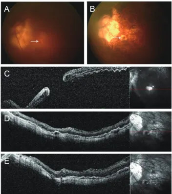
Fig. 1. Fundus photographs and optical coherence tomography.
(A) Fundus photograph of macular hole (arrow) with retinal de- tachment. (B) After 2 years, spontaneous resolution of macular hole (arrow) and associated retinal detachment were observed.
Atrophic scar change in the fovea was seen on fundus photo- graphs. (C) Macular hole with retinal detachment. (D,E) After 2 years, complete retinal reattachment was observed, without any retinoschisis or macular hole on horizontal- and vertical-view op- tical coherence tomography.
A
C
D
E
B
573 closure of MH with retinal reattachment in an eye with
high myopia and staphyloma was also reported by Yu et al.
[5]. They observed a very small MH (66 μm in diameter), the size of which may have influenced spontaneous clo- sure. Furthermore, the development of macular retinoschi- sis accompanied by complete retinal reattachment was ob- served, which was believed to reduce the traction force.
The present case varies from previously reported cases because of the presence of a large MH with complete reti- nal reattachment and no macular schisis. Although several factors that could have negatively affected prognosis, such as a large MH and the absence of macular schisis, were present in this case, complete retinal reattachment as well as MH closure occurred spontaneously. The underlying mechanism of spontaneous MH closure in this case despite the presence of these negative factors is not fully under- stood, but spontaneous release of vitreoretinal traction is believed to play a role.
In conclusion, we reported a case of spontaneous resolu- tion of MH with RD in a highly myopic eye, which is unique because of the large size of the MH, complete reti- nal reattachment, and the absence of macular schisis.
Soo Jin Lee, Yu Cheol Kim
Department of Ophthalmology, Dongsan Medical Center, Keimyung University School of Medicine, Daegu, Korea E-mail (Yu Cheol Kim): [email protected]
Conflict of Interest
No potential conflict of interest relevant to this article was reported.
References
1. Alkabes M, Pichi F, Nucci P, et al. Anatomical and visual outcomes in high myopic macular hole (HM-MH) without retinal detachment: a review. Graefes Arch Clin Exp Oph- thalmol 2014;252:191-9.
2. Min WK. Spontaneous reattachment of retinal detachment with macular hole in nonmyopic patients. Korean J Oph- thalmol 1995;9:66-8.
3. Tam BS, Kwok AK, Bhende P, Lam DS. Spontaneous reat- tachment of retinal detachment in a highly myopic eye with a macular hole. Eye (Lond) 2000;14(Pt 4):661-2.
4. Li Y, Jonas JB, Lu L. Spontaneous closure of highly myo- pic macular hole associated with retinal detachment. Acta Ophthalmol 2014;92:e408-10.
5. Yu J, Jiang C, Xu G. Spontaneous closure of a myopic mac- ular hole with retinal reattachment in an eye with high my- opia and staphyloma: a case report. BMC Ophthalmol 2014;14:111.