However, these methods still suffer from light microscopy given their morphological changes in response to various environmental conditions and insufficient taxonomic characteristics(Lehtimäki et al. 2000; Gugger et al. 2002;
Willame et al. 2006). Recently, molecular phylogenetic data and ultrastructural and morphological characteristics have been used together to describe cyanobacterial species (Komárek and Anagnostidis 2005; Komárek 2016).
In Korea, studies on cyanobacteria focus on taxa with high environmental impacts, such as the genus Microcystis, which produces toxins, and the genus Anabaena, which generates odorants and neurotoxins. Additionally, only 377 cyanobacterial species have been reported(NIBR 2019), which is less than 10% of the number of cyanobacteria re- ported worldwide(4,707 species, Guiry and Guiry 2020).
Thus, studies on the diversity of cyanobacteria species in
https://doi.org/10.11626/KJEB.2021.39.1.032
INTRODUCTION
Cyanobacteria are widely distributed worldwide and a major causative species of algae bloom in freshwater eco- systems(Robarts and Zohary 1987; Yunes et al. 2003; Ha- vens 2008; You et al. 2013). This algae blooming caused by cyanobacteria induces visual disturbance(Barnett 1984).
Some species produce off-flavor compounds, such as geo- smin and 2-MIB(Kim et al. 2015), and liver or neurotoxin substances, such as microcystin and anatoxin-a(Suffet et al. 1995; Zander and Pingert 1997). Taxonomic studies on cyanobacteria have emerged due to the harmful effects of these cyanobacteria on the environment(Komárek 2006; Ryu et al. 2018). Taxonomic studies on cyanobac- teria have been mainly based on morphological charac- teristics(Pfannkuche and Lochte 1993; Choi et al. 1998).
Note
New records of the genus Cyanobium and Cyanobium gracile (Synechococcales, Cyanophyceae) in Korean freshwater
Dae Ryul Kwon†, Bok Yeon Jo†, Seok Won Jang, Chang Soo Lee and Seung Won Nam*
Nakdonggang National Institute of Biological Resources, Sangju 37242, Republic of Korea
Korean J. Environ. Biol.
39(1) : 32-38(2021) ISSN 1226-9999(print) ISSN 2287-7851(online)
* Corresponding author Seung Won Nam Tel. 054-530-0843
E-mail. [email protected]
†These authors contributed equally to this work
Received: 30 June 2020 First Revised: 4 February 2021 Second Revised: 11 February 2021 Revision accepted: 4 March 2021
Abstract: Cyanobium is a genus of picoprokaryotic cyanophytes, which includes species worldwide. The present study investigated the morphology, ultrastructure, and molecular phylogeny of the unrecorded genus Cyanobium Rippka & Cohen-Bazire 1983 and species Cyanobium gracile Rippka & Cohen-Bazire 1983. A C. gracile culture from a freshwater sample collected from the Adongji pond was established by single- cell isolation. Morphological data were analyzed using light and transmission electron microscopy. C. gracile lives as solitary cells without gelatinous envelopes and is ovate, oval, or shortly rod-shaped. Thylakoids are laid along the cell walls, with three thylakoid membranes parallel to each other. Nucleoplasm was observed in the center of the cell.
Molecular phylogeny performed with data from 16S small subunit ribosomal DNA gene(SSU rDNA) sequences showed that the three strains of C. gracile, including the type strain(PCC6307) and a newly recorded strain(Adong101619), formed a distinct clade with a high supporting value(maximum-likelihood=100, pp=1.00). Based on morphology and molecular data, we report the newly recorded C. gracile in Korea.
Keywords: cyanobacteria, Cyanobium, phylogeny, ultrastructure, unrecorded
Korea are quite insufficient(Ryu et al. 2018).
The genus Cyanobium is an oval-shaped or short rod- shaped unicellular cyanobacteria(Rippka and Cohen- Bazire 1983; Komárek et al. 1999). This genus is often con- sidered morphologically similar to the genus Synechococcus (Nägeli 1849; Padisák et al. 1997). However, a difference is noted between the two genera in terms of DNA base com- position. The average GC content of Cyanobium is 66-71 moles%, whereas that of Synechococcus is 48-56moles%
(Rippka and Cohen-Bazire 1983).
The genus Cyanobium is an important primary producer in oligotrophic and mesotrophic environments(Jezberová and Komárková 2007). However, only 14 species have been reported worldwide(Guiry and Guiry 2020) due to the absence of distinct morphological characteristics and the low population density(Komárek et al. 1999). Addi- tionally, this genus and species have not yet been reported in Korea.
In this study, morphological and ultrastructural studies and phylogenetic analysis were performed to report the unrecorded genus Cyanobium and the unrecorded species Cyanobium gracile in Korea.
MATERIALS AND METHODS
1. Sampling and clonal culture of Cyanobium gracile
Cultures of C. gracile were established by single-cell iso- lation from freshwater samples collected at Adongji pond, Korea(35°58ʹ47.4ʺN, 126°46ʹ14.6ʺE) in October 2019.
The cultures were grown in BG11 medium at 25°C under a 14 : 10 light : dark cycle and a light intensity of 4,000lux provided by cool-white fluorescent lamps.
2. Light microscopy
Living C. gracile cells were studied using a Nikon ECL- IPSE Ni-U(Nikon, Japan) equipped with differential inter- ference contrast optics. Images were captured using a digi- tal camera(DS-Ri2, Nikon).
3. Transmission electron microscopy
For transmission electron microscopy, the cells were prefixed in a 1 : 1 mixture of 5%(V/V) glutaraldehyde and BG11 culture media for 1 h at 4°C. The glutaraldehyde- fixed cells were washed 3 times in BG11 culture media and
postfixed in 1%(W/V) OsO4 for 1 h at 4°C. The fixed cells were rinsed three times with distilled water. Dehydration was carried out at 4°C using a graded ethanol series of 50, 60, 70, 80, and 90% for 10 min each and three 10 min changes of pure ethanol. Pellets were then brought to room temperature and transferred through propylene oxide two times for 20 min each with 50% and 75% Spurr’s embed- ding resin(Spurr 1969) in propylene oxide for 1 h each and 100% overnight. On the following day, pellets were moved to new pure resin and polymerized at 70°C. Blocks were thin-sectioned on a PT-X ultramicrotome(RMC Pro- ducts, Boeckeler Instruments, Tucson, AZ). Sections of 70 nm thickness were collected on slot copper grids, stained with 3%(w/v) uranyl acetate and Reynold’s lead citrate (Reynolds 1963), and observed using a JEM-1400 Plus at Korea Basic Science Institute(KBSI) operated at 120kV (JEOL, Tokyo, Japan).
4. DNA extraction, amplification, and sequencing
Approximately 10 mL aliquots of culture media were ob- tained in the exponential growth phase. Cells were harves- ted by centrifugation(1,330×g, model 5415; Eppendorf, Hamburg, Germany) for 1 min at room temperature fol- lowed by washing three times with sterilized distilled water.
According to the manufacturer’s protocol, total genomic DNA was extracted from the pellet using InstaGenetm Matrix(BIO-RAD, CA, USA). Polymerase chain reaction (PCR) was performed using 27F/1492R universal primers to amplify 16S SSU rDNA(Edwards et al. 1989). PCR amplification was performed with a total volume of 30μL containing EF-Taq(SolGent, Daejeon, Korea), each dNTP, 10·Ex Taq Buffer, each primer, and 20 ng of template DNA.
The 16S SSU rDNA gene was amplified using a DNA Engine Tetrad 2 Peltier Thermal Cycler(BIO-RAD, CA, USA) with the following conditions: initial denaturation at 95°C for 2 min; 35 cycles each of 95°C for 2 min, 55°C for 1 min, and 72°C for 1 min; final extension at 72°C for 10 min; and holding at 4°C. According to the manufacturer’s pro tocol, the PCR products were purified using a Multi- screen filter plate(Millipore Corp., MA, USA). The puri- fied template was sequenced with PRISM BigDyeTM Ter- minator v3.1 Cycle Sequencing Kit(Applied Biosystems, CA, USA). The 16S SSU rDNA gene sequence alignment was edited using the Genetic Data Environment(GDE 2.2) program(Smith et al. 1994), and the aligned sequence was registered in GenBank(Accession Number MT644519).
5. Phylogenetic analyses
Sequence data of 14 strains(Table 1) were used for the analysis of MODELTEST v.3.7(Posada and Cradall 1998), and maximum likelihood(ML). ML analysis was perfor- med with RAxML v8.2.4(Stamatakis 2014) using the gen- eral time reversible plus gamma(GTR+G) model with random sequence addition 1,000 times followed by a heu- ris tic search using tree-bisection reconnection(TRB) branch swapping. Bayesian analysis was performed using MrBayes v3.2(Ronquist et al. 2012) to construct random inference trees with the GTR+G+I model 2,000,000 times. The phylogenetic tree was constructed every 1,000
cycles, and the burn-in point was graphically identified based on the likelihood score in the phylogenetic tree (Tracer v1.5; http://tree.bio.ed.ac.uk/software/tracer/).
RESULTS AND DISCUSSION 1. Taxonomic summary
Phylum Cyanobacteria Stanier ex Cavalier-Smith, 2002 Class Cyanophyceae Schaffner, 1909
Order Synechococcales Hoffmann, Komárek & Kastov- sky, 2005
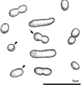
Fig. 1. Light micrographs of Cyanobium gracile Adong101619 showing symmetrical oval, ellipsoid(arrowhead), and dividing(arrow) cells. A.
×400. B.×1000.
A
Table 1. List of strains used in the molecular study and GenBank accession number
Species Strains Accession no. References
Cyanobium gracile Adong191016 MT644519 This study
Cyanobium gracile PCC6307 CP003495 Shih et al. 2013
Cyanobium gracile PCC9604 AF216944 Robertson et al. 2001
Prochlorococcus marinus AS9601 NC_008816 Kettler et al. 2007
Prochlorococcus marinus MIT9312 CP000111 Coleman et al. 2006
Prochlorococcus marinus MIT9515 CP000552 Kettler et al. 2007
Prochlorococcus marinus CCMP1375 AE017126 Dufresne et al. 2003
Synechococcus elongatus PCC6301 AP008231 Sugita et al. 2007
Synechococcus elongatus PCC7942 CP000100 Holtman et al. 2005
Synechococcus spongiarum 15L JYFQ01000001 Burgsdorf et al. 2015
Synechococcus spongiarum 142 JXUO01000104 Burgsdorf et al. 2015
Synechococcus spongiarum SH4 JENA01000091 Gao et al. 2014
Synechococcus sp. WH7803 CT971583 Doron et al. 2016
Synechococcus sp. RS9917 NZ_CH724158 Dufresne et al. 2008
New sequences are indicated in bold type.
B
Family Synechococcaceae Komárek & Anagnostidis, 1995
Genus Cyanobium Rippka & Cohen-Bazire, 1983 Cyanobium gracile Rippka & Cohen-Bazire 1983
Holotype. Type strain deposited at Pasteur Culture Col- lection(PCC), PCC6307.
Material examined. Freshwater was collected from the Adong ji pond, Adong-ri, Gaejeong-myeon, Gunsan-si, Jeolla buk-do, Republic of Korea(35°58ʹ47.4ʺN, 126°46ʹ 14.6ʺE) on October 16, 2019.
Diagnosis. Cells are a pale blue-green single cell. The cell shape is ovate, oval, or short rod-shaped without gelatinous
envelopes. Cells solitary or in twos after division, not in colonies. 1.08-3.87μm long and 0.75-1.51μm wide. The thylakoid membranes are stacked in three and arranged along the cell walls. Obligatory photoautotroph.
Distribution. North America(Smith 2010) and the Rep- ublic of Korea.
Voucher slide. Two slides of gelatin-embedded specimens were deposited at Nakdonggang National Institute of Bio- logical Resources, Korea(NNIBRCY894 and NNIBRCY 895).
2. Morphology and ultrastructure
Cyanobium gracile was pale blue-green and ovate-, oval-, or rod-shaped(Figs. 1, 2). Synechococcus species, similar to Cyanobium species in morphology, generally have a long cy- lindrical shape and are occasionally asymmetrical(Komá- rek et al. 1999), but C. gracile cells were observed to be symmetrical(Figs. 1, 2). When the cell divided by simple dichotomy, the cell was elongated rod-shaped or eight-sha- ped(Figs. 1B, 2). C. gracile cells were 1.08-3.87μm(n=75, mean=1.84±0.43μm) long and 0.75-1.51μm(n=75, mean=1.08±0.18μm) wide. Cells were larger than that of the type strain PCC6307(0.4-2.4×0.25-0.4μm, Komárek et al. 1999). The nucleolus was observed in the center of the cell of C. gracile. Peripheral thylakoid membranes were stacked in three, and each of the thylakoid membranes was arranged in parallel(Fig. 3). This thylakoid architecture is a typical characteristic of the genus Cyanobium(Gantt and Conti 1969; Komárek and Cepak 1998). Additionally, an electron opaque material, which was presumed to be poly- phosphate granules, was observed in the cytoplasm. These granules were distributed between the cell wall and the outer thylakoid membrane(Fig. 3).
Fig. 3. Transmission electron micrographs of Cyanobium gracile Adong101619 showing thylakoids arranged parallel to the cell walls. A.
Cross-sectional image. B. Longitudinal section image. CW, cell wall; N, nucleoplasm; P, polyphosphate granule; Ty, thylakoid membrane.
A B
Fig. 2. Schematic drawing showing symmetrical oval, ellipsoid (arrowhead) to shortly rod-shaped and dividing(arrow) cells.
3. Phylogeny
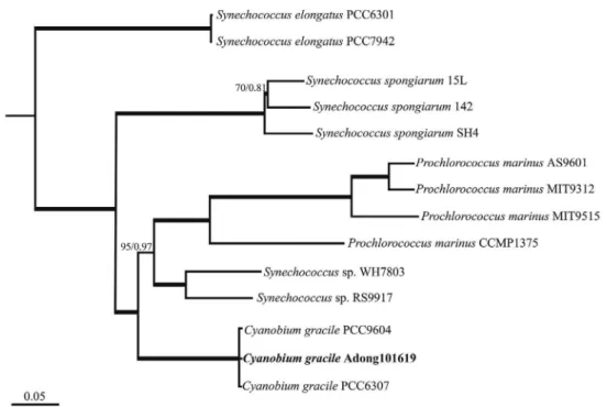
BLAST analysis indicated that the 16S SSU rDNA seq- uence of C. gracile showed 100% similarity to the refer- ence sequences of CP003495, NR_102447, AF216944, MT488300, DQ275599, and NR_114406. Bayesian and maximum likelihood(ML) analyses were performed with the 16S SSU rDNA sequence of C. gracile and 13 references (Fig. 4). In the phylogenetic tree, C. gracile formed a mono- phyletic clade with C. gracile PCC9604 and PCC6307 (ML=100, pp=1.00). In addition, C. gracile formed a sister group with four strains of Prochlorococcus marinus and two strains of Synechococcus sp.(ML=100, pp=1.00).
ACKNOWLEDGEMENTS
This research was funded by the Nakdonggang National Institute of Biological Resources(NNIBR202101103).
REFERENCES
Barnett RH. 1984. Research on control of taste and odor pro- ducing algae in surface reservoir. In: Proceedings of AWWA
WQTC. American Water Works Association-Water Quality Technology Conference. Denver, CO.
Burgsdorf I, BM Slaby, KM Handley, M Haber, J Blom, CW Mar- shall, JA Gilbert, U Hentschel and L Steindler. 2015. Lifestyle evolution in cyanobacterial symbionts of sponges. mBio.
6:1-14.
Choi SD, JT Yoon and YC Jo. 1998. Shell necrosis of Haliotis dis- cus hannai by Mastigocoleus sp.(Cyanophyta) in Korea. J.
Aquacult. 11:465-474.
Coleman ML, MB Sullivan, AC Martiny, C Steglich, K Barry, EF DeLong and SW Chisholm. 2006. Genomic islands and the ecology and evolution of Prochlorococcus. Science 311:1768-1770.
Doron S, A Fedida, MA Hernández-Prieto, G Sabehi, I Karunker, D Stazic, R Feingersch, C Steglich, M Futschik, D Lindell and R Sorek. 2016. Transcriptome dynamics of a broad host- range cyanophage and its hosts. ISME J. 10:1437-1455.
Dufresne A, M Salanoubat, F Partensky, F Artiguenave, IM Ax- mann, V Barbe, S Duprat, MY Galperin, EV Koonin, FL Gall, KS Makarova, M Ostrowski, S Oztas, C Robert, IB Rogozin, DJ Scanlan, NT Marsac, J Weissenbach, P Wincker, YI Wolf and WR Hess. 2003. Genome sequence of the cyanobacte- rium Prochlorococcus marinus SS120, a nearly minimal oxy- phototrophic genome. Proc. Natl. Acad. Sci. USA 100:10020- 10025.
Fig. 4. Maximum-likelihood(ML) tree of the genus Cyanobium based on 16S SSU rDNA sequence data. Bayesian posterior probability (pp) and maximum-likelihood(ML) bootstrap values are shown above the branches. The bold branches indicate strongly supported values (ML=100, pp=1.00). Scale bar=0.05 substitutions/site.
Dufresne A, M Ostrowski, DJ Scanlan, L Garczarek, S Mazard, BP Palenik, IT Paulsen, NT Marsac, P Wincker, C Dossat, S Ferriera, J Johnson, AF Post, WR Hess and F Partensky.
2008. Unraveling the genomic mosaic of a ubiquitous genus of marine cyanobacteria. Genome Biol. 9:1-16.
Edwards U, T Rogall, H Blöcker, M Emde and EC Böttger. 1989.
Isolation and direct complete nucleotide determination of entire genes. Characterization of a gene coding for 16S ribo- somal RNA. Nucleic Acids Res. 17:7843-7853.
Gantt E and SF Conti. 1969. Ultrastructure of blue-green algae. J.
Bacteriol. 97:1486-1493.
Gao ZM, Y Wang, RM Tian, YH Wong, ZB Batang, AM Al-Su- wailem, VB Bajic and PY Qian. 2014. Symbiotic adaptation drives genome streamlining of the cyanobacterial sponge symbiont Candidatus Synechococcus spongiarum”. mBio 5:1-14.
Gugger M, C Lyra, P Henriksen, A Coute, JF Humbert and K Sivonen. 2002. Phylogenetic comparison of the cyanobacte- rial genera Anabaena and Aphanizomenon. Int. J. Syst. Evol.
Microbiol. 52:1867-1880.
Guiry MD and GM Guiry. 2020. AlgaeBase. Worldwide electronic publication, National University of Ireland, Galway. http://
www.algaebase.org.
Havens KE. 2008. Cyanobacteria blooms: effects on aquatic ecosystems. pp. 733-747. In: Cyanobacterial Harmful Algal Blooms: State of the Science and Research Needs. Springer.
NY.
Holtman CK, Y Chen, P Sandoval, A Gonzales, MS Nalty, TL Thomas, P Youderian and SS Golden. 2005. High-throughput functional analysis of the Synechococcus elongatus PCC 7942 genome. DNA Res. 12:103-115.
Jezberová J and J Komárková. 2007. Morphological transforma- tion in a freshwater Cyanobium sp. induced by grazers. Envi- ron. Microbiol. 9:1858-1862.
Kettler GC, AC Martiny, K Huang, J Zucker, ML Coleman, S Ro- drigue, F Chen, A Lapidus, S Ferriera, J Johnson, C Steglich, GM Church, P Richardson and SW Chisholm. 2007. Patterns and implications of gene gain and loss in the evolution of Prochlorococcus. PLoS Genet. 3:e231.
Kim KY, JB Khan, IC Choi, SH Hong, JB Lee, SH Lee and JJ Lee. 2015. Temporal and spatial distribution of Geosmin and 2-MIB in the Daecheong Reservoir. Korean J. Environ. Agric.
34:14-20.
Komárek J. 2006. Cyanobacterial taxonomy: current problems and prospects for the integration of traditional and molecular approaches. Algae 21:349-375.
Komárek J and V Cepák. 1998. Cytomorphological characters supporting the taxonomic validity of Cyanothece(Cyanopro- karyota). Plant Syst. Evol. 210:25-39.
Komárek J, J Kopecký and V Cepák. 1999. Generic characters of the simplest cyanoprokaryotes Cyanobium, Cyanobacterium and Synechococcus. Cryptogam. Algol. 20:209-222.
Komárek J and K Anagnostidis. 2005. Bd. 19/2: Cyanoprokaryo- ta: teil 2: Oscillatoriales. Elsevier. München, Germany.
Komárek J. 2016. A polyphasic approach for the taxonomy of cyanobacteria: principles and applications. Eur. J. Phycol.
51:346-353.
Lehtimäki J, C Lyra, S Suomalainen, P Sundman, L Rouhiainen, L Paulin, M Salkinoja-Salonen and K Sivonen. 2000. Charac- terization of Nodularia strains, cyanobacteria from brackish waters, by genotypic and phenotypic methods. Int. J. Syst.
Evol. Microbiol. 50:1043-1053.
Nägeli C. 1849. Gattungen einzelliger Algen: physiologisch und systematisch bearbeitet. Friedrich Schulthess.
NIBR. 2019. National List of Species of Korea(Algae). National Institute of Biological Resources. Incheon, Korea.
Padisák J, L Krienitz, R Koschel and J Nedoma. 1997. Deep-layer autotrophic picoplankton maximum in the oligotrophic Lake Stechlin, Germany: origin, activity, development and erosion.
Eur. J. Phycol. 32:403-416.
Pfannkuche O and K Lochte. 1993. Open ocean pelago-benthic coupling: cyanobacteria as tracers of sedimenting salp fae- ces. Deep-Sea Res. Pt. I 40:727-737.
Posada D and KA Crandall. 1998. MODELTEST: testing the mod- el of DNA substitution. Bioinformatics 14:817-818.
Reynolds ES. 1963. The use of lead citrate at high pH as an electron-opaque stain for electron microscopy. J. Cell Biol.
17:208.
Rippka R and G Cohen-Bazire. 1983. The cyanobacteriales: a legitimate order based on the type strain Cyanobacterium stanieri ? pp. 21-36. In: Annales de l’Institut Pasteur/Microbi- ologie(Vol. 134, No. 1). Elsevier. Masson.
Robarts RD and T Zohary. 1987. Temperature effects on photosyn- thetic capacity, respiration, and growth rates of bloom-form- ing cyanobacteria. New Zeal. J. Mar. Fresh. 21: 391-399.
Robertson BR, N Tezuka and MM Watanabe. 2001. Phylogenetic analyses of Synechococcus strains(cyanobacteria) using sequences of 16S rDNA and part of the phycocyanin operon reveal multiple evolutionary lines and reflect phycobilin con- tent. Int. J. Syst. Evol. Micr. 51:861-871.
Ronquist F, M Teslenko, PVD Mark, D Ayres, A Darling, S Höhna, B Larget, L Liu, MA Suchard and JP Huelsenbeck. 2012.
MrBayes 3.2: efficient Bayesian phylogenetic inference and model choice across a large model space. Syst. Biol. 61:539- 542.
Ryu HS, RY Shin, K Seo, JH Lee and K Kim. 2018. Succession of cyanobacterial species and taxonomical characteristics of Dolichospermum spp.(Nostocales, Cyanophyceae) in the
Weir Regions of the Nakdong River. J. Korean Soc. Water Environ. 34:503-513.
Shih PM, D Wu, A Latifi, SD Axen, DP Fewer, E Talla, A Calteau, F Cai, NT Marsac, R Rippka, M Herdman, K Sivonen, T Cour- sin, T Laurent, L Goodwin, M Nolan, KW Davenport, CS Han, EM Rubin, JA Eisen, T Woyke, M Gugger and CA Kerfeld.
2013. Improving the coverage of the cyanobacterial phylum using diversity-driven genome sequencing. Proc. Natl. Acad.
Sci. USA 110:1053-1058.
Smith SW, R Overbeek, CR Woese, W Gilbert and PM Gillevet.
1994. The Genetic Data Environment: an expandable GUI for multiple sequence analysis. Comput. Appl. Biosci. 10:671- 675.
Smith TE. 2010. Revised list of algae from Arkansas(USA) and new additions. Int. J. Algae 12:230-256.
Spurr AR. 1969. A low-viscosity epoxy resin embedding medium for electron microscopy. J. Ultrastruct. Res. 26:31-43.
Stamatakis A. 2014. RAxML version 8: a tool for phylogenetic analysis and post-analysis of large phylogenies. Bioinformat- ics 30:1312-1313.
Suffet IH, J Mallevialle and E Kawczynski. 1995. Advances in Taste -and -Odor Treatment and Control. American Water
Works Association.
Sugita C, K Ogata, M Shikata, H Jikuya, J Takano, M Furumi- chi, M Kanehisa, T Omata, M Sugiura and M Sugita. 2007.
Complete nucleotide sequence of the freshwater unicellular cyanobacterium Synechococcus elongatus PCC 6301 chro- mosome: gene content and organization. Photosynth. Res.
93:55-67.
You KA, MS Byeon, SJ Youn, SJ Hwang and DH Rhew. 2013.
Growth characteristics of blue-green algae(Anabaena spiroi- des) causing tastes and odors in the North-Han River, Korea.
Korean J. Ecol. Environ. 46:135-144.
Yunes JS, NT Cunha, LP Barros, LAO Proença and JM Monser- rat. 2003. Cyanobacterial neurotoxins from Southern Brazil- ian freshwaters. Comments Toxicol. 9:103-115.
Willame R, C Boutte, S Grubisic, A Wilmotte, J Komárek and L Hoffmann. 2006. Morphological and molecular characteriza- tion of planktonic cyanobacteria from Belgium and Luxem- bourg 1. J. Phycol. 42:1312-1332.
Zander AK and P Pingert. 1997. Membrane-based extraction for detection of tastes and odors in water. Water Res. 31:301- 309.