광나무의 주성분, Ursolic acid와 Oleanolic acid의 항노화 효능
홍 용 덕†⋅유 대 성*⋅남 미 희⋅김 현 정⋅박 시 준*⋅신 송 석†⋅천 종 우*⋅박 영 호
(주)아모레퍼시픽 기술연구원, *(주)에이씨티 기술연구소
(2012년 4월 12일 접수, 2012년 6월 4일 수정, 2012년 6월 10일 채택)
Excellent Anti-aging Effects of Ursolic acid and Oleanolic acid Present in Ligustrum lucidum
Yong Deog Hong†, Dae Sung Yoo*, Mi Hee Nam, Hyeon Chung Kim Si Jun Park*, Song Seok Shin†, Jong Woo Cheon*, and Young Ho Park Amorepacific Corporation R&D Center, 314-1, Bora-dong, Giheung-gu, Yongin-si,
Gyeonggi-do 446-729, Korea, *R&D Center, ACT Co., Ltd.
(Received April 12, 2012; Revised June 4, 2012; Accepted June 10, 2012)
요 약: 광나무
(L. lucidum)는 ursolic acid와 oleanolic acid를 다량 포함하고 있다. 본 연구에서는 광나무 열매, 줄기, 잎 세 부위 추출물의 항주름 효능을 평가하였다. 광나무 추출물은 human skin fibroblasts에서 독성이 없을 뿐만 아니라 MMP-1 과 MMP-2의 발현을 감소시키고 COL1A1의 발현을 증가시켰다. 이들 추출물은 모두 농도 의존적으로 COL1A1의 발현을 증가시켰으며 MMP-1과 MMP-2의 발현을 감소시켰다. 광나무 세 부위 추출물 가운데, 열매 부위에 가장 많은 양의 ursolic acid 와 oleanolic acid가 함유되어 있었으며 가장 강한 COL1A1 upregulating 효과와 MMP-1 과 MMP-2 downregulating 효과를 나타냈다. 이처럼 항주름 효능을 보이는 광나무 열매 추출물은 기능성 화장품 소재 로 개발될 수 있는 가능성이 있다.
Abstract:
Ligustrum lucidum (L. lucidum)contains large quantities of ursolic acid and oleanolic acid. In this study, we evaluated anti-wrinkle effects of three parts of
L. lucidumextracts. We found that
L. lucidumextracts were not only innocuous to human skin fibroblasts but also significantly decreased the expression of both MMP-1 and MMP-2 and increased the expression of COL1A1. Among the three parts of
L. lucidumextracts (i.e., fruit, cane, and leaf extracts), the fruit extract was found to contain the greatest amounts of ursolic acid and oleanolic acid.
These three parts of
L. lucidumextracts increased the expression of COL1A1 and decreased the expression of MMP-1 and MMP-2 in a dose-dependent manner. Especially, the fruit extract of
L. lucidumhad the greatest upre- gulating effect on COL1A1 and the greatest downregulating effect on MMP-1 and MMP-2 in a non-toxic and dose-dependent manner. These results suggest that
L. lucidum, especially the fruit, could be used as active in- gredients for functional cosmetics.
Keywords: l
igustrum lucidum, ursolic acid, oleanolic acid, COL1A1, MMP-1, and MMP-2
1. Introduction
1)
Aging involves atrophic changes that impair physical and psychological functions with age. In general, skin
† 주 저자(e-mail: [email protected], [email protected])
aging has many different manifestations, including xe- roderma, changes in skin tone, changes in skin elas- ticity, vasodilation, and wrinkles[1-3]. Among these manifestations, wrinkles may be classified as signs of intrinsic aging or photoaging.
Wrinkles are influenced by age, external environ-
(1)
(2)
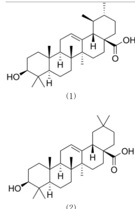
Figure 1. Structure of ursolic acid(1) and oleanolic acid(2).
ment, and UV irradiation. Skin damage by these fac- tors promotes several aging processes, including de- creased skin elasticity and wrinkle formation[4]. Espe- cially, UV-induced skin damage is characterized by skin inflammation, DNA damage, pigmentation, and in severe cases, skin cancer[5,6].
The mechanism of photoaging involves decreased collagen synthesis and overexpression of MMPs by UV irradiation, which results in aging processes such as wrinkle formation[7,8].
MMPs are zinc-dependant endopeptidases, which break down collagen and other proteins of the ex- tracellular matrix (ECM) and basement membranes.
UV-induced MMPs degrade the connective tissue of the skin, thereby impairing the structural integrity of the dermis. Such accumulative ECM protein damage acts as a major contributor to the formation of wrinkles, reduction of elasticity, and drooping of skin[9].
Collagen is the most abundant protein in the body, making up approximately 25 % of the whole body pro- tein content. Bundles of collagen molecules called
‘collagen fibers’ have a distinctive structure of triple helices that provide great strength and elasticity.
Collagen fibers are a major component of the ECM, supporting most of the connective tissue in animals.
Strong, mature type I collagen fibers are formed from triple-stranded, rope-like type I procollagen molecules by enzymatic processes outside the cell. Type I pro- collagen molecules contain 2 pro-alpha-1 chains and 1 pro-alpha-2 chain that are encoded by COL1A1 (collagen, type I, alpha 1) and COL1A2 (collagen, type I, alpha 2), respectively[10].
The fruits of Ligustrum lucidum are commonly used for their tonic effects in Chinese medicine[11]. Previous studies have found volatile components, including tri- terpenes, flavonoids, secoiridoid glucosides, and phenolic compounds, in this plant and constituents such as phe- nylethanoids, monoterpenes, and secoiridoid glucosides in other species of the genus Ligustrum[12-15].
A recent study reported that ursolic acid and ole- anolic acid have anti-wrinkle activity[26,27]. The fruits of L. lucidum contain large quantities of ursolic acid and oleanolic acid(Figure 1). Therefore, we examined
L. lucidum as a potential anti-wrinkle cosmetic ingre- dient. Among the three parts of L. lucidum extracts (i.e., fruit, cane, and leaf extracts), the fruit extract was found to contain the greatest amounts of ursolic acid and oleanolic acid. We used powdered L. lucidum fruits and examined their anti-aging efficacy by per- forming in vitro efficacy tests on fibroblasts.
In this study, we confirm the anti-aging effects of ur- solic acid and oleanolic acid present in powdered L. lu- cidum fruits by examining the in vitro expression of genes such as COL1A1, MMP-1, and MMP-2.
2. Materials and Methods
2.1. Extraction and Drying of L. lucidum Fruits
A total of 200 g of the fruit was refluxed with 7-fold 95 % aqueous ethanol at 50 ~ 60 ℃ for 3 hours. After 3 h, the solution was diluted to 1/10, and recrystalliza- tion was achieved by adding 10-fold distilled water with agitation. Filtration of the recrystallized powder and drying was performed at 60 ℃ for 24 h, after which the powder was removed (quantity: 5.18 g;
yield : 2.59 %).
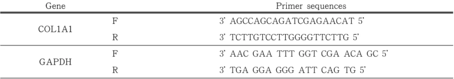
Table 1. Primers for Genes Investigated Using RT-PCR Analysis
Gene Primer sequences
COL1A1 F 3' AGCCAGCAGATCGAGAACAT 5'
R 3' TCTTGTCCTTGGGGTTCTTG 5'
GAPDH F 3' AAC GAA TTT GGT CGA ACA GC 5'
R 3' TGA GGA GGG ATT CAG TG 5'
* F : Forward, R : Reverse 2.2. HPLC Analysis
HPLC was performed using the Waters 2424 alliance separation module (Waters, U.S.A.). Chromatography was performed using the Prostar210/Prepstar218 series with an RP-C18 column (250 nm × 4.6 nm, 5 µm;
Mightysil) and a wavelength of 203 nm. The mobile phase contained 9 : 1 acetonitrile and 0.1 % phosphoric acid.
2.3. Cell Culture
Human fibroblast HS683 cells were cultured in RPMI1640 with 10 % fetal bovine serum (FBS) and 100 U/mL penicillin/streptomycin at 37 ℃ in a humidi- fied atmosphere with 5 % CO2. FBS, penicillin/strep- tomycin, and trypsin-EDTA were provided by Lonza (Walkersville, MD).
2.4. Cell Viability Assay
Cell viability and the extent of proliferation were as- sessed by conventional MTT (3-[4,5-dimethylthiazol- 2-yl]-2,5-diphenyl tetrazolum bromide) assay (Zund et al. 1999). Briefly, HS683 cells (5 × 4 cells/well) were incubated with various concentrations of test com- pounds for the indicated times. After this, the cells were incubated with MTT solution (0.5 mg/mL) for an additional 3 h at 37 ℃. Absorbance of the samples was measured at 490 nm with a microplate reader manufactured by Molecular Devices Corp (Menlo Park, CA).
2.5. mRNA Analysis by Semiquantitative Reverse- Transcription Polymerase Chain Reaction (RT-PCR) To determine the cytokine mRNA expression levels, total RNA was extracted from LPS-treated RAW264.7
cells with TRIzol Reagent (Gibco BRL), according to the manufacturer’s instructions. Total RNA was stored at -70 ℃ until use. Analysis of mRNA was perform- edusing semiquantitative RT-PCR according to the manufacturer's instructions (Bioneer, Daejeon, Korea).
The results were expressed as the ratio of the optical density at 280 nm to GAPDH mRNA concentration.
Alternatively, the results were normalized to GAPDH mRNA. The primers (Bioneer) used for RT-PCR are listed in Table 1.
2.6. DPPH Free Radical-Scavenging Activity
1,1-Diphenyl-2-picrylhydrazyl (DPPH) free radical- scavenging activity was evaluated using the method by Blois (1958) with minor modifications. DPPH solution (0.1 mM in ethanol) was added to the same volume of sample solution and allowed to react for 10 min at room temperature ; optical density was then measured at 565 nm by using a microplate reader manufactured by Molecular Devices Corp.
2.7. Statistical Analysis
Means ± S.D of the means were calculated ; stat- istical analysis of results was performed using Student's t-test for independent samples.
3. Results and Discussion
3.1. Comparison of Ursolic Acid and Oleanolic Acid Contents in Natural Products
Thin-layer chromatography was performed to first determine the content of active components. We iden- tified three natural products containing ursolic acid and oleanolic acid, namely, L. lucidum, Sambucus williamsii,
Table 2. Contents of Ursolic Acid and Oleanolic Acid Containing Three Natural Products
Ligustrum lucidum Sambucus williamsii Epilobium angustifolium L.
Oleanolic acid 20.1 % 0.69 % -
Ursolic acid 4.08 % 0.99 % 0.11 %
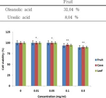
Table 3. HPLC Analysis of Ursolic Acid and Oleanolic Acid in Three Parts Extract of Ligustrum lucidum
Fruit Cane Leaf
Oleanolic acid 31.04 % 18.1 % 5.1 %
Ursolic acid 8.04 % 2.1 % 0.3 %
Figure 2. Cytotoxicity of three L. lucidum extracts in human fibroblasts, *p < 0.05 and **p < 0.01 compared to control.
and Epilobium angustifolium L. Next, we performed HPLC quantitative analysis of these natural products containing ursolic acid and oleanolic acid. We confirmed that L. lucidum contains the largest quantities of ursolic acid and oleanolic acid among these three natural prod- ucts (Table 2).
In sequence, we analyzed the content of ursolic acid and oleanolic acid in 3 extracts of L. lucidum, namely, the fruit, cane, and leaf. Our analysis showed that the extract of L. lucidum fruit contained the largest quan- tities of ursolic acid and oleanolic acid among these three L. lucidum extracts (Table 3).
3.2. Cytotoxicity of The Three L. lucidum Extracts in Human Fibroblasts
To determine the cytotoxicity of the three L. luci- dum extracts in human fibroblasts, we measured the
cell viability of human fibroblasts by MTT assay. Our results showed that none of the extracts had a cyto- toxic effect at concentrations up to 0.5 % ; however, minimal cytotoxic effects were observed at concen- trations of 1 % and 2 % (approximately 10 % ex- tinction of cells) (Figure 2).
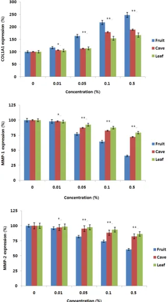
3.3. Effect of The Three L. lucidum Extracts on The Expression of COL1A1, MMP-1, and MMP-2 genes Extrinsic and intrinsic skin aging commonly increases wrinkling, sagging, and laxity[16]. Extrinsic aging is generally related to photoaging and is caused by re- peated exposure to UV light. UV irradiation induces the synthesis of MMPs in fibroblasts. Upregulation of MMPs usually enhances the degradation of dermal col- lagen during UV-induced skin aging[17]. MMPs, a family of zinc-dependent endoproteinases, play a key role in remodeling ECM structures during wound heal- ing[18], dermal photoaging[19], and several patholo- gies such as carcinogenesis[20]. For example, MMP-1 initiates the cleavage of fibrillar collagen (Type I and III in the skin) at a single site within its central triple helix. Once cleaved by MMP-1, collagen is further de- graded by the elevated activities of other MMPs [21].
For this reason, we examined the expression of COL1A1, MMP-1, and MMP-2 genes involved in pho- toaging and the subsequent formation of wrinkles, and investigated the changes in the expression of these genes by RT-PCR in human fibroblasts treated with the three L. lucidum extracts. We found that all three L. lucidum extracts increased the expression of COL1A1 and decreased the expression of MMP-1 and
Figure 3. Effect of the three L. lucidum extracts on the expression of COL1A1, MMP-1, and MMP-2 genes, *p
< 0.05 and **p < 0.01 compared to control.
Figure 4. DPPH free radical-scavenging activity of the three L. lucidum extracts, *p < 0.05 and **p < 0.01 compared to control.
MMP-2 in a dose-dependent manner. Especially, the fruit extract of L. lucidum had the greatest upregulat- ing effect on COL1A1 and the greatest downregulating effect on MMP-1 and MMP-2. Therefore, we suggest that the fruit extract of L. lucidum has an excellent anti-wrinkle efficacy and may be a suitable ingredient for anti-aging cosmetics (Figure 3).
3.4. DPPH Free Radical-scavenging Activity of The Three L. lucidum Extracts
UV radiation is just one of the environmental factors
that may induce injury to human skin. Free radicals and ROS are defined as any molecular species contain- ing 1 or more unpaired electrons that are capable of in- dependent existence. These unstable states make them highly reactive and capable of initiating chain reactions, which result in the production of new free radicals.
Moreover, they can interfere and alter protein func- tions, increase lipid peroxidation, damage genes, and promote diseases and aging [9]. Figure. 4 shows the DPPH free radical-scavenging activity of the three L.
lucidum extracts. The three L. lucidum extracts showed dose-dependent scavenging activity against DPPH free radicals. Especially, the fruit extract of L.
lucidum showed the greatest scavenging activity against DPPH free radicals.
4. Conclusions
MMPs are enzymes that are directly responsible for the degradation of ECM components such as collagen, gelatin, and elastin. MMP-1, an interstitial collagenase, belongs to a subfamily of MMPs that can specifically degrade the collagen triple helix. MMP-2 is a gelatinase that degrades denatured collagen, gelatin, and elastin.
Thus, UV irradiation induces the degradationof ECM components, such as collagen, by increasing the pro- duction of MMPs in the skin. Therefore, the regulation of MMP activities may protect the skin from UV-in- duced damage. This is the rationale for most bioassays
searching for agents able to protect the skin from UV-induced overexpression of MMPs.
Many plant extracts have been examined for the presence of potential MMP inhibitors. For example, green tea [22], blackberry [23], Kaempferia pandurata [24], and Pothomorphe umbellata extracts [25] have demonstrated inhibitory effects on MMP activities.
In this study, we found that L. lucidum extracts were not only innocuous to human skin fibroblasts but also significantly decreased the expression of both MMP-1 and MMP-2 and increased the expression of COL1A1. L. lucidum was shown to have anti-wrinkle and anti-oxidant activities. HPLC analysis showed that L. lucidum contained more ursolic acid and oleanolic acid than S. williamsii and E. angustifolium L. Among the three L. lucidum extracts (i.e., fruit, cane, and leaf extracts), the fruit extract was found to contain the greatest amounts of ursolic acid and oleanolic acid.
These three L. lucidum extracts increased the ex- pression of COL1A1 and decreased the expression of MMP-1 and MMP-2 in a dose-dependent manner.
Especially, the fruit extract of L. lucidum had the greatest upregulating effect on COL1A1 and the great- est downregulating effect on MMP-1 and MMP-2 in a non-toxic and dose-dependent manner. The three L.
lucidum extracts also showed a dose-dependent scav- enging activity against DPPH free radicals. Especially, the fruit extract of L. lucidum showed the greatest scavenging activity against DPPH free radicals.
These results indicate that L. lucidum, Especially the fruit, appears to contain useful cosmetic ingredients having anti-wrinkle effects. However, it is necessary to conduct further in vivo studies and clinical tests with different dosages of L. lucidum extracts. Thus, L. luci- dum may constitute an excellent anti-aging ingredient.
References
1. I. M. Freedberg, A. Z. Eisen, K. W. Wolff, K. F.
Austen, L. A. Goldsmith, and S. I. Katz, Derma- tology in General Medicine, McGraw-Hill 6th ed, 165 (2003).
2. K. H. Han, K. H. Cho, D. Y. Noh, H. C. Eun, and
J. I. Youn. Histological changes in the skin with in- nate ageing, Kor. J. Dermatol., 36(6), 971 (1998).
3. K. H. Cho, M. K. Lee, S. J. Jo, K. H. Kim, K. C.
Park, and H. C. Eun. Histologic changes in the skin with photoaging, Kor. J. Dermatol., 41(6), 754 (2003).
4. E. S. Yang, R. H. Hong, and S. M. Kang, The ef- fect of genistein on the proliferation and type I pN collagen synthesis in aged normal human fibro- blasts, Kor. J. Microbiol. Biotechnol., 35(4), 316 (2007).
5. J. Y. Seo, E. K. Kim, S. H. Lee, K. C. Park, K. H.
Kim, H. C. Eun, and J. H. Chung, Enhanced ex- pression of cylooxygenase-2 by UV in aged human skin in vivo, Mechanisms of Ageing and Develop- ment, 124, 903 (2003).
6. J. A. Nichols and S. K. Katiyar, Skin photo- protection by natural polyphenols: anti-inflamma- tory, antioxidant and DNA repair mechanisms, Arch. Dermatol. Res., 302(2), 71 (2009).
7. A. Hashem, K. Y. Jun, E. Y. Lee, S. Y. Lim, H. Y.
Park Choo, and Y. J. Kwon, A rapid and sensitive screening system for human type Ⅰ collagen with the aim of discoverying potent anti-aging or anti- fibrotic compounds, Mol. Cells OS, 26(6), 625 (2008).
8. W. M. Yang, H. M. Kim, M. S. Chang, W. S. Park, W. N. Kim, S. W. Kim, D. G. Choi, H. C. Lee, Y.
K. Kim, and S. K. Park, Effects of ethanol extract of Nelumbo nucifera leaves on anti-oxidation and type I procollagen expression in CCD-986sk cells, The Korean Journal of Oriental Medical Prescrip- ton, 14(2), 67 (2006).
9. B. C. Lee, S. Y. Lee, H. J. Lee, G. S. Sim, J. H.
Kim, J. H. Kim, Y. H. Cho, D. H. Lee, H. B. Pyo, T. B. Choe, D. C. Moon, Y. P. Yun, and J. T. Hong, Anti-oxidative and photo-protective effects of cou- marins isolated from fraxinus chinensis, Arch Pharm Res, 30, 10, 1293 (2007).
10. G. A. Di Lullo, S. M. Sweeney, J. Korkko, L.
Ala-Kokko, and J. D. San Antonio, Mapping the li- gand-binding sites and disease associated mutations on the most abundant protein in the human, type
I collagen; J. Biol. Chem. OTT, 4223 (2002).
11. M. L. Li, M. L. Lui, and W. H. Feng, Advances in the research on the fruits of ligustrum lucidum, Zhongguo Zhongyao Zhazhi, 19(8), 504 (1994).
12. P. Garibodi, F. Jommi, and L. Verotta, Secoiridoids from oleaeuropaea, Phytochemistry, 25, 865 (1986).
13. Z. D. He, Y. Q. Liu, and C. R. Yang, Glycosides from ligustrumpurpurascens, Acta Botanica Yan- nanica, 14(3), 328 (1992).
14. Z. D. He, S. Ueda, M. Akaji, T. Fujita, K. Inoue, and C. R. Yang, Monoterpenoid and phenylethanoid glycosides from ligustrum pedunculare, Phytoche- mistry, 36(3), 709 (1994).
15. J. Tian, H. J. Zhang, and H. D. Sun. Four new gly- cosides, ligurobustosides A, B, C and D from ligus- trum robustum, Chinese Chemical Letters, 7(4), 341 (1996).
16. G. Jenkins, Molecular mechanisms of skin ageing, Mechanisms of Ageing and Development, 123, 801 (2002).
17. G. J. Fisher, S. C. Datta, H. S. Talwar, Z. Q. Wang, J. Varani, S. Kang, and J. J. Voorhees, Molecular basis of sun-induced premature skin aging and reti- noid antagonism, Nature, 379, 335 (1996).
18. Z. Werb, ECM and cell surface proteolysis: regu- lating cellular ecology, Cell 14, 439 (1997).
19. V. M. Kahari and U. Saarialho-Kere, Matrix metal- loproteinases in skin, Experimental Dermatology, 6, 199 (1997).
20. S. Curran and G. I. Murray, Matrix metallopro- tei- nases in tumour invasion and metastasis, Journal of Pathology, 189, 300 (1999).
21. M. D. Sternlicht and Z. Werb, How matrix metal- loproteinase regulate cell behavior, Annual Review of Cell and Developmental Biology, 17, 463 (2001).
22. S. K. Katiyar and C. A. Elmets, Green tea poly- phenolic antioxidants and skin photoprotection, Inter- national Journal of Oncology, 18, 1307 (2001).
23. M. Herrmann, S. Grether-Beck, I. Meyer, H. Fran- ke, H. Joppe, J. Krutmann, and G. Vielhaber, Black- berry leaf extract: a multifunctional anti-aging ac- tive, International Journal of Cosmetic Science, 10, 41 (2007).
24. J. S. Shim, E. J. Choi, C. W. Lee, H. S. Kim, and J. K. Hwang, Matrix metalloproteinase-1 inhibitory activity of Kaempferia pandurata Roxb, Journal of Medicinal Food, 12, 601 (2009).
25. C. D. Ropke, V. V. da Silva, C. Z. Kera, D. V.
Miranda, R. L. de Almeida, T. C. Sawada, and S.
B. Barros, In vitro and in vivo inhibition of skin matrix metalloproteinases by pothomorphe umbella- ta root extract, Photochemistry and Photobiology, 82, 439 (2006).
26. Y. S. Lee, D. Q. Jin, S. M. Beak, E. S. Lee, and J.
A. Kim, Inhibition of ultraviolet-A-modulated sig- naling pathway by asiatic acid and ursolic acid in HaCaT human keratinocytes, European Journal of Pharmacology, 476, 173 (2003).
27. M. Niladri, K. N. Neelesh, A. Karim, K. S. Birendra, and K. M. Pulok, Exploring targets erecta Linn flower for the elastase, hyaluronidase and MMP-1 inhibitory activity, Journal of Ethnophamacology, 137, 1300 (2011).