Since Kelman first introduced phacoemulsification in 1967, there have been numerous developments in the removal of the cataracts. Recently introduced, AquaLase
Ⓡis one of the methods in which to remove a cataract using the Infinity Vision System (Alcon Laboratories, Fort Worth, Texas). It uses water-jet pulses to liquefy the lens nucleus. The AquaLase
Ⓡhandpiece warms a balanced salt solution up to 57℃ and then creates a micropulse (4 µl, 50 pulses per second) for extracting the cataract.
1The theoretically proposed advantages of AquaLase
Ⓡfor removing a cataract are reduced thermal risk (no incisional burn) and reduced posterior capsular rupture.
1,2The MicroFlow
Ⓡsystem was developed by Bausch &
Lomb Surgical with a grooved outer surface that allows for an increased rate of fluid flow into the eye, keeping the
needle cool. The MicroFlow
Ⓡsystem delivers improved performance with advanced fluids and chamber stability. The expected advantages of the MicroFlow
Ⓡsystem are expanded chamber depth, improved cutting ability for a variety of lens densities and reduced fluid volume resulting in less endothelial cell loss. The MicroFlow
Ⓡsystem was considered the appropriate control group to compare with AquaLase
Ⓡ.
The purpose of our study was to compare any changes in surgically induced astigmatism (SIA), corneal endothelial cell damage, postoperative recovery of visual acuity and change of corneal thickness after cataract surgery performed by using the AquaLase
Ⓡand phacoemulsification in the MicroFlow
Ⓡsystem.
Materials and Methods
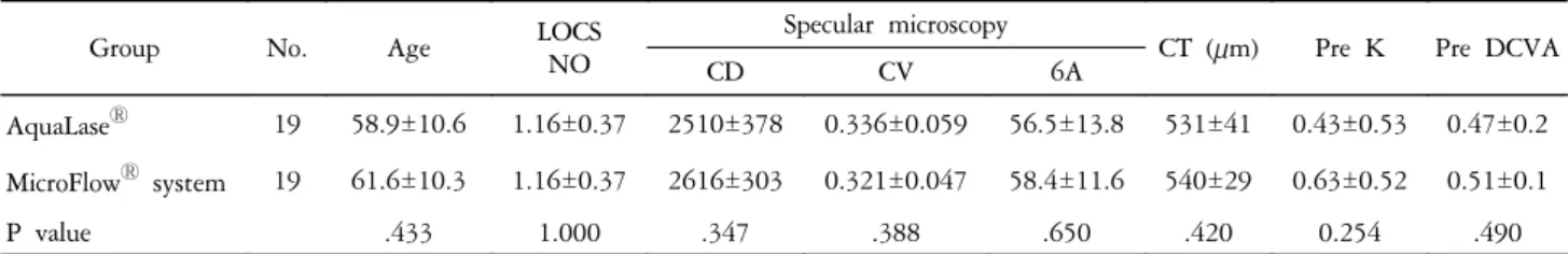
In this prospective study, 19 patients (19 eyes) with cataracts (Lens Opacities Classification System (LOCS), version III
3nuclear grade below two) were included. The cataract was graded as the 4 grading scales of LOCS III using a biomicroscope. Therefore LOCS III nuclear grade below 2 means that the cataract had less than moderate
Aqualase Ⓡ with Phacoemulsification Using MicroFlow Ⓡ System
Hyun-Wook Ryu, MD
1, Shin-Hae Park, MD
1, Choun-Ki Joo, MD, PhD
1,2Department of Ophthalmology and Visual Science1, Kangnam St. Mary's Hospital, College of Medicine, The Catholic University of Korea, Seoul, Korea
Korean Eye Tissue and Gene Bank Related to Blindness
2, Seoul, Korea
Purpose: To compare the outcomes after phacoemulsification performed with the AquaLase
Ⓡand phacoemulsification in MicroFlow
Ⓡsystem, including surgically induced astigmatism (SIA), corneal endothelial cell damage and postoperative recovery of visual acuity.
Methods: The cataracts of Lens Opacities Classification System, version III (LOCS III) nuclear grade below 2 were subjected in this study. Nineteen eyes underwent cataract operation using AquaLase
Ⓡ(Alcon Laboratories, Fort Worth, Texas, U.S.A.). A control group (19 eyes) used the MicroFlow
Ⓡsystem (Millenium, Stortz, U.S.A.) and was selected by matching age, sex, systemic disease, corneal astigmatism and corneal endothelial cell density. All the surgeries were performed by the same operator. SIA, corneal endothelial cell loss, visual acuity, and corneal thickness were evaluated postoperatively.
Results: SIA in the group using AquaLase
Ⓡwas less than that of the group using MicroFlow
Ⓡsystem (P=0.022) at 2 months postoperatively. Evaluation of corneal endothelial cell loss, recovery of visual acuity and corneal thickness found no statistically significant differences between the two groups.
Conclusions: Cataract surgery using AquaLase
Ⓡinduces less surgically induced astigmatism in mild to moderate cataracts.
Korean Journal of Ophthalmology 21(3):137-141, 2007
Key Words: AquaLase, Corneal endothelial cell damage, Surgically induced astigmatism
Received: February 12, 2007 Accepted: August 3, 2007
Reprint requests to Choun-Ki Joo, MD, PhD. Department of Ophthal- mology Kangnam St. Mary's Hospital, College of Medicine, The Catholic University of Korea. 505 Banpo-dong, Seocho-gu, Seoul, 137-040, Korea. Tel: 82-2-590-2755, Fax: 82-2-599-7405, E-mail:
[email protected]
nucleosclerosis. Nineteen eyes underwent cataract operation using AquaLase
Ⓡand the control group (19 eyes) was selected by matching age, sex, systemic disease, grade of nuclear color and opacification, axis and magnitude of corneal astigmatism and corneal endothelial cell density. The opposite eye of each of the test patients was not included in the control group. One surgeon (C.K.J.) performed all surgical procedures in October of 2005. Preoperative examinations included visual acuity, intraocular pressure, slit lamp examination, fundus examination, A & B-scan, pachymetry, keratometry and specular microscopy.
Keratometry was measured using a manual keratometer (Topcon DM-4, Japan) and corneal thickness was measured using ultrasound pachymeter (Humphrey Instrument Inc.) preoperatively and at 1 week and 2 months postoperatively.
Decimal visual acuity was measured preoperatively and at 1 day, 1 week, and 2 months postoperatively. Corneal endothelial cell density was measured using a specular microscope (Noncon Robo-CA SP-8000, JAPAN) preoperatively and at 2 months postoperatively. In addition, the coefficient of variation of cell size, and percentage of hexagonal cells were assessed. All endothelial parameters were measured in the central cornea. Surgically induced astigmatism was analyzed by the Law of Cosines and Sines
4method at 1 week and 2 months postoperatively.
After topical anesthesia, a self-sealing 3.2 mm temporal clear corneal incison was made by a diamond knife. After hydrodissection and hydrodelineation, the lens nucleus was divided by an Akahoshi prechopper. The lens was liquified using the AquaLase
Ⓡof the Infinity Vision System (Alcon Laboratories, Fort Worth, Texas). In the control group the lens was phacoemulsified using a bevel-down phaco tip with the Millenium
Ⓡ(Stortz, U.S.A.) unit. Surgical settings of the AquaLase
Ⓡand Millenium
Ⓡunit were presented in Table 1.
All patients had implantation of a foldable acrylic IOL. No corneal suturing was done.
SPSS software was used for statistical analysis. An unpaired t-test was used to test for interindividual differences and Pearson's bivariate correlation was used to examine the correlations between all of the numerical variables. A p value less than 0.05 was considered statistically significant and all
p values reported were 2-sided.
Results
The mean age was 58.9±10.6 years in the AquaLase
Ⓡgroup and 61.6±10.3 years in the MicroFlow
Ⓡsystem group (p>0.05). The mean LOCS Ⅲ classification nuclear grade was 1.16±0.37 in AquaLase
Ⓡgroup and 1.16±0.37 in the control group (p>0.05). The mean preoperative magnitude of corneal astigmatism was 0.43±0.53 diopters in AquaLase
Ⓡgroup and 0.63±0.52 diopters in the control group (p>0.05).
The mean preoperative endothelial cell density was 2510±
378/mm
2in AquaLase
Ⓡgroup and 2616±303/mm
2in the control group (p>0.05). None of these preoperative factors were statistically significant between the two groups. Table 2 summarizes the preoperative patient data.
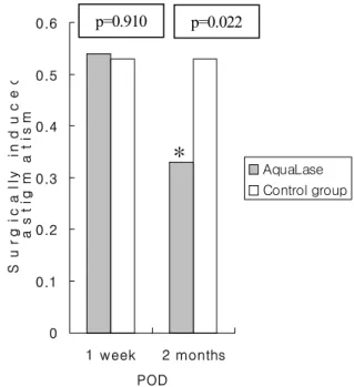
The mean SIA at 1 week postoperatively was 0.54±0.34 diopters in the AquaLase
Ⓡgroup and 0.53±0.37 diopters in the control group (p=0.910). The mean SIA at 2 months postoperatively was 0.33±0.20 diopters in the AquaLase
Ⓡgroup and 0.53±0.28 diopters in the control group (p=0.022, Fig. 1). The mean corneal endothelial cell loss at 2 months postoperatively was 4.5±24 % in the AquaLase
Ⓡgroup and 10.2±10.8 % in the control group (p=0.417, Fig. 2A). The mean coefficient of variation at 2 months postoperatively was 0.332±0.050 in the AquaLase
Ⓡgroup and 0.339±0.045 in the control group (p=0.686, Fig. 2C). The mean percentage of hexagonal cells at 2 months postoperatively was 52.1±
8.70 in the AquaLase
Ⓡgroup and 54.6±10.0 in the control group (p=0.461, Fig. 2D).
Table 1. Surgical settings used in the study with the AquaLase
Ⓡand MicroFlow
Ⓡsystem
AquaLase
ⓇMicroFlow
Ⓡsystem
Magnitude 80 -
Power - 20%
Aspiration flow 36 ml/min (linear) -
Vacuum 370 mmHg 200 mmHg
Bottle height 95 cm 105 cm
Mode Pulse (50 PPS, 80 msec) Pulse (50PPS, 80msec)
PPS=pulse per second. Fig. 1. Mean surgically induced astigmatism after operation.
(* : statistically significant difference) 0
0.1 0.2 0.3 0.4 0.5 0.6
1 week 2 months POD
Su r g ic a ll y in d u c e d as t ig m at is m
AquaLase Control group
*
p=0.910 p=0.022
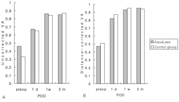
The uncorrected decimal visual acuity at 1 day, 1 week and 2 months postoperatively was 0.67±0.21, 0.86±0.22, 0.85±0.17 in the AquaLase
Ⓡgroup and 0.65±0.30, 0.84±
0.23, 0.87±0.13 in the control group (p>0.05, Fig. 3A). The distance-corrected decimal visual acuity at 1 day, 1 week and 2 months postoperatively was 0.82±0.28, 0.93±0.16, 0.95
±0.09 in the AquaLase
Ⓡgroup and 0.87±0.16, 0.95±0.10, 0.94±0.10 in the control group (p>0.05, Fig. 3B). The corneal thickness at 1 week and 2 months postoperatively was 545±52 µm, 533±42 µm in the AquaLase
Ⓡgroup and 551±31 µm, 540±29 µm in the control group (p>0.05, Table 3).
A statistically significant difference was found between the two groups only in the SIA at 2 months postoperatively (p=0.022). However, there were no correlations between any of the numerical variables (p>0.05).
Discussion
The AquaLase
Ⓡhandpiece warms a balanced salt solution up to 57℃ and the warmed saline is mixed with a peripheral irrigation fluid, causing the temperature of the mixed fluids to stabilize at 32℃ (R. Dimalanta, ASCRS Symposium on Cataract, IOL and Refractive Surgery, San Francisco, CA, April 12-16, 2003). Therefore, during the procedure, peripheral tissues are not damaged from the effect of temperature extremes. Furthermore, experimental measurement of internal wound temperature has shown that no incision heat is generated even at full power.
1So the advantage of the AquaLase
Ⓡinclude lower corneal endothelial cell damage and no incisional burn compared with preexisting ultrasound phacoemulsification.
2However, there has not been objective research examining theses advantages except for the research about corneal endothelial cell damage after lens extraction using the fluid-based system in human cadaver eyes.
5The MicroFlow
Ⓡsystem developed by Bausch & Lomb Surgical has high performance with advanced fluids and chamber stability and the proposed advantage that reduced fluid volume results in less endothelial cell loss. The MicroFlow
Ⓡsystem was used as the operative instrument for the control group in the comparison with AquaLase
ⓇWe predicted that the fluid-base system in AquaLase
Ⓡwould induce less endothelial cell damage and wound burn compared with phacoemulficiation in the MicroFlow
Ⓡsystem because it produces negligible heat. But the results show that only development of SIA was significantly different between the two groups (p=0.022) and there were no significant differences in the endothelial cell density, recovery of visual acuity or the change of corneal thickness.
The mean corneal endothelial cell loss at 2 months postoperatively was 4.5±24% in the AquaLase
Ⓡgroup and 10.2±10% in the control group and there was no significant difference between two groups (p>0.05, Fig. 2A). This result is supported by similar results by Sandoval et al.
5They reported that the mean total damaged endothelial cells was 60.2±24.1/mm
2in the AquaLase
Ⓡgroup and 60.4±
42.6/mm
2in the ultrasound phacoemulsification group and there were no significant differences between the two groups in Human cadaver eyes (p>0.05). However, there was a high level of variability in cell loss for the AquaLase
Ⓡgroup, Table 2. Patient data (Mean±S.D).
Group No. Age LOCS
NO
Specular microscopy
CT (µm) Pre K Pre DCVA
CD CV 6A
AquaLase
Ⓡ19 58.9±10.6 1.16±0.37 2510±378 0.336±0.059 56.5±13.8 531±41 0.43±0.53 0.47±0.2 MicroFlow
Ⓡsystem 19 61.6±10.3 1.16±0.37 2616±303 0.321±0.047 58.4±11.6 540±29 0.63±0.52 0.51±0.1
P value .433 1.000 .347 .388 .650 .420 0.254 .490
No.=Number; LOCS=Lens Opacity Classification System; NO=nuclear opacity; CD=cell density; CV=coefficient of variation;
6A=hexagonality; CT=corneal thickness; Pre K=preoperative keratometry; Pre DCVA=preoperative distance-corrected visual acuity.
Fig. 2. Endothelial cell loss (A), endothelial cell density (B), coefficient of variation of cell area (C), and percentage of hexagonal cells (D) in the AquaLase
Ⓡgroup and control group at 2 months postoperatively. No statistically significant differences were found between the two groups.
0 0.05 0.1 0.15 0.2 0.25 0.3 0.35 0.4
Coefficient of variation
A B
C D
0 5 10 15 20 25 30
RateofEndothelial cell loss(%
0 500 1000 1500 2000 2500 3000 3500
Endothelial celldensity(cells/mm2
0 10 20 30 40 50 60
Percentageofhexagonal cells(%
p = 0.417
p = 0.461 p = 0.832
p = 0.686
AquaLase Control group
which was not explained clearly, so it is necessary to further evaluate this result.
The surgical factors related with postoperative corneal edema are trauma, inflammation and surgical skills. The power and overall time required for phacoemulsification are also associated with corneal edema. Lundberg et al.
6have reported that the increase in pachymetry at 1 day postoperatively is strongly associated with a clinically significant corneal endothelial cell loss. Considering that recovery of visual acuity at 1 day postoperatively in the AquaLase
Ⓡgroup was not significantly different from that in the control group (p=0.39, Fig. 3), we assume that corneal edema at 1 day postoperatively is likely to be similar in the two groups. At 1 week and 2 months postoperatively, corneal thickness was increased by 2.6±4.3%, 0±1.9% in the AquaLase
Ⓡgroup and by 2.0±4.9%, 0±3.4% in the control group. There were no statistically significant differences between the two groups at 1 week and 2 months postoperatively (p>0.05, Table 3).
These results may indicate that remarkable improvements with phacoemulsification in the MicroFlow
Ⓡsystem, surgical skill and ophthalmic viscosurgical devices has enabled us to decrease many complications, including corneal endothelial cell damage in cataract surgery. Also, patients with LOCS Ⅲ
nuclear grade more than 3 were not included in this study.
Hayashi et al.
7reported that firmness of the nucleus was the most significant risk factor for endothelial injury because of mechanical contact with the nuclear fragments. And Walkow et al.
8reported that phacoemulsification time was a significant factor for central endothelial cell loss and it was highly correlated with the relative intensity of the phacoemulsification. In the case of a relatively soft nuclear cataract, it needs less time and less power to phacoemulsify the nucleus so that minimal mechanical trauma occurs. So there is no sufficient reason to develop statistically significant corneal endothelial cell damage. Bourne et al.
9have reported that no significant difference in overall corneal endothelial cell loss was found between modern phacoemulsification and extracapsular cataract surgery which did not produce heat. In their report, significant decreases in the corneal endothelial cell count were found only in the case of severe nucleosclerosis removed by phacoemulsification. Although AquaLase
Ⓡhas the advantage of less heat production compared to phacoemulsification in the MicroFlow
Ⓡsystem, AquaLase
Ⓡhas shown limited ability in producing significant differences in corneal endothelial cell damage compared with the control group for extracting mild to moderate cataract.
The mean SIA at 2 months postoperatively was 0.33±0.20 Fig. 3. Uncorrected visual acuity (VA) (A) and distance-corrected visual acuity (b) between AquaLase
Ⓡgroup and
control group after operation. There were no statistically significant differences between the two groups (p>0.05).
0 0.1 0.2 0.3 0.4 0.5 0.6 0.7 0.8 0.9 1
preo p 1 d 1w 2 m
POD
U n co r r ec t e d V A
0 0.1 0.2 0.3 0.4 0.5 0.6 0.7 0.8 0.9 1
preo p 1 d 1 w 2 m POD
Distance-correctedVA
AquaLase Control group