생체간이식에서의 담도 문합에 대한 단일기관 경험: 담도-담도 문합술
연세대학교 의과대학 외과학교실
남진훈ㆍ양석정ㆍ이재근ㆍ주동진ㆍ한대훈ㆍ최기홍ㆍ최진섭
Single Center Experience of Biliary Reconstruction in Living Donor Liver Transplantation: Duct-to-Duct Anastomosis
Jin Hoon Nam, M.D., Seok Jeong Yang, M.D., Jae Geun Lee, M.D., Dong Jin Joo, M.D., Dai Hoon Han, M.D., Gi Hong Choi, M.D. and Jin Sub Choi, M.D.
Department of Surgery, Yonsei University College of Medicine, Seoul, Korea
Background: Duct-to-duct anastomosis is the most common biliary reconstruction method in living donor liver transplantation.
However, biliary complications can frequently occur. The objective of this study was to examine biliary complications and related risk factors of patients with living donor liver transplantation during the last 12 years in our institution.
Methods: Surgical outcomes of 252 consecutive patients with duct-to-duct anastomosis for biliary reconstruction in living donor liver transplantation between December 2000 and July 2012 were analyzed retrospectively.
Results: Among the 252 patients, there were 65 cases (25.8%) of biliary complications. Before 2010, the incidence of biliary compli- cations was 30.4% (56 of 184 cases). After 2011, the incidence was significantly (P<0.05) decreased to 13.2% (nine out of 68 cases).
The complication rate of anastomosis of two separated bile ducts of graft to recipient two separated bile ducts using cystic duct and common bile duct of recipient was 50% (10 out of 20), which was relatively higher compared to that of single to single duct anastomosis (47 out of 191, 24.6%) or multiple duct to single duct anastomosis (eight out of 41, 19.5%).
Conclusions: Duct to duct anastomosis between two separated bile ducts of a graft to two separated bile ducts of a recipient, the most common biliary reconstruction method, was associated with higher rate of biliary complications. Complications related to biliary reconstruction of living donor liver transplantation was gradually decreased. Standardization of bile duct anastomosis might lead to sequential reduction of biliary complications in living donor liver transplantations.
Key Words: Liver transplantation, Bile ducts, Anastomosis, Postoperative complications 중심 단어: 간이식, 담도, 문합술, 수술후 합병증
Received September 22, 2015 Revised January 12, 2016 Accepted February 4, 2016 Corresponding author: Jin Sub Choi
Department of Surgery, Yonsei University College of Medicine, 50-1 Yonsei-ro, Seodaemun-gu, Seoul 03722, Korea
Tel: 82-2-2228-2122, Fax: 82-2-2313-8289 E-mail: [email protected]
서 론
간이식은 급성 및 만성 간부전 환자의 생명을 구할 수 있는 유일한 방법이며 간암에 대한 우수한 치료성적 등으 로 점차 그 적응증이 확대되고 있다(1,2). 동양에서는 이러 한 간이식의 수요를 뇌사 공여자만으로는 충족시킬 수가 없어 생체 간이식이 활발하게 진행되고 있다(3). 자발적 기증에 의한 생체 부분 간이식은 대기 시간을 조절할 수
있고, 냉허혈시간(cold ischemic time, CIT)이 짧다는 장점 이 있으나, 대부분 전간을 이용하는 사체 간이식에 비해 작고 짧은 혈관 및 담도를 연결해야 하는 문제가 있다(4,5).
생체 간이식에서의 담도-담도 문합은 1998년 Wachs 등 (6)이 보고한 이후, 담도-장 문합술과 비교하여 생리적인 경로가 유지되고 담도에 대해 진단이나 치료 목적의 내시 경적 접근이 가능하며, 수술 시간이 더 적게 소요되어 많 은 기관에서 기본 술식으로 이용되고 있다. 그러나 담도 문합부의 누출이나 협착, 담도석, 수혜자 담도 유두부의 기능 장애 등의 합병증은 보고자에 따라 다르지만 5.3%∼
40% 정도로 발생하여 사체 간이식에 비해 발생 빈도가 높 고, 수혜자의 삶의 질을 저해하는 주요한 요인으로 지적되 고 있다(7-9).
담도 합병증은 공여 간의 해부학적 특성, 수혜자 담도 상태 및 수술 술기와 연관되어 있으며, 특히 담도-담도 문 합 술기의 개선을 위해 여러 연구가 있었다. 담도-담도 문 합술에 대한 Wilson 등(10)의 단일섬유(monofilament) 봉 합사의 유용성, Soejima 등(11)의 봉합 방식에 따른 협착 발생의 차이, Rouch 등(12)에 의한 담도 스텐트 삽입에 대 한 연구 결과, 간문판 루핑술(hilar plate looping technique) 의 유용성 보고 등을 통한 결과를 바탕으로 본원에서도 담 도 문합술의 많은 변화와 술기 개선이 있었다(10-13).
이에 본 기관의 최근 12년간 시행된 생체 간이식 후 발 생한 담도 합병증 및 관련 인자를 분석하여 수술 술기의 적정성을 평가하고 개선을 도모하고자 본 연구를 시행하 였다.
대상 및 방법
1. 연구 대상 및 방법
2000년 12월부터 2012년 7월까지 본원에서 생체 간이식 수술을 받은 총 278명의 환자 중 18세 이하의 소아, 수술 후 조기 사망하였거나 분석을 위한 정보가 부족한 경우 및 담도-담도 문합술을 시행하지 않은 26예를 제외한 우측간 을 이식편으로 사용한 252명에 대해 의무기록을 바탕으로 후향적 분석을 하였다.
담도 합병증 발생과 관련 인자를 파악하기 위하여 환자 들의 성별, 나이, Model for End-Stage Liver Disease (MELD) 점수, 공여 간 용적, 이식편 대비 수혜자 체중분율(graft versus recipient weight ratio, GRWR), 수술 시간, 수술 중 수혈, 담도 문합술의 세부사항 및 수술 후 재원기간에 대 한 자료를 수집하였으며, 담도 합병증은 담도 문합 부위 협착과 담도 누출로 구분하여 조사하였다.
또한 담도 문합술 변화에 따른 담도 합병증의 발생률을 알고자 연도별 생체 간이식과 담도 합병증의 발생빈도를 조사하였으며, 담도 문합 방식에 따른 담도 합병증과 그 발생시기를 분석하였다.
통계분석은 SPSS ver. 22 (IBM Co., Armonk, NY, USA) 를 이용하여 연속형 변수는 독립 t-test를 통하여 비교하였 고, 범주형 변수의 비교는 chi-square test 또는 Fisher exact test를 이용하였으며, P 값이 0.05 미만인 경우를 통계적으 로 유의한 것으로 정의하였다.
2. 수술 방법
2000년에서 2005년까지의 총 50예의 생체 간이식 수술 중 26예에서 담도 스텐트 혹은 T tube를 삽입하였으나, 이 에 대한 이득이 없고 스텐트 관련 합병증 유발 가능성 때 문에 2006년 이후로는 스텐트 삽입을 하지 않고 있다. 현 재 본 기관의 생체 간이식 중 담도 문합술은 담도-담도 문 합이 기본 술식이다. 담도-담도 문합술은 비흡수성 단일 섬유 봉합사인 Prolene 6-0 (Prolene, Ethicone, Cincinnati, OH, USA)을 이용하여 담도 절단면에서 1 mm 부위를 약 1∼2 mm 간격으로 후면부는 연속 봉합, 전면부는 단속 봉 합을 시행하였다. 담도-담도 문합술의 세부적 술기는 다음 과 같다. 우선, 담도-담도 문합술 후 문합한 담도의 길이가 길어서 꺾이거나, 문합 후 담도에 장력이 가해질 정도로 문합부가 짧지 않도록 이식편의 담도와 수여자의 총담관 사이의 거리를 가늠하여 문합을 시행할 수여자의 총담관 위치를 확인한 후 절단면 주변의 담도 주위 혈관을 결찰한 다. 이때 절단면의 근위부 및 이식편의 담도 주변의 허혈 손상을 막기 위하여 전기소작기 사용은 하지 않았다. 공여 간과 수혜자 담도의 직경 차이가 클 경우에는 수혜자 총담 관의 점막층을 이식편 담도의 전층에 덮어 씌우는 형식으 로 문합하고 있다. 한편, 이식편의 담도가 두 개인 경우 담 도 간의 거리가 5 mm 이하로 가까울 때에는 수혜자의 총 담관에 연결을 하고 있다. 단, 5 mm 이상의 거리를 두고 있는 이식편의 두 개 이상의 담도는 과거 수혜자의 총담관 및 담낭관에 각각 따로 문합하였으나, 현재에는 거리가 멀 경우 담도-공장 문합술을 일차적으로 고려하여 시행하고 있다.
또한, 2010년부터 공여자 수술 시 이식편의 담도 주변 혈관을 최대한 보존하여 추후 담도 협착을 예방하기 위해, 담도 주변을 박리하지 않고, 간문판에서 우측 글리손지의 전면부와 후면부에 작은 절개창을 가하여 담도를 확보하 는 간문판 루핑술을 모든 생체 간이식에 적용하고 있다(13).
Table 1. Clinical feature of the patients with duct-to-duct anastomosis
Variable Value
Sex (male/female) 191 (75.8)/61 (24.2)
Age (yr) 51.17±8.886
Original disease
Cirrhosis, HBV 88 (34.9)
Cirrhosis, HCV 6 (2.4)
Cirrhosis, HBV+HCV 1 (0.4)
Cirrhosis, alcoholic 16 (6.3)
Cirrhosis, cryptogenic 2 (0.8)
Cirrhosis, autoimmune 3 (1.2)
Primary biliary cirrhosis 4 (1.6) Metabolic liver disease 3 (1.2)
Acute liver failure 7 (2.8)
Malignant, HCC 120 (47.6)
Malignant, others 1 (0.4)
Others 1 (0.4)
MELD score 14.98±8.03
Graft weight (g) 766.79±135.2
GRWR (%) 1.27±0.31
OP time (hr) 11.97±2.14
Intraoperative transfusion (unit)
Red blood cells 7.71±9.21
Fresh frozen plasma 6.98±7.22
Platelet concentrate 3.13±6.31
Duration of hospital stay 32.97±27.58 Data are presented as number (%) or mean±SD.
Abbreviations: HBV, hepatitis B virus; HCV, hepatitis C virus;
HCC, hepatocellular carcinoma; MELD, Model for End-Stage Liver Disease; GRWR, graft versus recipient weight ratio; OP,
operation. Fig. 1. Annual Incidence of living donor liver transplantation.
3. 담도 합병증 진단
담도 합병증은 담즙 누출, 담도 협착, 담도 담석 및 외부 요인에 의한 담즙 배액 장애가 있으며, 담즙 누출 및 협착 은 담도 문합 부위에 발생한 경우와 담도 비문합 부위에 발생하는 경우로 구분된다(14). 담즙 누출은 수술 후 5일 이후에도 배액관에 담즙이 고이는 경우로 복부 컴퓨터 단 층 촬영 후 필요 시 내시경적 역행성 담도 조영술이나 경 피적 담도 조영술을 시행하여 담즙 누출 부위를 확인하였 다. 담도 협착은 외래 추적 관찰 중 지속적인 가려움증을 호소하거나, 혈액검사상 폐쇄성 황달 소견을 보이는 경우, 소화 불량과 발열 등으로 담도염 소견이 있을 시에 컴퓨터 단층 촬영을 시행하여 담도 협착이 의심되면 내시경적 역 행성 담도 조영술이나 경피적 담도 조영술을 시행하여 담 도 문합부의 협착과 담도 비문합 부위 협착을 구분하였다.
결 과
1. 대상 환자 분석
연구 대상에 포함된 총 252예의 담도-담도 문합술을 받 은 생체 간이식 환자 중 남자는 191명, 여자는 61명으로 남녀 비는 1:0.31이었으며, 평균 연령은 51.17세이었다. 원 인 질환으로는 간세포암이 47.6%로 가장 많았으며, B형 간 염에 의한 간경화가 34.9%로 두 번째를 차지하였다. 대상 환자의 평균 MELD 점수는 14.98±8.03이었으며 GRWR 은 1.27±0.31이었다(Table 1).
본 연구 대상의 추적 관찰 기간의 중앙값 43.2개월(범 위; 3∼135) 중 발생된 담도 합병증은 총 65건으로 25.8%
이었다.
2. 연도별 수술 및 합병증 비율 분석
본원에서 2001년부터 2012년까지 생체 간이식 수술 시 행 건수는 매해 점차 증가 추세를 보이고 있다(Fig. 1).
2010년 이전과 2011년 이후의 두 군으로 분류하여 분석한 결과 담도 합병증 발생률이 2010년 이전 군에서 184명 중 56건(30.4%)과 2011년 이후 군에서 68명 중 9건(13.2%)으 로 통계학적으로 유의한 차이를 보였으며(P=0.006), 수술 후 3개월 이내의 조기 담도 합병증도 2011년 이후에는 7건 (10.3%)으로, 2010년 이전에 수술을 받은 군에서 발생한 46건(25%)에 비하여 유의하게 빈도가 감소하였다(P=0.014) (Table 2, Fig. 2).
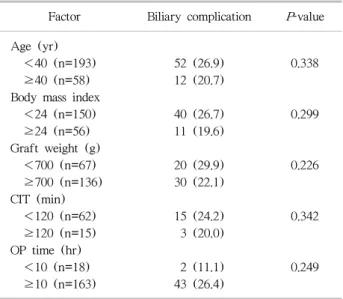
3. 수혜자와 공여자 요인에 따른 분석
공여자 관련 인자로 공여자의 연령, 체질량 지수(body mass index, BMI), 공여 간 용적, CIT 및 총 수술 시간과
Fig. 2. Annual biliary complication after living donor liver transplantation.
Table 3. Donor factors associated with biliary complications Factor Biliary complication P-value Age (yr)
<40 (n=193) 52 (26.9) 0.338
≥40 (n=58) 12 (20.7)
Body mass index
<24 (n=150) 40 (26.7) 0.299
≥24 (n=56) 11 (19.6)
Graft weight (g)
<700 (n=67) 20 (29.9) 0.226
≥700 (n=136) 30 (22.1)
CIT (min)
<120 (n=62) 15 (24.2) 0.342
≥120 (n=15) 3 (20.0)
OP time (hr)
<10 (n=18) 2 (11.1) 0.249
≥10 (n=163) 43 (26.4)
Data are presented as number (%).
Abbreviations: CIT, cold ischemic time; OP, operation.
Table 2. Comparison of biliary complication by operation period Until 2010
(n=184)
Since 2011
(n=68) P-value Biliary complication 56 (30.4) 9 (13.2) 0.006 Early developed
biliary complication (within 3 mo)
46 (25.0) 7 (10.3) 0.014
Data are presented as number (%).
Table 4. Recipient factors associated with biliary complications Factor Biliary complication P-value Age (yr)
<50 (n=90) 26 (28.9) 0.403
≥50 (n=162) 39 (24.1)
Body mass index
<24 (n=149) 43 (28.9) 0.195
≥24 (n=102) 22 (21.6)
Child-Pugh score
A (n=123) 30 (24.4) 0.763
B or C (n=115) 30 (26.1)
MELD core
<25 (n=207) 52 (25.1) 0.876
≥25 (n=38) 10 (26.3)
GRWR (%)
<1.0 4 (14.8) 0.189
≥1.0 47 (26.6)
Data are presented as number (%).
Abbreviations: MELD, Model for End-Stage Liver Disease; GRWR, graft versus recipient weight ratio.
수혜자와 관련된 연령, BMI, Child-Pugh 점수, MELD 점 수, GRWR 등은 담도 합병증의 발생에 유의한 차이를 보 이지 않았다(Tables 3, 4).
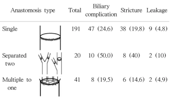
4. 문합 담도수와 방법에 따른 분석
공여 간에 두 개 이상의 담도구가 있을 시 문합 방식의 차이에 따른 담도 합병증을 비교 분석해 보았을 때 공여 간의 담도를 분리하여 수혜자 담도, 즉 담낭관 및 총담관 에 각각 문합한 경우(separated two)가 담도 합병증 비율이 50%로 가장 높았고, 단일 담도 문합(single to single)에서 는 24.6%였으며, 담도 성형술을 통해 공여 간의 여러 담도 구를 하나로 연결하여 수혜자의 단일 담도에 문합하는 경 우(multiple to one)에서 19.5%의 담도 합병증 발생률을 보 였다(Table 5).
고 찰
뇌사자의 장기 기증이 상대적으로 부족한 우리나라에 서는 전체 간이식의 3분의 2 이상이 생체 제공자에 의한
부분 간이식으로 이루어지고 있다(15,16). 그러나, 생체 간 이식은 공여자의 안정성을 고려해야 할 뿐만 아니라, 복잡 한 간 내 혈관과 담도를 박리한 후 재건해야 하기에 사체 간이식에 비해 수술 합병증 발생률이 높다(5). 특히, 수술 현미경의 도입과 혈관 문합술의 발달로 간이식 후 혈관 합 병증은 많이 감소하였으나, 사체 간이식의 17.3%에 비해
Table 5. Biliary complication according to the type of anastomosis
Anastomosis type Total Biliary
complication Stricture Leakage
Single 191 47 (24.6) 38 (19.8) 9 (4.8)
Separated two
20 10 (50.0) 8 (40) 2 (10)
Multiple to one
41 8 (19.5) 6 (14.6) 2 (4.9)
Data are presented as number (%).
28.7%의 높은 담도 합병증 발생률은 생체 간이식의 취약 점이라 할 수 있다(8,17). 생체 간이식의 담도 합병증이 높 은 이유는 사체 간이식에 비해 이식편의 담도 직경이 작 고, 다양한 해부학적 변이로 2개 이상의 담도 개구부가 나 타나는 경우가 흔하여 담도 문합이 어려운 경우가 많으며, 담관 절제 시 과도한 박리로 인해 허혈성 담도 협착이 발 생할 수 있기 때문이다(18).
초창기 생체 간이식 후 담도 재건은 담도 공장 문합술을 시행하였으나, 현재는 생리적인 담즙 배액 경로를 유지하 도록 수혜자 총담관과 이식편의 간 내 담도를 연결하는 담 도-담도 문합술이 일차적으로 고려되는 기본 술식으로 자 리 잡고 있다(19). 이러한 담도-담도 문합은 수혜자 수술 중 추가적인 장 절제가 없어 수술을 단순화 할 수 있고, 상행성 담도염의 위험성이 낮으며, 추후 내시경적 담도 접 근이 가능하다는 장점이 있으나, 이식편에서 여러 개의 담 도구가 나타나거나 4 mm 이하의 작은 담도에서는 수술 후 담도 합병증이 발생할 가능성이 매우 높다(20,21). 따라 서, 담도 합병증을 예방하기 위한 첫 번째 관문은 수술 전 간 내 담도의 정확한 해부학적인 변이를 파악하여 가능하 면 단일 개구부의 4 mm 이상의 담도를 얻을 수 있는 공여 자를 선택하는 것과 수술 중에 정확한 절단면을 얻는 것이 다(22,23). 특히, 자기공명영상은 수술 중 담관 조영술에 필적할 만큼 상세한 간 내 담도의 해부학적 구조를 수술 전에 확인할 수 있게 하여 적절한 공여자 선택과 수술 계 획 수립에 필수적인 역할을 하고 있다(24). 수술 중에는 대부분의 병원에서 전통적으로 담관 조영술을 시행하고 있다. 공여자 수술 중 담관 조영술은 최적의 담도 개구부 를 얻을 수 있도록 하여 담도 합병증을 통계적으로 유의하 게 감소시키는 것으로 알려져 있다(25,26). 그러나 수술 중 담관 조영술은 상당한 수술 시간을 소비하면서도, 자기공
명영상과는 달리 다양한 각도에서의 촬영이 불가능하여 3차원적인 절단면을 얻을 수 없는 단점이 있어 최근에는 담도 내 지침기를 삽입하여 수술 중 실시간으로 담도 절단 부위를 결정하여 담도 조영술을 생략하는 방법으로 담도 합병증을 줄일 수 있었다는 보고도 있다(27).
또한, 담도 문합부의 적절한 혈류 공급은 담도 협착을 예방하기 위한 중요한 요인으로, 공여자 수술 시에는 이식 편 담도 주위 혈관에 손상을 주지 않기 위하여 담도를 직 접 박리하지 않고 간문판에서 글리손지를 걸어 담도를 확 보하는 일명 간문판 루핑술을 통해 담도 합병증의 유의한 감소가 확인된 바 있다(13). 수혜자의 간 적출 시에도 간문 판 상부에서 담도 절단을 하여 수혜자 담도의 혈류를 보존 하고, 담도-담도 문합 시에 긴장이 없게 하면 담도 합병증 을 줄일 수 있다고 보고된 바 있다(28).
본 연구에 포함된 총 12년 동안 시행한 담도-담도 문합 의 합병증 발생률은 25.8%로 Duailibi와 Ribeiro(8)가 메타 분석을 통하여 보고한 28.7%와 유사하다. 이러한 담도 합 병증은 공여자의 연령, BMI, 수술 시간 및 CIT나 이식편의 중량과는 연관이 없었으며 수혜자 측에서도 통계학적으로 유의한 관련인자는 발견되지 않았다(Tables 3, 4). 담도 문 합 상황과 관련하여 단일 담도 간의 문합이나, 다수의 담 도구가 나오더라도 담도 성형술 등을 통해 하나로 연결하 여 문합할 수 있는 경우에는 비교적 담도 합병증의 발생이 낮으나 두 개 이상의 담도를 담낭관 및 총담관을 이용하여 따로 문합하는 경우에는 담도 합병증 발생률이 50%까지 상승하였다(Table 5). 수술 기간으로 보면, 본원에서 생체 간이식의 초창기라 볼 수 있는 2001년에서 2010년까지는 담도 합병증이 184명 중 56건(30.4%)으로 비교적 많이 발 생하였다. 이 기간은 생체 간이식 수술 전 혹은 수술 중 담도에 대한 평가 및 수술 술기의 표준화가 부족하여, Li 등(29)이 주장한 생체 간이식의 학습 곡선상에 있었다고 볼 수 있다. 그러나 이후 점차 수술 표준화가 형성되었고, 당시 빈번히 발생하는 담도 합병증을 예방하고자 본원에 서는 담도 주위 미세 혈관을 보존하고자 간문판 루핑술을 부분적으로 도입하였으며, 수혜자의 담도 절제 시에도 가 능한 간문부 상부에서 담도를 박리하여 총담관의 혈류를 보존하도록 하여 담도 합병증이 점차 감소하는 것을 확인 하였다(13). 그리고 2011년부터는 모든 생체 간이식 공여 자 수술 시에 간문판 루핑과 함께, 수술 중 담관 조영술을 생략하는 대신 공여자 간절제술 시에 담낭관을 통해 담도 에 지침기를 삽입하여 간문부 담도 합류 부위의 총담관에 서 최소 2 mm 우측 부위를 실시간으로 확인하여 절단함으 로써 이식편의 담도 구멍의 수를 최소화하면서 공여자 담도
협착을 예방하도록 하여 수술 후 담도 합병증이 평균 13.2%
로 통계적으로 유의하게 감소하였다(P=0.006) (Table 2)(27).
본 연구는 후향적 연구로서 수술 상황과 술기에 대한 세부 사항의 기술이 부정확하고, 술기의 능숙도를 평가할 수 없다는 제한점과 함께 담도 장문합과의 비교를 하지 못 한 단점이 있다. 그러나, 본 연구를 통해 국내 단일 기관에 서의 생체 간이식의 담도 합병증에 대한 결과를 보고하고 타 기관들과의 공유를 통해 추가적인 연구가 진행된다면 생체 간이식 환자에서의 담도 합병증의 개선에 대한 더욱 의미 있는 결과를 도출해 낼 수 있을 것으로 본다.
결 론
생체 간이식에서의 담도-담도 문합술의 경우 합병증 발 생률은 본 연구에서 12년 동안 25.8%였으며, 앞선 2001년 에서 2010년까지의 기간보다 2011년 이후에 합병증 발생 률이 유의하게 감소하였다. 담도 문합술에서의 여러 술식 들의 표준화와 담도 혈류 보존술식 등 수술 술기의 개선을 통해 생체 간이식에서의 담도 문합 후 합병증 발생률을 낮 출 수 있을 것이다.
REFERENCES
1) Murray KF, Carithers RL Jr: AASLD. AASLD practice guide- lines: evaluation of the patient for liver transplantation.
Hepatology 2005;41:1407-32.
2) Vitale A, Morales RR, Zanus G, Farinati F, Burra P, Angeli P, et al. Barcelona Clinic Liver Cancer staging and transplant survival benefit for patients with hepatocellular carcinoma:
a multicentre, cohort study. Lancet Oncol 2011;12:654-62.
3) de Villa VH, Lo CM, Chen CL. Ethics and rationale of liv- ing-donor liver transplantation in Asia. Transplantation 2003;75(3 Suppl):S2-5.
4) Kaido T, Uemoto S. Does living donation have advantages over deceased donation in liver transplantation? J Gastroenterol Hepatol 2010;25:1598-603.
5) Wan P, Yu X, Xia Q. Operative outcomes of adult living donor liver transplantation and deceased donor liver trans- plantation: a systematic review and meta-analysis. Liver Transpl 2014;20:425-36.
6) Wachs ME, Bak TE, Karrer FM, Everson GT, Shrestha R, Trouillot TE, et al. Adult living donor liver transplantation using a right hepatic lobe. Transplantation 1998;66:1313-6.
7) Akamatsu N, Sugawara Y, Hashimoto D. Biliary recon- struction, its complications and management of biliary complications after adult liver transplantation: a systematic
review of the incidence, risk factors and outcome. Transpl Int 2011;24:379-92.
8) Duailibi DF, Ribeiro MA Jr. Biliary complications following deceased and living donor liver transplantation: a review.
Transplant Proc 2010;42:517-20.
9) Zimmerman MA, Baker T, Goodrich NP, Freise C, Hong JC, Kumer S, et al. Development, management, and reso- lution of biliary complications after living and deceased donor liver transplantation: a report from the adult-to-adult living donor liver transplantation cohort study consortium.
Liver Transpl 2013;19:259-67.
10) Wilson BJ, Marsh JW, Makowka L, Stieber AC, Koneru B, Todo S, et al. Biliary tract complications in orthotopic adult liver transplantation. Am J Surg 1989;158:68-70.
11) Soejima Y, Taketomi A, Yoshizumi T, Uchiyama H, Harada N, Ijichi H, et al. Biliary strictures in living donor liver transplantation: incidence, management, and technical evolution. Liver Transpl 2006;12:979-86.
12) Rouch DA, Emond JC, Thistlethwaite JR Jr, Mayes JT, Broelsch CE. Choledochocholedochostomy without a T tube or internal stent in transplantation of the liver. Surg Gynecol Obstet 1990;170:239-44.
13) Ju MK, Choi GH, Joo DJ, Hur KH, Choi J, Kim MS, et al.
Use of the hilar plate looping technique for bile duct dis- section in living donor liver transplantation significantly reduces recipient biliary complications. Transplant Proc 2010;42:4161-3.
14) Simoes P, Kesar V, Ahmad J. Spectrum of biliary complica- tions following live donor liver transplantation. World J Hepatol 2015;7:1856-65.
15) Lee SG. Current status of liver transplantation in Korea.
Korean J Gastroenterol 2005;46:75-83. (이승규, 국내 간이식 의 현황과 전망. 대한소화기학회지 2005;46:75-83.) 16) Korean Network for Organ Sharing (KONOS). Statistics of
organ transplantation [Internet]. Seoul: KONOS; c2014 [cited 2016 Mar 2]. Available from: http://www.konos.go.kr.
17) Yang Y, Yan LN, Zhao JC, Ma YK, Huang B, Li B, et al.
Microsurgical reconstruction of hepatic artery in A-A LDLT:
124 consecutive cases without HAT. World J Gastroenterol 2010;16:2682-8.
18) Seehofer D, Eurich D, Veltzke-Schlieker W, Neuhaus P.
Biliary complications after liver transplantation: old prob- lems and new challenges. Am J Transplant 2013;13:253-65.
19) Sugawara Y, Makuuchi M. Living donor liver trans- plantation: present status and recent advances. Br Med Bull 2005;75-76:15-28.
20) Yazumi S, Chiba T. Biliary complications after a right-lobe living donor liver transplantation. J Gastroenterol 2005;
40:861-5.
21) Hwang S, Lee SG, Sung KB, Park KM, Kim KH, Ahn CS, et al. Long-term incidence, risk factors, and management of biliary complications after adult living donor liver transplantation. Liver Transpl 2006;12:831-8.
22) Ayuso JR, Ayuso C, Bombuy E, De Juan C, Llovet JM, De Caralt TM, et al. Preoperative evaluation of biliary anatomy in adult live liver donors with volumetric mangafodipir trisodium enhanced magnetic resonance cholangiography.
Liver Transpl 2004;10:1391-7.
23) Ju MK, Kim MS, Choi GH, Chang HK, Ahn HJ, Kim YS, et al. The efficacy of pre-transplant radiologic evaluation for graft volume and anatomy in living donor liver transplantation. J Korean Soc Transplant 2007;21:128-34.
(주만기, 김명수, 최기홍, 장혜경, 안형준, 김유선, 등. 생체 간이식 공여자의 수술 전 이식간 용적과 해부학적 구조에 대한 영상학적 검사의 유용성. 대한이식학회지 2007;21:128-34.)
24) Park S, Choi GS, Jung J, Cho G, Shin E, Lim C, et al. Clinical efficacy of pretransplant magnetic resonance cholangiog- raphy of donor for living donor liver transplantation. J Korean Soc Transplant 2010;24:311-5. (박승완, 김형철, 조규 석, 송옥평, 신응진, 임철완, 등. 생체간이식 수술에서 이식 전
제공자 자기공명담관조영술의 임상적 유용성. 대한이식학회지 2010;24:311-5.)
25) Taketomi A, Morita K, Toshima T, Takeishi K, Kayashima H, Ninomiya M, et al. Living donor hepatectomies with procedures to prevent biliary complications. J Am Coll Surg 2010;211:456-64.
26) Takatsuki M, Eguchi S, Yamanouchi K, Hidaka M, Soyama A, Kanematsu T. Technical refinements of bile duct division in living donor liver surgery. J Hepatobiliary Pancreat Sci 2011;18:170-5.
27) Testa G, Malago M, Porubsky M, Marinov M, Sankary H, Oberholzer J, et al. Hilar early division of the hepatic duct in living donor right hepatectomy: the probe-and-clamp technique. Liver Transpl 2006;12:1337-41.
28) Lee KW, Joh JW, Kim SJ, Choi SH, Heo JS, Lee HH, et al. High hilar dissection: new technique to reduce biliary complication in living donor liver transplantation. Liver Transpl 2004;10:1158-62.
29) Li C, Mi K, Wen T, Yan L, Li B, Yang J, et al. A learning curve for living donor liver transplantation. Dig Liver Dis 2012;44:597-602.