Metaplastic carcinoma is a rare form of breast carcino- ma. It accounts for less than 5% of breast cancers (1-3), and it may have a poorer prognosis than most other breast malignancies (4, 5). Some authors have reported a metaplastic carcinoma of the breast with the mammo- graphic and sonographic features (3, 6), however, typi- cal magnetic resonance imaging (MRI) findings are un- known. The MRI findings of a metaplastic carcinoma of the breast were evaluated in this study. To our knowl- edge, this is the first report describing the MRI findings of a metaplastic carcinoma of the breast.
Case Report
A 44-year-old woman noticed a palpable mass in the
right breast for 2 months. On a physical examination, a large, hard and fixed mass was noticed in the right breast. The mammographic images showed an ovoid shaped large dense mass with an ill-defined margin at the upper portion of the right breast with enlarged and dense ipsilateral axillary lymph nodes; there was also a loss of the fatty hilum (Fig. 1). An ultrasound study with a 5-12.5 MHz probe revealed a microlobulated hetero- geneous hypoechoic mass which had central multifocal irregular anechoic areas (Fig. 2). A dynamic MRI was performed using a 1.5 T unit (Magnetom Vision Plus, Siemens, Erlangen, Germany). The patient was exam- ined in the prone position using a double breast coil.
The sequences were as follows: axial nonenhanced spin- echo T1 and T2-weighted image, a T1-weighted three-di- mensional fast low-angle shot (3D FLASH) sequence be- fore and six times after the contrast injection. Axial T1- weighted images were obtained using a 3D FLASH se- quence (TR=16 ms, TE=4 ms, 20°flip angle, 3-mm slice thickness with no slice gap, 224×256 in-plane ma- trix) that covered the whole breast. The field of view was 340 mm. For dynamic contrast enhancement, gadopentetate dimeglumine (0.15 mmol per Kg of body weight) was injected in a bolus pattern for a duration of
J Korean Radiol Soc 2004;50:277-280
─ 277 ─
MR Findings of Metaplastic Carcinoma of the Breast: Case Report1
Soo Ah Im, M.D., Hak Hee Kim, M.D.2, Song Yee Han, M.D., Youn Soo Lee, M.D.3
We report the dynamic magnetic resonance imaging findings (MRI) of a metaplastic carcinoma of the breast. A 44-year-old woman presented with a 2-month history of a mass in the right breast. The tumor showed a central irregular necrosis that was de- picted upon low signal intensity on a T1-weighted image and high signal intensity on a T2-weighted image. The periphery of the tumor showed strong enhancement on a contrast enhanced T1-weighted image. The time-signal intensity curve revealed an ear- ly strong contrast enhancement and a delayed washout pattern.
Index words :Metaplastic carcinoma Breast
Magnetic resonance (MR), image display
1Department of Radiology, College of Medicine, The Catholic University of Korea
2Department of Radiology, Asan Medical Center, University of Ulsan College of Medicine
3Department of Clinical Pathology, College of Medicine, The Catholic University of Korea
Received August 14, 2003 ; Accepted January 10, 2004
Address reprint requests to : Hak Hee Kim, M.D., Department of Radiology, Asan Medical Center, University of Ulsan College of Medicine, 388-1 Poongnap-dong, Songpa-gu, Seoul 138-040, Korea.
Tel. 82-2-3010-4390 Fax. 82-2-476-0090 E-mail: [email protected]
10 s. Post-contrast T1-weighted images were obtained six times with no delay at the same slice position and lo- cation. Acquisition time for each T1-weighted 3D FLASH sequence was about 50 s and total acquisition time was about 350 s. The images were postprocessed by the subtraction method. The axial nonenhanced spin- echo MR images demonstrated a tumor, which had a central irregular low signal intensity surrounded by an intermediate signal intensity on the T1-weighted image (Fig. 3A), and a high signal intensity surrounded by pe- ripheral intermediate signal intensity on the T2-weight- ed image (Fig. 3B). The subtraction images revealed the lesion to be an intensive enhancement at the peripheral solid portion (Fig. 3C). The time-signal intensity curve revealed an early strong contrast enhancement and a de- layed washout pattern (Fig. 3D).
Using 14-gauge US-guided core biopsy, the histopatho- logic diagnosis was a squamous type of metaplastic car- cinoma. A modified radical mastectomy with an axillary node dissection was performed. The tumor measured up to 4.5×3.5×3.5 cm, and an axial cut surface of it showed gross prominent necrotic areas throughout the mass (Fig. 4). The areas of high signal intensity on the T2-weighted images were well correlated with patholog- ic necrosis. The axillary lymph nodes (3/21) were in- volved. There has been no recurrence or any distant
metastasis in the 2 years since the treatment.
Discussion
Breast carcinomas that undergo metaplastic changes are rare and they occur in less than 5% of the breast car- cinomas. Tumors are considered to be metaplastic carci- nomas if the metaplastic component constitutes at least 25% of the tumor and if it is merged into the foci of an infiltrative ductal carcinoma that is not otherwise speci- fied (3). Metaplastic foci have squamous cells; spindle cells; both squamous and spindle cells; or heterologous elements, including muscle, cartilage, or bone (3).
Several reports have described a metaplastic carcino- ma of the breast, and these reports concentrate mainly the clinicopathologic, mammographic and sonographic features (1-7). We have looked for reports regarding the MRI findings for this tumor and they are not to be found in the medical literature. The mammographic appear- ance of a metaplastic breast carcinoma has been de- scribed as a well-circumscribed mass or an irregular or spiculated mass (5), an ill-defined, round mass with an associated architectural distortion (6), a predominantly circumscribed, noncalcified mass with a spiculated por- tion (3, 7) or a rapidly growing, palpable mass that has a high density (8). Patterson et al. (3) reported that the
Soo Ah Im, et al: MR Findings of Metaplastic Carcinoma of the Breast
─ 278 ─ Fig. 1. Mediolateral oblique mammogram of the right breast shows an ill-defined ovoid mass (arrows) in the upper portion of the right breast. There are noted the enlarged and dense ip- silateral axillary lymph nodes with the loss of the fatty hilum (arrowheads).
Fig. 2. Sonography of the right breast reveals a microlobulated heterogeneously hypoechoic mass with the central irregular anechoic necrotic areas.
metaplastic portions of these tumors were associated with a circumscribed or lobular appearance on mammo- grams, whereas the invasive ductal carcinoma portions, if not otherwise specified, had irregular margins on mammograms. On sonography, the mass showing an in- homogeneous echo pattern with solid and cystic compo- nents was confirmed to relate it with the hemorrhagic or cystic necrosis finding upon pathology (6, 8). These find- ings are similar to those in our case. In addition, a histopathologic examination of this tumor at the same section on MRI demonstrated a good radiological and pathological correlation. The peripheral solid portion showed intensive and fast contrast enhancement with a delayed washout pattern that is often observed in carci- nomas of the breast (9). This peripheral pattern is consis- tent with typical invasive ductal carcinoma upon
J Korean Radiol Soc 2004;50:277-280
─ 279 ─
A B
C D
Fig. 3A. Axial nonenhanced, T1-weighted MR image demonstrates about a 5 cm-sized tumor (arrows) with a central irregular low signal intensity surrounded by an intermediate signal intensity on the T1-weighted spin echo sequence.
B. On T2-weighted spin echo image, the tumor shows a central high signal intensity surrounded by peripheral intermediate signal intensity.
C. A Dynamic 3D FLASH subtraction image shows a strong-enhanced high signal intensity at the periphery of the tumor.
D. The time-signal intensity curve reveals an early strong contrast enhancement and delayed wash out.
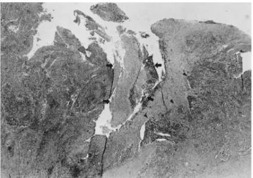
Fig. 4. Microphotograph shows an extensive necrosis (arrows) in the squamous metaplastic area (arrowheads) (HE, ×20).
histopathology. The central necrotic areas of the mass corresponded to the squamous metaplastic area upon histopathology (6, 8).
We experienced that MRI findings of metaplastic car- cinoma of the breast were well correlated with the sono- graphic findings. These MRI findings demonstrated the central necrotic portion, the peripheral solid portion and the depicted characteristic signals on spin echo T1 and T2 weighted images and 3D dynamic enhanced images.
References
1. Kline TS, Kline IK. Metaplastic carcinoma of the breast-diagnosis by aspiration biopsy cytology: report of two cases and literature re- view. Diagn Cytopathol 1990;6:63-67
2. Rosen PP, Oberman HA. Atlas of tumor pathology: tumors of the mammary gland. Washington, DC: Armed Forces Institute of Pathology, 1993:194-203
3. Patterson SK, Tworek JA, Roubidoux MA, Helvie MA, Oberman HA. Metaplastic carcinoma of the breast: mammographic appear- ance with pathologic correlation. AJR Am J Roentgenol 1997;169:
709-712
4. Taylor DB, Adamson R, Minchin DE, Reading L. Carcinoma of the breast with sarcomatous metaplasia. Australas Radiol 1994;38:262- 264
5. Brenner RJ, Turner RR, Schiller V, Arndt RD, Giuliano A.
Metaplastic carcinoma of the breast: report of three cases. Cancer 1998;82:1082-1087
6. Park JM, Han BK, Moon WK, Choe YH, Ahn SH, Gong G.
Metaplastic carcinoma of the breast: mammographic and sono- graphic findings. J Clin Ultrasound 2000;28:179-186
7. Oberman HA. Metaplastic carcinoma of the breast. A clinico- pathological study of 29 patients. Am J Surg Pathol 1987;11:918-929 8. Gu¨nhan-Bilgen I, Memis¸A, U¨stu¨n EE, Zekioglu O, Ozdemir N.
Metaplastic carcinoma of the breast: clinical, mammographic, and sonographic findings with histopathologic correlation. AJR Am J Roentgenol 2002;178:1421-1425
9. Heywang-Ko¨brunner SH, Haustein J, Pohl C, et al. Contrast-en- hanced MR imaging of the breast: comparison of two different doses of gadopentetate dimeglumine. Radiology 1994;191:639-646 Soo Ah Im, et al: MR Findings of Metaplastic Carcinoma of the Breast
─ 280 ─
대한영상의학회지 2004;50:277-280
이형성 유방암의 자기공명영상소견: 증례 보고1
1가톨릭대학교 의과대학 진단방사선과학교실
2울산대학교 의과대학 서울아산병원 진단방사선과학교실
3가톨릭대학교 의과대학 해부병리학교실
임수아・김학희2・한송이・이연수3
저자들은 이형성 유방암의 자기공명영상소견을 보고하고자 한다.
44세 여자환자가 우측유방에 2개월 전부터 종괴가 만져져서 내원하였다. 유방 자기공명영상검사에서 종양 내부에 불규칙한 모양의 T1 강조영상에서 저신호강도, T2 강조영상에서 고신호강도를 보이는 괴사성 혹은 낭성변형을 나타 내었으며 종양변연부는 조영증강 후 영상에서 강하게 조영증강이 되는 소견을 나타내었다. 시간-신호강도 커브는 조 기에 강하게 조영 증강이 되고 서서히 조영제가 빠져나가는 양상을 보여 악성임을 시사하였다.