ABSTRACT
Cardiac magnetic resonance (CMR) imaging provides accurate anatomic information and advanced soft contrast, making it the reference standard for assessing cardiac volumes and systolic function. In this review, we summarize the recent advances in CMR sequences. New technical development has widened the use of CMR imaging beyond the simple characterization of myocardial scars and assessment of contractility. These novel CMR sequences offer comprehensive assessments of coronary plaque characterization, myocardial fiber orientation, and even metabolic activity, and they can be readily applied in clinical settings. CMR imaging is able to provide new insights into understanding the pathophysiologic process of underlying cardiac disease, and it can help physicians choose the best treatment strategies. Although several limitations, including the high cost and time- consuming process, have limited the widespread clinical use of CMR imaging so far, recent advances in software and hardware technologies have made the future more promising.
Keywords: Cardiology; Magnetic resonance imaging; Cardiac magnetic resonance
INTRODUCTION
Cardiac magnetic resonance (CMR) imaging provides accurate anatomic information along with advanced soft contrast, which enables superior discrimination of lesions compared to other imaging modalities. Currently, CMR imaging is considered the reference standard for quantification of the cardiac chamber size and systolic function of the heart.
1)Further, tissue characterization techniques including late gadolinium enhancement (LGE), T1 mapping, T2 mapping, and T2-star mapping provide insight into the pathophysiological changes of the myocardium.
2)3)The drawbacks for the clinical application of CMR imaging have been the long acquisition time, relatively low spatial resolution, and high costs. However, new technical advances in hardware and software components accelerated the image acquisition time, and the introduction of 3T magnetic resonance imaging (MRI) has further improved the spatial resolution and diagnostic accuracy of CMR imaging in the assessment of the myocardium
Review Article
Received: Jul 25, 2018 Revised: Sep 25, 2018 Accepted: Oct 23, 2018 Correspondence to Hyuk-Jae Chang, MD, PhD Division of Cardiology, Severance
Cardiovascular Hospital, Yonsei-Cedars-Sinai Integrative Cardiovascular Imaging Research Center, Yonsei University College of Medicine, Yonsei University Health System, 50-1, Yonsei-ro, Seodaemun-gu, Seoul 03722, Korea.
E-mail: [email protected]
Copyright © 2019. The Korean Society of Cardiology
This is an Open Access article distributed under the terms of the Creative Commons Attribution Non-Commercial License (https://
creativecommons.org/licenses/by-nc/4.0) which permits unrestricted noncommercial use, distribution, and reproduction in any medium, provided the original work is properly cited.
ORCID iDs Yibin Xie
https://orcid.org/0000-0002-0333-567X Hyuk-Jae Chang
https://orcid.org/0000-0002-6139-7545 Funding
This study was supported by Leading Foreign Research Institute Recruitment Program through the National Research Foundation of Korea funded by the Ministry of Science, ICT &
Future Planning (grant No. 2012027176).
Sang-Eun Lee, MD, PhD
1,2,3, Christopher Nguyen, PhD
3,4,5, Yibin Xie , PhD
3, Zixin Deng, PhD
3, Zhengwei Zhou, PhD
3, Debiao Li, PhD
3, and
Hyuk-Jae Chang , MD, PhD
1,21
Division of Cardiology, Severance Cardiovascular Hospital, Yonsei University College of Medicine, Yonsei University Health System, Seoul, Korea
2
Integrative Cardiovascular Imaging Center, Yonsei University Health System, Seoul, Korea
3
Biomedical Imaging Research Institute, Cedars-Sinai Medical Center, Los Angeles, CA, USA
4
Cardiovascular Research Center, Massachusetts General Hospital, Charlestown, MA, USA
5
Harvard Medical School, Boston, MA, USA
Recent Advances in Cardiac Magnetic
Resonance Imaging
Conflict of Interest
The authors have no financial conflicts of interest.
Author Contributions
Conceptualization: Chang HJ, Lee SE;
Supervision: Chang HJ, Li D; Writing - original draft: Lee SE; Writing - review & editing: Chang HJ, Lee SE, Nguyen C, Xie Y, Deng Z, Zhou Z, Li D.
and coronary arteries.
1)4)5)Consequently, CMR imaging has become one of the routine clinical imaging investigations used for assessing patients with various cardiac diseases.
In this review, we summarize the recent advances in CMR imaging techniques that can readily be applied in clinical practice.
IMAGING AND CHARACTERIZATION OF THE MYOCARDIUM
T1 and T2 mapping
CMR imaging is optimized for characterization of the myocardium through its excellent soft-tissue contrast and various different imaging parameters, and it can provide insight into disease etiologies and the extent and stage of the diseases.
6)7)Recently, more sophisticated CMR imaging techniques that can provide myocardial tissue characterization beyond the presence of myocardial fibrosis have been introduced.
8)Native T1 mapping techniques and extracellular volume (ECV) fraction quantification can go beyond LGE imaging and identify more diffuse interstitial fibrosis.
9)10)Native T1 value is a composite signal of myocytes and ECV, and is influenced by the filed strength, with higher values at 3T than at 1.5T.
11)The native T1 mapping does not require gadolinium-based contrast agents and hence is feasible in patients with renal impairment.
12)The native (non-contrast) T1 mapping can detect pathophysiological process including diffuse interstitial fibrosis, myocardial edema, protein deposition, and other T1-altering substances including iron and lipid.
13)In conditions that causes myocardial edema (e.g. acute myocardial infarction) or an increase of interstitial space (e.g. interstitial fibrosis caused by infarction or cardiomyopathies), the native T1 value increases. In case of lipid (e.g. Anderson-Fabry disease) or iron overload, the native T1 value decreases. Therefore, changes in myocardial T1 reflects various cardiac and systemic conditions.
12)ECV expansion is a main feature of heart failure, and ECV is a marker of myocardial remodeling.
14)For the calculation of the ECV fraction, contrast-enhanced T1 mapping is used.
12)Gadolinium-based contrast agents are distributed throughout the interstitial space and shorten T1 relaxation times proportional to the local concentration of gadolinium.
Therefore, myocardial segments with fibrosis and scar exhibit shorter T1 relaxation times after the contrast administration. Calculation of the ECV requires measurement of myocardial and blood T1 before and after the contrast enhancement. In healthy individuals, normal ECV values of 25.3±3.5% in 1.5T have been reported.
15)An increase in ECV is mostly caused by excessive collagen deposition and is thus represents myocardial fibrosis, and ECV reduces in the presence of thrombus or fat/lipomatous metaplasia.
Both native T1 mapping and ECV can help the differential diagnosis of patients with chest
pain, and also can provide information for the distinction of acute from chronic conditions.
9)Native T1 values of chronic myocardial infarction is lower and less extensive compared with
acute stage, and the ECV of chronic myocardial infarction is significantly elevated than
normal but lower than acute conditions. In recent studies, native T1 was also an important
predictor of clinical outcomes in patients with CAD.
10)14)Native T1 and ECV can also be
applied in the assessment of non-ischemic cardiomyopathies. Native T1 was mildly elevated
in patients with hypertrophic cardiomyopathy and was highest in segments that subsequently
demonstrated LGE.
16)Native T1 was able to accurately discriminate hypertrophic cardiomyopathy from hypertensive heart disease,
17)and was better in discriminating cardiomyopathies from the normal hearts.
18)The T2 mapping can accurately and reliably detect areas of myocardial edema, a hallmark of acute inflammation,
8)19)and has been shown to be useful in acute myocardial infarction,
20)myocarditis,
21)stress cardiomyopathy,
21)and cardiac allograft rejection.
22)For this, conventional T2-weighted short-τ inversion recovery (T2-STIR) has been most commonly used and helpful in differentiating acute infarction from chronic infarction.
23)However, as it depends on regional differences in myocardial signal intensity, its interpretation is qualitative and subjective.
24)25)T2 mapping is more reproducible than T2-STIR,
26)and it can overcome this limitation of classic T2-STIR.
27)28)T2 values can be directly quantified in vivo, as a surrogate for myocardial water content.
29)Normal T2 values acquired with steady-state free precession MRI have been reported to be 52.18±3.4 ms at 1.5T and 45.1 ms at 3T.
30)31)Studies have shown that T2 values are increased in the state of advanced heart failure, representing decompensation and decreases after treatment.
28)In a recent study, T2 mapping was superior compared with T1 mapping and ECV for the assessment of the myocarditis in patients with recent-onset heart failure with reduced ejection fraction.
27)Recently, a novel MRI sequence named CMR multitasking that enables simultaneous T1 mapping, T1/T2 mapping, and time-resolved T1 mapping and hence markedly reduces the total acquisition time has been introduced.
32)CMR multitasking uses a continuous acquisition approach, which conceptualizes the physiological motions and other dynamic processes as multiple time dimensions, making efficient quantitative CMR imaging possible without the use of electrocardiogram gating and breath holds.
Late gadolinium enhancement
The LGE technique is one of the most established methods in characterization of the myocardium using CMR imaging so far.
2)7)LGE is a T1-weighted technique that can detect and quantify areas of myocardial injury and cardiac infiltration in various cardiac diseases, and it has become the gold standard method for detecting myocardial scars. LGE uses the pattern and velocity of distribution and accumulation of the gadolinium-chelate contrast agent within the extracellular compartment of the myocardium.
Because the LGE pattern follows the pathophysiological process of the underlying disease, it helps identify the etiology of heart failure. In ischemic heart disease, the injury starts in the subendocardium and progresses into the transmural myocardium as ischemia continues.
33)Thus, the LGE pattern in patients with CAD starts as subendocardial and extends
transmurally, whereas that in patients with dilated cardiomyopathy demonstrates a linear mid-wall pattern in the interventricular septum.
33)34)Importantly, the presence and extent of LGE are significant prognostic indicators associated
with an increased risk of future cardiac events, regardless of the underlying etiology or
pattern of fibrosis.
35-37)Further, LGE can assess viability of the myocardium in ischemic and
non-ischemic cardiac disease; thereby, it has the potential to guide the clinical decision.
37-40)The main limitation of LGE is that it is a marker of irreversible replacement fibrosis, the final
pathophysiologic process of all cardiac diseases, and therefore is limited in assessing the
early changes of cardiac diseases.
41)42)Further, LGE is mostly useful in cardiac diseases which
results in regional differences within the myocardium.
37)Assessment of myocardial fiber orientation
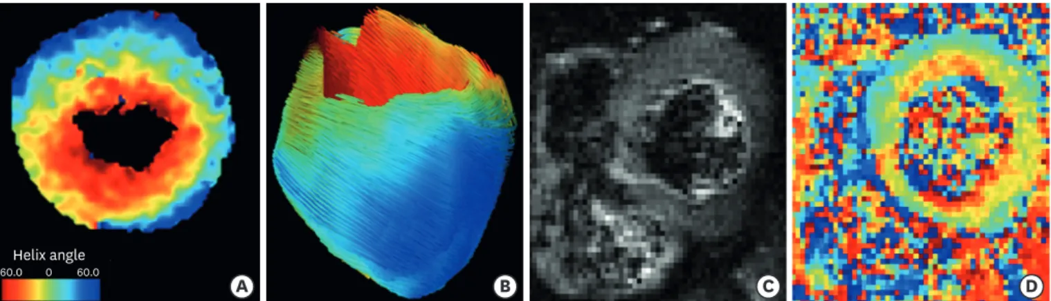
CMR imaging can characterize the property of the myocardium and directly visualize the myocardial fiber architecture using diffusion tensor imaging (DTI).
43)44)Previous preclinical studies have demonstrated the ability of DTI to evaluate therapeutic responses of the myocardial microstructure, including stem cell or surgical treatment, in animal and human models.
45-49)Recently, technical advances have made the in vivo acquisition of DTI using clinical MR scanners possible in large animals and humans.
50-52)The DTI technique has been evaluated most frequently in hypertrophic cardiomyopathy and myocardial infarction.
45)53)Novel DTI was also feasible using a 3T clinical scanner.
54)The clinical application of DTI has been limited because the ability of DTI to delineate the microstructure of the myocardium has been histologically validated only using destructive 2D methods that perturb the original myocardial fiber orientation.
55)56)The reliability of DTI was recently validated in normal and ischemic heart failure mice models, and the myocardial fiber orientation calculated by DTI was directly compared with histology using a non-destructive 3D method with the tissue-clearing technique.
57)The correlations were significant in the normal and diseased models, showing the capability and accuracy of DTI for mapping the myocardial microstructure.
58)Based on these observations, the clinical application of DTI using the clinical MRI system for characterizing the myocardial fiber architecture seems possible, and it can provide additional information to conventional viability characterizations (Figure 1).
Assessment of metabolic activity of the myocardium
To maintain its continuous contraction, the heart has a high rate of adenosine triphosphate (ATP) production and utilization.
59)Previous studies have shown that cardiac dysfunction is associated with decreased myocardial ATP.
60)61)Therefore, characterization of cardiac metabolism can be potentially useful in assessing disease progression and understanding the pathophysiology,
59)and the direct measurement of metabolites involved in ATP synthesis can be used to assess myocardial ischemia, oxygenation, remodeling, and viability.
62)The chemical exchange saturation transfer (CEST) technique has been developed to quantify metabolites and macromolecules.
63-65)In a recent study, CEST was able to map the creatine distribution within the myocardium to evaluate the metabolic activity in an animal model,
66)and an optimized in vivo cardiac CEST sequence that maps free creatine in myocardial tissue, with a markedly reduced scan time by 10-fold and improved motion registration and signal
A B C D
Helix angle
−60.0 0 60.0