Recent Update of Advanced Imaging for Diagnosis of Cardiac Sarcoidosis:
Based on the Findings of Cardiac Magnetic Resonance Imaging and Positron Emission Tomography
This is an Open Access article distributed under the terms of the Creative Commons Attribution Non-Commercial License (http://creativecommons.org/licenses/
by-nc/4.0/) which permits unrestricted non-commercial use, distribution, and reproduction in any medium, provided the original work is properly cited.
Received: December 27, 2018 Revised: March 5, 2019 Accepted: March 26, 2019 Correspondence to:
Eun Ju Chun, M.D., Ph.D.
Department of Radiology, Seoul National University Bundang Hospital, 82 Gumi-ro 173beon- gil Bundang-gu, Seongnam-si, Gyeonggi-do 13620, Korea.
Tel. +82-31-787-7618 Fax. +82-31-787-4011 E-mail: [email protected]
Copyright © 2019 Korean Society of Magnetic Resonance in Medicine (KSMRM)
Review Article
Sarcoidosis is a multisystem disease characterized by noncaseating granulomas.
Cardiac involvement is known to have poor prognosis because it can manifest as a serious condition such as the conduction abnormality, heart failure, ventricular arrhythmia, or sudden cardiac death. Although early diagnosis and early treatment is critical to improve patient prognosis, the diagnosis of CS is challenging in most cases. Diagnosis usually relies on endomyocardial biopsy (EMB), but its diagnostic yield is low due to the incidence of patchy myocardial involvement. Guidelines for the diagnosis of CS recommend a combination of clinical, electrocardiographic, and imaging findings from various modalities, if EMB cannot confirm the diagnosis.
Especially, the role of advanced imaging such as cardiac magnetic resonance (CMR) imaging and positron emission tomography (PET), has shown to be important not only for the diagnosis, but also for monitoring treatment response and prognostication.
CMR can evaluate cardiac function and fibrotic scar with good specificity. Late gadolinium enhancement (LGE) in CMR shows a distinctive enhancement pattern for each disease, which may be useful for differential diagnosis of CS from other similar diseases. Effectively, T1 or T2 mapping techniques can be also used for early recognition of CS. In the meantime, PET can detect and quantify metabolic activity and can be used to monitor treatment response. Recently, the use of a hybrid CMR-PET has introduced to allow identify patients with active CS with excellent co-localization and better diagnostic accuracy than CMR or PET alone.
However, CS may show various findings with a wide spectrum, therefore, radiologists should consider the possible differential diagnosis of CS including myocarditis, dilated cardiomyopathy (DCM), hypertrophic cardiomyopathy, amyloidosis, and arrhythmogenic right ventricular cardiomyopathy. Radiologists should recognize the differences in various diseases that show the characteristics of mimicking CS, and try to get an accurate diagnosis of CS.
Keywords: Cardiac sarcoidosis; Magnetic resonance imaging;
Positron-emission tomography
Suyon Chang1, Won Woo Lee2, Eun Ju Chun3
1Department of Radiology, Seoul St. Mary's Hospital, College of Medicine, The Catholic University of Korea, Seoul, Korea
2Department of Nuclear Medicine, Seoul National University Bundang Hospital, Seongnam-si, Korea
3Department of Radiology, Seoul National University Bundang Hospital, Seongnam-si, Korea
INTRODUCTION
Sarcoidosis is a systemic disease with unknown etiology that is characterized by the presence of noncaseating, nonnecrotic granulomas in the involved organs (1).
Although the most involved organ is related to the lung, other susceptible sites include the heart, skin, eyes, reticuloendothelial system, kidneys, and central nervous systems (1). Cardiac sarcoidosis (CS) occurs with an incidence of 5-39% depending on detection method (2). Although clinical manifestations of CS have a wide range from no symptom to sudden cardiac death, CS is considered to have a poor prognosis (3). However, recent studies demonstrated early diagnosis and early initiation of corticosteroid therapy seem to improve the prognosis of CS (4, 5). Therefore, there is a need for accurate tool for clinicians to predict CS. Nevertheless, the diagnosis of CS is challenging due to the low yield of endomyocardial biopsy (EMB) (6), and no gold standard diagnostic criterion exists until now. This review will demonstrate the diagnosis of CS with a focus on advanced cardiovascular imaging, including cardiac magnetic resonance (CMR) and positron emission tomography (PET), and also summarize the differential
diagnosis of other CS mimics.
Epidemiology and Histopathologic Conditions
Sarcoidosis is a worldwide disease, with a prevalence of about 4.7 to 64 in 100,000; the highest rates are reported in northern Europeans and in the patient demographic of African Americans, particularly in women (7). Most patients are aged 25-45 years, and a second incidence peak occurs in women older than 50 years in Europe and Japan (8).
Elderly-onset sarcoidosis is much more common in women, and shows higher rates of change in general health and extrapulmonary manifestations (9).
Although clinical findings of cardiac involvement have been reported in 5-10% of patients with sarcoidosis, autopsy has revealed CS ranging from 20-30% (10, 11).
However, recent study reported 40% of patients with sarcoidosis had noted the presence of CS on advanced imaging, such as with CMR or PET (12). In this case, it is noted that with the continual improvement of technique of imaging, the prevalence of CS seems to be increased.
The histologic hallmark of sarcoidosis is the incidence
Table 1. JMHW 2006 Revised Guidelines for Diagnosis of CS Histologic diagnosis group
CS is confirmed when EMB specimens show noncaseating epithelioid cell granulomas with histologic clinical diagnosis of extracardiac sarcoidosis
Clinical diagnosis group
Although EMB specimens do not show noncaseating epithelioid cell granulomas, extracardiac sarcoidosis was diagnosed histologically or clinically and satisfies the following criteria*
Major criteria Advanced AV block
Basal thinning of the interventricular septum Positive gallium uptake in the heart Depressed LVEF (< 50%)
Minor criteria
Abnormal ECG findings: ventricular arrhythmias (VT, multifocal or frequent PVC), complete RBBB, axis deviation, or abnormal Q wave Abnormal echocardiographic findings: regional abnormal wall motion or morphologic abnormality (ventricular aneurysm, wall thickening)
Nuclear medicine findings: perfusion defect detected with thallium 201 (201Th) or technetium 99m (99mTc) myocardial scintigraphy MR imaging findings: delayed (late) gadolinium-induced enhancement of myocardium EMB findings: interstitial fibrosis or monocyte infiltration of more than moderate grade
*1) More than two major criteria are satisfied or one major and 2) more than two minor criteria are satisfied.
Reprinted with permission from John Wiley and Sons (20).
AV = atrioventricular; CS = cardiac sarcoidosis; EMB = endomyocardial biopsy; LVEF = left ventricular ejection fraction; MR = magnetic resonance; PVC = premature ventricular contraction; RBBB = right bundle branch block; VT = ventricular tachycardia
of noncaseating epithelioid granulomas composed of macrophages and T lymphocytes, which differs from Mycobacterium tuberculosis infection in that sarcoidosis has the effect of minimal necrosis (13). Granulomas in sarcoidosis are thought to form around and isolate poorly degraded antigens as a means of preventing antigen dissemination and further tissue damage. If the antigen persists, it has been assumed that the disease progresses by inducing a chronic immune response. Because granuloma is very rarely confirmed by EMB, it is found that the histologic findings of granulomas are not specific for diagnosis of CS, but it could support diagnosing or excluding other diseases (14). Therefore, CS is conventionally diagnosed by the appropriate combination of clinical, physiologic, and advanced imaging findings.
In an autopsy study for CS, gross scars due to cardiac involvement of sarcoidosis, were most commonly found in the interventricular septum (31.5%), followed by the inferior wall, anterior left ventricle, right ventricle and lateral left ventricle (15). In the ventricle, subepicardial scars were most common, followed by midmyocardial and in the subendocardial area (15). Atrial and valvular involvements were rarely occurred.
Clinical Manifestations of CS
Although clinical features of CS depend on the location, extent, and activity of the disease, the main clinically critical manifestations are shown to be conduction abnormalities; ventricular arrhythmias which can lead to sudden death; and heart failure in the patient (16).
Conduction abnormalities (23-30%), which range from second to third degree atrioventricular (AV) block and bundle branch block, occur due to an involvement of the basal interventricular septum (17). Ventricular tachycardia (VT) has been reported in 23% of CS patients, when re- entry circuit involves the fibrous scar (17). Heart failure occurs with systolic or diastolic dysfunction, and it is often associated with extensive cardiomyopathy (18).
Diagnostic Criteria
The most widely adopted set of clinical criteria for guiding diagnosis were established by the Japanese Ministry of Health and Welfare (JMHW), and this document is originally published in 1993 (19), and was updated in 2006 (20) (Table 1). According to this committee, CS can be histologically diagnosed if noncaseating granulomas are confirmed in
Table 2. Heart Rhythm Society (HRS) Expert Consensus Recommendations on Criteria for Diagnosis of CS*
Histologic diagnosis based on myocardial tissue
Presence of noncaseating granuloma in myocardial tissue specimen with no alternative cause identified (including negative organismal stain results, if applicable)
Clinical diagnosis based on invasive and noninvasive studies Histologic diagnosis of extracardiac sarcoidosis
In addition to histologic diagnosis, one or more of the following:
- Steroid- and/or immunosuppressive-responsive cardiomyopathy or heart block - Unexplained reduced LVEF (< 40%)
- Unexplained sustained (spontaneous or induced) VT - Mobitz type II second- or third-degree heart block
- Patchy uptake at dedicated cardiac PET (in a pattern consistent with CS) - LGE at cardiac MR imaging (in a pattern consistent with CS)
- Positive gallium 67 (67 Ga) uptake (in a pattern consistent with CS)
In addition, other causes of cardiac manifestations have been reasonably excluded
*In collaboration with international representatives from the American College of Cardiology, American College of Chest Physicians, American Heart Association, Asia Pacific Heart Rhythm Society, European Heart Rhythm Association, and World Association for Sarcoidosis and Other Granulomatous Disorders (WASOG).
Reprinted with permission from Elsevier (24).
CS = cardiac sarcoidosis; LGE = late gadolinium-induced enhancement; LVEF = left ventricular ejection fraction; MR = magnetic resonance; PET = positron emission tomography; VT = ventricular tachycardia
EMB, or it can be clinically diagnosed if the patient with extracardiac sarcoidosis satisfies the combination of major and minor criteria. However, there are several limitations to these criteria. Evidently, it is noted that EMB has a low sensitivity for the diagnosis of CS, particularly because of the patchy nature of the noncaseating granulomatous infiltration of the myocardium and limitations of sampling errors (7). In addition, Gallium-67 scintigraphy was included as a major criterion, which is no longer performed at most centers due to its limited diagnostic accuracy (21). In the updated version, late gadolinium enhancement (LGE) in CMR was added as a minor criterion for the clinical diagnosis, but PET was not included in the diagnostic criteria. In line with this theory, the lower diagnostic performances of JMHW criteria have been reported compared to CMR or PET (22), or autopsy studies (23).
In 2014, the international guideline for the diagnosis
of CS was first reported by experts in the field chosen by the Heart Rhythm Society (HRS) in collaboration with multiple other societies (24). In its most positive context, if extracardiac sarcoidosis is histologically proven, “cardiac involvement is considered probable” if it satisfies one or more of the following criteria: (a) treatment-responsive cardiomyopathy, (b) conduction defects at ECG, and (c) abnormal findings at nuclear medicine or CMR imaging (Table 2). “Cardiac involvement is considered probable,” was defined as more than 50% likelihood (25). In this sense, abnormal cardiac accumulation on 18F-FDG PET was newly added in this guideline as well to determine the extent of this condition in a patient. However, it may be limited to diagnose the presence of isolated CS, in the absence of a diagnostic EMB. Therefore, the aforementioned limitations of these criteria lead to a significant heterogeneity in diagnosis algorithms for CS up until this time.
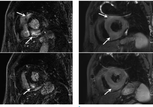
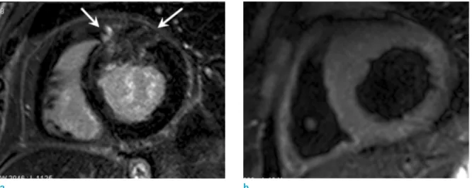
Fig. 1. Acute cardiac sarcoidosis (CS) in a 54-year-old man who presented as an acute atypical chest pain. Short-axis late gadolinium enhanced (LGE) MR images (a) show multifocal patchy enhancement in the anteroseptal and inferoseptal wall of basal LV level (upper row) and inferoseptal wall of midventricuar LV level (lower row). Notice the enhancement is located in the non-coronary vascular territory and preserved subendocardial layer. Short-axis T2-weighted MR images (b) show high signal intensity (arrows) in the corresponding regions with the enhancement area, which is suggested of the myocardial edema.
a b
Cardiac MRI
To begin with, CMR imaging offers a multi-dimensional assessment of CS, allowing for a non-invasive detection of scar, biventricular function and edema using LGE, bright- blood steady-state free precession (SSFP) imaging and T2- weighted imaging, respectively (26). Recently, T1 and T2 mapping are reported to be useful for early detection of CS, because it is possible to detect myocardial inflammation by directly relating to the altered magnetization properties (27).
Therefore, in a recent study for assessing CS using various cardiac tests, CMR was reported to be the most valuable in the diagnosis and prognosis of CS in a general sarcoidosis population demographic (28).
LGE imaging is considered the most useful sequence in the diagnosis of CS. While there is no specific pattern of LGE in the CMR that diagnoses CS, the most typical patterns include subepicardial and midwall LGE along the basal septum and/or inferolateral wall (16) (Fig. 1). When the septum is involved, it is noted that there is often contiguous involvement which includes the right ventricular insertion points, which is often present. The LGE patterns reported in various studies are summarized in the Table 3. In this sense, the diagnostic performances of CMR for diagnosis of CS are reported as sensitivity of 75-100% and specificity of 76.9-
78% (26). It is noted that CMR was reported to be more than twice sensitive compared to JMHW criteria (1993) for detecting cardiac involvement in a study with 81 patients with extracardiac sarcoidosis (26% vs. 12%) (29).
LGE may predict the adverse patient events including ventricular arrhythmias and sudden death (29, 30). A large extent of LGE was also reported as a marker for the
Table 3. The Reported Common Distribution Patterns of Late Gadolinium Enhancement of Myocardium in Patients with Cardiac Sarcoidosis
Authors Distribution pattern
Birnie et al. (16) Patchy and multifocal pattern Basal segments, particularly of the septum and lateral wall
Midmyocardium and epicardium, transmural
Youssef et al. (17) Basal septum
Jeudy et al. (26) Transmural, subepicardium or midmyocardium
Perez et al. (33) Linear in the subepicardium, transmural, or nodular with patchy distribution
Usually septum, basal, lateral segment and papillary muscles
Manoushagian et al. (39) Basal and lateral segment
Fig. 2. Chronic CS in a 74-year-old woman who complained dyspnea. Short-axis SSFP MR cine image (a) shows decreased wall motion and decreased wall thickness of inferolateral wall (arrows) at the basal LV level. Short-axis LGE MR image (b) shows transmural enhancement at thinned inferolateral wall (arrows) and multifocal patchy enhancement in the inferoseptal wall (arrowheads).
a b
absence of left ventricle (LV) functional improvement, and a high incidence of adverse outcome in patients with CS after steroid therapy (31). However, LGE alone cannot differentiate between the onset of an active inflammatory and the chronic fibrotic phase of CS in a patient (32). It is noted that edema or infiltration in the early inflammatory phase exhibits as an identified increased wall thickness with wall motional abnormality on cine images, and increased T2 signal intensity at the involved area (16, 33) (Fig. 1). However, the sensitivity of these findings was not high, because a number of technical challenges include myocardial signal loss caused by through-plane motion and inhomogeneous signal intensity (26). In the onset of the chronic phase, the thinning of the ventricular wall with global or regional ventricular dysfunction is also noted (34) (Fig. 2). In most cases, it is shown that these two phases
overlap (34).
CMR can also detect right-sided ventricular dysfunction which can be caused by elevated right heart pressures from pulmonary sarcoidosis or right ventricle (RV) involvement of CS. As it is shown, the right ventricular involvement of CS has been reported in 42% of autopsy specimens (35).
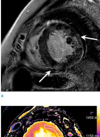
Recently, T1- or T2 mapping techniques are used for the recognition of early cardiac involvement of systemic sarcoidosis (Fig. 3). Greulich et al. (36) showed that CMR mapping provided an incremental value in detecting subclinical myocardial involvement in systemic sarcoidosis when there was no abnormal LGE and LV function was normal. Myocardial edema and inflammation induce an increase in T1, T2, and ECV, but that T2-weighted imaging, including T2 mapping, may be more specific for active myocardial inflammation and edema (37). In other words,
b c
a
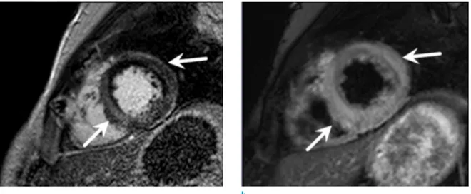
Fig. 3. Suspected CS in a 36-year-old man who complained orthopnea. Short-axis LGE (a) shows subtle enhancement at midventricular inferoseptal wall and anterolateral wall (arrows). Pre-contrast T1 map (b) and post-contrast T1 map (c) shows increased native T1 value (1320 msec) and increased extracellular volume (35.3%) than normal control value, which is helpful for the early diagnosis of a cardiac involvement of sarcoidosis.
it is shown that the native T1 is increased not only by acutely injured myocardium but also by the incidence of fibrosis in chronic myocardial changes (38). In the study by Puntmann et al. (27), it showed that patients with systemic sarcoidosis had reported to have higher myocardial native T1 and T2 and lower ejection fraction. Among these, the native T1 was the strongest discriminator between the patient’s health and the onset of the disease. There was also a significant reduction of native T1 and T2 in the patients who underwent treatment, suggesting information on the central role of inflammation in the case of a myocardial injury. This study suggests the potential role of CMR for determining a treatment guide and the proper monitoring in the case of CS.
18
F-FDG PET
18F-FDG PET is a noninvasive molecular imaging technique that is highly sensitive to the metabolically active processes.
18F-FDG PET uses glucose analogs to identify areas of increased inflammation (39). In the inflammatory cells, the case of an overexpression of the glucose transporter and overproduction of the glycolytic enzyme leads to an increase in glucose metabolism (26, 33). Areas with cardiac inflammation have increased glucose metabolism and increased activity in 18F-FDG PET (39). In Patients with CS, Focal or patch FDG uptake is usually seen throughout the
myocardium (21, 26) (Fig. 4).
When myocardial uptake of 18F-FDG is evaluated in CS, it is noted that the physiologic myocardial uptake of 18F-FDG always presents such a problem that special preparatory processes have been recommended. Long fasting (more than 18 hours) before the PET study (40), low dose intravenous unfractionated heparin immediately before the 18F-FDG injection (41), and special diet (high fat, low carbohydrate, and protein permitted) (42) have been used to overcome challenges with managing this process. All of these methods are intended to increase insulin resistance of the patients, whereby myocardial energy requirement is shifted to fatty acid from glucose. There are many debates about the most useful method of myocardial suppression of 18F-FDG uptake.
If all the 3 methods are applied together, the effects may be the best to be applied for the benefit of the patient.
Myocardial perfusion imaging is commonly performed with 18F-FDG PET to detect the area of myocardial scar (33, 39). In this case, the single-photon emission computed tomography (SPECT) using Technetium-99m labeled radiopharmaceuticals or Thallium-201 or PET-perfusion using Rubidium-82 can be used (39). It is noted that an increased FDG uptake with normal perfusion indicates the onset of the early stage of disease. Increased FDG uptake with decreased perfusion represents active CS with myocardial damage. The absence of FDG uptake with abnormal perfusion suggests the end stage fibrous disease (26).
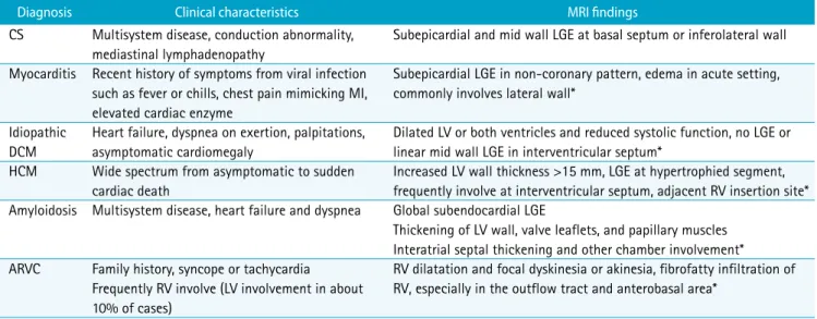
Table 4. Differential Diagnosis of CS
Diagnosis Clinical characteristics MRI findings
CS Multisystem disease, conduction abnormality, mediastinal lymphadenopathy
Subepicardial and mid wall LGE at basal septum or inferolateral wall Myocarditis Recent history of symptoms from viral infection
such as fever or chills, chest pain mimicking MI, elevated cardiac enzyme
Subepicardial LGE in non-coronary pattern, edema in acute setting, commonly involves lateral wall*
Idiopathic DCM
Heart failure, dyspnea on exertion, palpitations, asymptomatic cardiomegaly
Dilated LV or both ventricles and reduced systolic function, no LGE or linear mid wall LGE in interventricular septum*
HCM Wide spectrum from asymptomatic to sudden cardiac death
Increased LV wall thickness >15 mm, LGE at hypertrophied segment, frequently involve at interventricular septum, adjacent RV insertion site*
Amyloidosis Multisystem disease, heart failure and dyspnea Global subendocardial LGE
Thickening of LV wall, valve leaflets, and papillary muscles Interatrial septal thickening and other chamber involvement*
ARVC Family history, syncope or tachycardia Frequently RV involve (LV involvement in about 10% of cases)
RV dilatation and focal dyskinesia or akinesia, fibrofatty infiltration of RV, especially in the outflow tract and anterobasal area*
* Own characteristic findings for differential point from other diseases
ARVC = arrhythmogenic right ventricular cardiomyopathy; CS = cardiac sarcoidosis; DCM = dilated cardiomyopathy; HCM = hypertrophic cardiomyopathy; LGE = late gadolinium-induced enhancement; LV = left ventricle; RV = right ventricle
According to the meta-analysis of 7 diagnostic studies, the overall range of reported sensitivities and specificities of 18F-FDG PET for diagnosis of CS was 79-100% and 38-
100%, respectively. The pooled estimates for 18F-FDG PET yielded 89% sensitivity (95% CI, 79-96%) and 78%
specificity (95% CI, 68-86%) (21).
a b
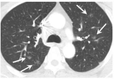
Fig. 4. Multimodality imaging in a 59-year-old woman with systemic sarcoidosis. Non-ECG gated chest CT images with mediastinal window setting (a) show enlarged lymph nodes along the mediastinum (arrowheads) and lung window setting (b) show scattered several lung nodules in both lung (arrows). Four-chamber LGE MR image (c) shows subepicardial enhancement (arrowheads) at the basal septal wall and midventricular to basal lateral wall. The FDG PET images (d, e) show increased uptake in mediastinal lymph nodes (arrowheads in d) and septal and lateral wall of the LV (arrowheads in e) which are at a corresponding region with enhancement on the LGE MR image (arrowheads in c).
e
c d
The prevailing discipline notes that PET may have a role as a prognostic indicator of CS. One retrospective study on CS patients with implantable cardioverter-defibrillators suggested that active inflammation detected on the PET scan, may be a potential marker for the incidence and presence of VT risk (43). In addition, a larger cohort study with 118 patients found that the presence of both focal FDG uptake and myocardial perfusion defects on PET was
associated with increased risk of death or VT. This study also identified focal FDG uptake on RV as a marker for a higher cardiac event rate (43). Therefore, the quantitation of myocardial inflammation using standardized uptake values (SUVs) is an important advantage of PET. Recent studies suggested that PET with FDG quantitation can be useful to predict cardiac events and monitor the response to immunosuppressive treatment (44).
a b
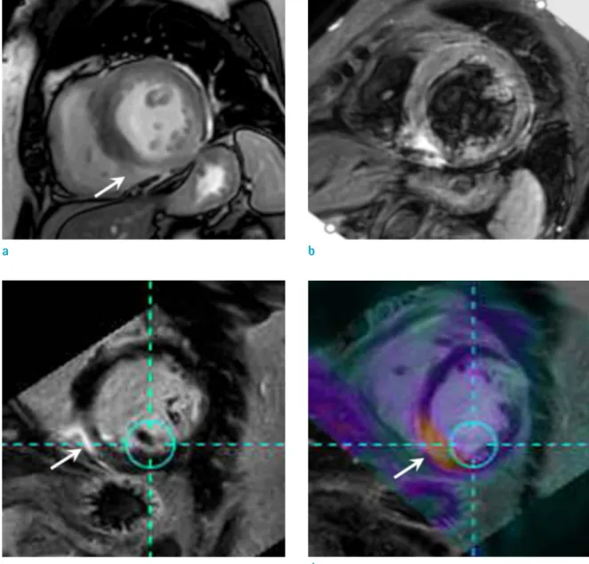
Fig. 5. Conjoined imaging of PET and MR in a 59-year-old woman with cardiac sarcoidosis. Short-axis SSFP cine image (a) shows hypokinesia at the region of the midventricular inferoseptal wall (arrow), with decreased EF as 48%. Short-axis T2- weighted image (b) shows high signal intensity at inferoseptal wall of the LV (arrow), suggesting edema. The LGE MR image (c) shows subepicardial enhancement at concordant area, indicating fibrosis. Conjoined PET-MR image (d) reveals increased uptake (arrow) at the enhanced area of the inferoseptal wall in LGE MR image, which is well presented in the active inflammatory phase of CS (c and d images are LGE MR image and PET image matching the same level).
c d
Recent studies reported other tracers such as 68Ga- DOTANOC or 18F-fluoromisonidazole (FMISO) may be useful for CS detection (45, 46), but further study is required to establish the benefits of these agents.
Combined Use of Multimodality Findings (Hybrid Imaging _ PET and CMR)
It is emphasized that a comparison of diagnostic performances between PET and CMR is limited at this time. Only one study with 8 patients reported that PET may be more sensitive than CMR, and CMR may be more specific than PET for detecting CS. However, the authors noted that the distribution of the findings were different from the two modalities, as each study reflects different pathologic process (47). While LGE identifies fibrosis, FDG- PET identifies and finds areas with active inflammation in those cases. Therefore, these two modalities could be used as complementary tools. Recently, the use of hybrid imaging combined with PET-MR has been applied to CS diagnosis (Fig. 5). This new method has the strengths of both imaging techniques and can evaluate cardiac function and burden of scar/fibrosis at one time (39). Recent studies with suspected CS patients showed that hybrid PET-MR could demonstrate both myocardial injury patterns by LGE and disease activity by PET simultaneously with accurate co-localization (48).
The hybrid PET-MR has several advantages over the use of CMR or PET in isolation: an improved diagnostic sensitivity and prognostic utility, while reducing radiation exposure (48, 49). Right ventricular PET abnormalities and presence of LGE were independent predictors of adverse events.
Abnormalities found on both the CMR and PET was the strongest predictor of major adverse cardiac events (49).
The hybrid PET-MR can identify patients with active CS and differentiate these patients from those without active disease, and review patients with false-positive FDG uptake due to incomplete myocardial suppression (48). Patients with MR+/PET+ (characteristic LGE aligning exactly with increased FDG uptake) are considered to have active CS.
MR+/PET- (characteristic LGE but no increased FDG) is thought to represent inactive CS with residual scar. In the meantime, MR-/PET- (neither characteristic LGE nor increased FDG) is considered negative. In the case of MR-/
PET+ (increased FDG uptake without LGE), global, focal, or focal-on-diffuse uptake can be observed. Global uptake is considered more likely to represent false positive activity, as CS is known to be localized, not considered to be characteristically seen as diffuse. Focal and focal-on-diffuse uptakes are generally considered positive findings of CS, but there is controversy in their interpretation and other possible causes should be excluded (50). In this case, focal or focal-on-diffuse MR-/PET+ pattern may represent early CS without LGE.
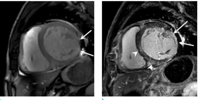
Fig. 6. Acute myocarditis in a 28-year-old man who presented with the syncope and elevated cardiac enzyme. Short- axis LGE images (a) show multifocal patchy enhancement at the subepicardial layer of anterolateral and inferoseptal wall with preserved subendocardial layer. T2-weighted images (b) show high signal intensity in the same regions (arrows) with enhanced area. The condition resembles CS, therefore clinical presentation and other blood tests should be carefully considered.
a b
Differential Diagnosis
As it is seen that the imaging finding of CS has a wide spectrum, the clinical context should be carefully considered in those cases. It is possible that a differential diagnosis of CS includes myocarditis, DCM, hypertrophic cardiomyopathy, amyloidosis, and arrhythmogenic right ventricular cardiomyopathy (26).
Myocarditis is caused by inflammation, mainly viral infection, resulting in myocardial damage. Thus, myocarditis
can be preceded in the patient by noting the patient’s experience of viral symptoms such as fever, chilling, or acute chest pain. The CMR with LGE and T2-weighted imaging provides the increased diagnostic accuracy for myocarditis (51) (Fig. 6). Evidently, the non-coronary patterns of LGE are considered to be similar with CS. Although both diseases demonstrate subepicardial LGE, it is noted that myocarditis more frequently involves the lateral wall, while CS is more frequently affected at the location region of the interventricular septum (52).
DCM is caused by various causes including myocarditis, alcoholics or toxins, although the most DCM cases are idiopathic. As LV dilation and reduced systolic function can be observed in CS, it can be misdiagnosed as idiopathic DCM (53). Upon review, the idiopathic DCM typically shows no LGE or midwall LGE in the interventricular septum, while CS more commonly demonstrates transmural or subepicardial LGE (54) (Fig. 7).
Hypertrophic cardiomyopathy is characterized by increased LV wall thickness (>15 mm in end-diastole).
At this juncture, it is seen that CS can also demonstrate thickened myocardium due to granulomatous infiltration.
Hypertrophic cardiomyopathy usually shows LGE in anterior and posterior RV insertion site to interventricular septum, and it does not show edema on T2 weighted images, because it is related with fibrosis, not associated with inflammation (Fig. 8). On the other hand, CS usually shows subepicardial LGE and increased T2 signal intensity on inflammatory phase (54).
Amyloidosis shows thickening of LV wall, valve leaflets,
Fig. 8. Hypertrophic cardiomyopathy in a 58-year-old woman with recurrent ventricular tachycardia. Short-axis LGE MR image (a) shows multifocal patchy enhancement (arrows) at hypertrophied anteroseptal wall, around the anterior RV insertion sites. However, the T2-weighted image (b) show no evidence of abnormally increased signal intensity.
a b
Fig. 7. Dilated cardiomyopathy in a 54-year-old woman who complained of dyspnea and a progressed heart failure.
Short-axis LGE MR image shows dilated LV chamber and linear enhancement at the midwall region of the interventricular septum (arrows).
and papillary muscles, which is characterized by the incidence of global subendocardial LGE (Fig. 9). At that time, CS may also involve global myocardium, which may mimic amyloidosis. However, atrial thickening and atrial LGE are more commonly noted in amyloidosis than CS (55).
It is worthy to note the differentiation of arrhythmogenic right ventricular cardiomyopathy (ARVC) and CS which is considered important, because the former needs genetic screening while the latter requires initiating an immunosuppressive therapy. However, the current diagnostic criteria for ARVC cannot reliably distinguish these two disease entities (56). In those cases, the RV involvement of CS can cause RV dilatation and focal dyskinesia or akinesia, and ARVC may involve left ventricle. The presence of septal scar and mediastinal lymphadenopathy favors a diagnosis of CS, especially when associated with the incidence of extracardiac sarcoidosis (57).
When diagnosing CS through PET, the possibility of false positives and other diseases should be considered. In case of focal FDG uptake, it is necessary to exclude other possible causes affecting the diagnosis, including ischemic heart diseases and hypertrophic cardiomyopathy (50). Especially, because FDG uptake in the lateral wall has been reported also in healthy people, this finding may represent false
positive (47, 58). Focal-on-diffuse uptake also requires careful interpretation, since potentially false positive case with such FDG uptake was reported in the past (59).
In conclusion, CS can manifest as a serious condition such as conduction abnormality, heart failure, ventricular arrhythmia, or sudden cardiac death, although some patients remain asymptomatic. Early recognition and early initiation of therapy is very important to improve the prognosis for the patient. However, the diagnosis of CS is challenging because of low sensitivity of the pathological analysis, as confirmed by EMB. Therefore, the diagnosis of CS is recommended to best be concluded by the utilization of a combination of clinical, electrocardiographic, and advanced imaging of CMR and PET. The CMR can evaluate the patient’s cardiac function and fibrotic scar, whereas a PET can detect active inflammation, and thus can be used to monitor treatment response. In addition, CMR with LGE sequence is useful for distinguishing CS by the distinctive enhancement pattern in patients with suspected CS.
Therefore, radiologists should be aware of the characteristic findings of CS in advanced imaging modalities and try to accurately diagnose the CS.
Acknowledgments
This work was supported by Korean study group of Cardiovascular Magnetic Resonance (KCMR) in the Korean Society of Magnetic Resonance in Medicine (KSMRM).
REFERENCES
1. Statement on sarcoidosis. Joint Statement of the American Thoracic Society (ATS), the European Respiratory Society (ERS) and the World Association of Sarcoidosis and Other Granulomatous Disorders (WASOG) adopted by the ATS Board of Directors and by the ERS Executive Committee, February 1999. Am J Respir Crit Care Med 1999;160:736- 755
2. Schatka I, Bengel FM. Advanced imaging of cardiac sarcoidosis. J Nucl Med 2014;55:99-106
3. Silverman KJ, Hutchins GM, Bulkley BH. Cardiac sarcoid:
a clinicopathologic study of 84 unselected patients with systemic sarcoidosis. Circulation 1978;58:1204-1211 4. Lynch JP 3rd, Hwang J, Bradfield J, Fishbein M, Shivkumar
K, Tung R. Cardiac involvement in sarcoidosis: evolving concepts in diagnosis and treatment. Semin Respir Crit Care Med 2014;35:372-390
5. Yazaki Y, Isobe M, Hiroe M, et al. Prognostic determinants Fig. 9. Cardiac amyloidosis in a 68-year-old man with
exertional dyspnea. Short-axis LGE image shows global subendocardial enhancement (arrows) at whole LV wall. Subtle enhancement is also noted in RV free wall (arrowheads).
of long-term survival in Japanese patients with cardiac sarcoidosis treated with prednisone. Am J Cardiol 2001;88:1006-1010
6. Ardehali H, Howard DL, Hariri A, et al. A positive endomyocardial biopsy result for sarcoid is associated with poor prognosis in patients with initially unexplained cardiomyopathy. Am Heart J 2005;150:459-463
7. Hillerdal G, Nou E, Osterman K, Schmekel B. Sarcoidosis:
epidemiology and prognosis. A 15-year European study. Am Rev Respir Dis 1984;130:29-32
8. Morimoto T, Azuma A, Abe S, et al. Epidemiology of sarcoidosis in Japan. Eur Respir J 2008;31:372-379
9. Varron L, Cottin V, Schott AM, Broussolle C, Seve P.
Late-onset sarcoidosis: a comparative study. Medicine (Baltimore) 2012;91:137-143
10. Perry A, Vuitch F. Causes of death in patients with sarcoidosis. A morphologic study of 38 autopsies with clinicopathologic correlations. Arch Pathol Lab Med 1995;119:167-172
11. Iwai K, Tachibana T, Takemura T, Matsui Y, Kitaichi M, Kawabata Y. Pathological studies on sarcoidosis autopsy. I.
Epidemiological features of 320 cases in Japan. Acta Pathol Jpn 1993;43:372-376
12. Mehta D, Lubitz SA, Frankel Z, et al. Cardiac involvement in patients with sarcoidosis: diagnostic and prognostic value of outpatient testing. Chest 2008;133:1426-1435
13. Rosen Y. Pathology of sarcoidosis. Semin Respir Crit Care Med 2007;28:36-52
14. Uemura A, Morimoto S, Hiramitsu S, Kato Y, Ito T, Hishida H.
Histologic diagnostic rate of cardiac sarcoidosis: evaluation of endomyocardial biopsies. Am Heart J 1999;138:299-302 15. Tavora F, Cresswell N, Li L, Ripple M, Solomon C, Burke
A. Comparison of necropsy findings in patients with sarcoidosis dying suddenly from cardiac sarcoidosis versus dying suddenly from other causes. Am J Cardiol 2009;104:571-577
16. Birnie DH, Nery PB, Ha AC, Beanlands RS. Cardiac sarcoidosis. J Am Coll Cardiol 2016;68:411-421
17. Youssef G, Beanlands RS, Birnie DH, Nery PB. Cardiac sarcoidosis: applications of imaging in diagnosis and directing treatment. Heart 2011;97:2078-2087
18. Sekhri V, Sanal S, Delorenzo LJ, Aronow WS, Maguire GP.
Cardiac sarcoidosis: a comprehensive review. Arch Med Sci 2011;7:546-554
19. Hiraga HHM, Iwai K. Guidelines for diagnosis of cardiac sarcoidosis: study report on diffuse pulmonary diseases (in Japanese). Tokyo: The Japanese Ministry of Health and Welfare, 1993:23-24
20. Soejima K, Yada H. The work-up and management of patients with apparent or subclinical cardiac sarcoidosis:
with emphasis on the associated heart rhythm abnormalities. J Cardiovasc Electrophysiol 2009;20:578- 583
21. Youssef G, Leung E, Mylonas I, et al. The use of 18F-FDG PET in the diagnosis of cardiac sarcoidosis: a systematic review and metaanalysis including the Ontario experience.
J Nucl Med 2012;53:241-248
22. Blankstein R, Osborne M, Naya M, et al. Cardiac positron emission tomography enhances prognostic assessments of patients with suspected cardiac sarcoidosis. J Am Coll Cardiol 2014;63:329-336
23. Kouranos V, Wells AU, Sharma R, Underwood SR, Wechalekar K. Advances in radionuclide imaging of cardiac sarcoidosis. Br Med Bull 2015;115:151-163
24. Birnie DH, Sauer WH, Bogun F, et al. HRS expert consensus statement on the diagnosis and management of arrhythmias associated with cardiac sarcoidosis. Heart Rhythm 2014;11:1305-1323
25. Hulten E, Aslam S, Osborne M, Abbasi S, Bittencourt MS, Blankstein R. Cardiac sarcoidosis-state of the art review.
Cardiovasc Diagn Ther 2016;6:50-63
26. Jeudy J, Burke AP, White CS, Kramer GB, Frazier AA.
Cardiac sarcoidosis: the challenge of radiologic-pathologic correlation: from the radiologic pathology archives.
Radiographics 2015;35:657-679
27. Puntmann VO, Isted A, Hinojar R, Foote L, Carr-White G, Nagel E. T1 and T2 mapping in recognition of early cardiac involvement in systemic sarcoidosis. Radiology 2017;285:63-72
28. Kouranos V, Tzelepis GE, Rapti A, et al. Complementary role of CMR to conventional screening in the diagnosis and prognosis of cardiac sarcoidosis. JACC Cardiovasc Imaging 2017;10:1437-1447
29. Patel MR, Cawley PJ, Heitner JF, et al. Detection of myocardial damage in patients with sarcoidosis. Circulation 2009;120:1969-1977
30. Greulich S, Deluigi CC, Gloekler S, et al. CMR imaging predicts death and other adverse events in suspected cardiac sarcoidosis. JACC Cardiovasc Imaging 2013;6:501- 511
31. Ise T, Hasegawa T, Morita Y, et al. Extensive late gadolinium enhancement on cardiovascular magnetic resonance predicts adverse outcomes and lack of improvement in LV function after steroid therapy in cardiac sarcoidosis. Heart 2014;100:1165-1172
32. Crouser ED, Ono C, Tran T, He X, Raman SV. Improved detection of cardiac sarcoidosis using magnetic resonance with myocardial T2 mapping. Am J Respir Crit Care Med 2014;189:109-112
33. Perez IE, Garcia MJ, Taub CC. Multimodality imaging in
cardiac sarcoidosis: is there a winner? Curr Cardiol Rev 2016;12:3-11
34. Orii M, Imanishi T, Akasaka T. Assessment of cardiac sarcoidosis with advanced imaging modalities. Biomed Res Int 2014;2014:897956
35. Roberts WC, McAllister HA, Jr., Ferrans VJ. Sarcoidosis of the heart. A clinicopathologic study of 35 necropsy patients (group 1) and review of 78 previously described necropsy patients (group 11). Am J Med 1977;63:86-108 36. Greulich S, Kitterer D, Latus J, et al. Comprehensive
cardiovascular magnetic resonance assessment in patients with sarcoidosis and preserved left ventricular ejection fraction. Circ Cardiovasc Imaging 2016;9:e005022
37. Ferreira VM, Piechnik SK. Seeing beyond the obvious:
subclinical cardiac sarcoidosis revealed by cardiovascular magnetic resonance mapping. Circ Cardiovasc Imaging 2016;9:e005592
38. Hinojar R, Foote L, Arroyo Ucar E, et al. Native T1 in discrimination of acute and convalescent stages in patients with clinical diagnosis of myocarditis: a proposed diagnostic algorithm using CMR. JACC Cardiovasc Imaging 2015;8:37-46
39. Manoushagian SJ, Lakhter V, Patil PV. Multimodality imaging in the diagnosis and management of cardiac sarcoidosis. J Nucl Cardiol 2017;24:29-33
40. Langah R, Spicer K, Gebregziabher M, Gordon L.
Effectiveness of prolonged fasting 18f-FDG PET-CT in the detection of cardiac sarcoidosis. J Nucl Cardiol 2009;16:801-810
41. Ishimaru S, Tsujino I, Takei T, et al. Focal uptake on 18F-fluoro-2-deoxyglucose positron emission tomography images indicates cardiac involvement of sarcoidosis. Eur Heart J 2005;26:1538-1543
42. Harisankar CN, Mittal BR, Agrawal KL, Abrar ML, Bhattacharya A. Utility of high fat and low carbohydrate diet in suppressing myocardial FDG uptake. J Nucl Cardiol 2011;18:926-936
43. Betensky BP, Tschabrunn CM, Zado ES, et al. Long- term follow-up of patients with cardiac sarcoidosis and implantable cardioverter-defibrillators. Heart Rhythm 2012;9:884-891
44. Ahmadian A, Brogan A, Berman J, et al. Quantitative interpretation of FDG PET/CT with myocardial perfusion imaging increases diagnostic information in the evaluation of cardiac sarcoidosis. J Nucl Cardiol 2014;21:925-939 45. Gormsen LC, Haraldsen A, Kramer S, Dias AH, Kim WY,
Borghammer P. A dual tracer (68)Ga-DOTANOC PET/CT and (18)F-FDG PET/CT pilot study for detection of cardiac sarcoidosis. EJNMMI Res 2016;6:52
46. Manabe O, Hirata K, Shozo O, et al. (18)F-fluoromisoni-
dazole (FMISO) PET may have the potential to detect cardiac sarcoidosis. J Nucl Cardiol 2017;24:329-331 47. Ohira H, Tsujino I, Ishimaru S, et al. Myocardial imaging
with 18F-fluoro-2-deoxyglucose positron emission tomography and magnetic resonance imaging in sarcoidosis. Eur J Nucl Med Mol Imaging 2008;35:933-941 48. Dweck MR, Abgral R, Trivieri MG, et al. Hybrid magnetic
resonance imaging and positron emission tomography with fluorodeoxyglucose to diagnose active cardiac sarcoidosis.
JACC Cardiovasc Imaging 2018;11:94-107
49. Wicks EC, Menezes LJ, Barnes A, et al. Diagnostic accuracy and prognostic value of simultaneous hybrid 18F-fluorodeoxyglucose positron emission tomography/
magnetic resonance imaging in cardiac sarcoidosis. Eur Heart J Cardiovasc Imaging 2018;19:757-767
50. I s h i d a Y, Yo s h i n a g a K , M i y a g a w a M , e t a l . Recommendations for (18)F-fluorodeoxyglucose positron emission tomography imaging for cardiac sarcoidosis:
Japanese Society of Nuclear Cardiology recommendations.
Ann Nucl Med 2014;28:393-403
51. Friedrich MG, Sechtem U, Schulz-Menger J, et al.
Cardiovascular magnetic resonance in myocarditis: a JACC white paper. J Am Coll Cardiol 2009;53:1475-1487
52. Yilmaz A, Ferreira V, Klingel K, Kandolf R, Neubauer S, Sechtem U. Role of cardiovascular magnetic resonance imaging (CMR) in the diagnosis of acute and chronic myocarditis. Heart Fail Rev 2013;18:747-760
53. Yazaki Y, Isobe M, Hiramitsu S, et al. Comparison of clinical features and prognosis of cardiac sarcoidosis and idiopathic dilated cardiomyopathy. Am J Cardiol 1998;82:537-540 54. Vignaux O. Cardiac sarcoidosis: spectrum of MRI features.
AJR Am J Roentgenol 2005;184:249-254
55. Moraes GL, Higgins CB, Ordovas KG. Delayed enhancement magnetic resonance imaging in nonischemic myocardial disease. J Thorac Imaging 2013;28:84-92; quiz 93-95 56. Marcus FI, McKenna WJ, Sherrill D, et al. Diagnosis
of arrhythmogenic right ventricular cardiomyopathy/
dysplasia: proposed modification of the task force criteria.
Circulation 2010;121:1533-1541
57. Steckman DA, Schneider PM, Schuller JL, et al. Utility of cardiac magnetic resonance imaging to differentiate cardiac sarcoidosis from arrhythmogenic right ventricular cardiomyopathy. Am J Cardiol 2012;110:575-579
58. Bartlett ML, Bacharach SL, Voipio-Pulkki LM, Dilsizian V.
Artifactual inhomogeneities in myocardial PET and SPECT scans in normal subjects. J Nucl Med 1995;36:188-195 59. Tahara N, Tahara A, Nitta Y, et al. Heterogeneous
myocardial FDG uptake and the disease activity in cardiac sarcoidosis. JACC Cardiovasc Imaging 2010;3:1219-1228