• 검색 결과가 없습니다.

Severe Aortic Coarctation in a 75-Year-Old Woman: Total Simultaneous Repair of Aortic Coarctation and Severe Aortic Stenosis

N/A
N/A
Protected

Academic year: 2022

Share "Severe Aortic Coarctation in a 75-Year-Old Woman: Total Simultaneous Repair of Aortic Coarctation and Severe Aortic Stenosis"

Copied!
3
0
0

로드 중.... (전체 텍스트 보기)

전체 글

(1)

62 Copyright © 2012 The Korean Society of Cardiology Korean Circulation Journal

Introduction

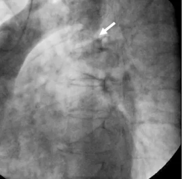
Aortic coarctation is a congenital cardiovascular malformation that should be diagnosed and corrected early in life.

1)

Survival of a patient to more than 70 years of age is extremely unusual in cases of uncorrected coarctation. There are a few case reports of patients who were first diagnosed with coarctation at a very late age,

2-4)

and their management remains controversial. We report a case of a wo- man who was first diagnosed with coarctation of the aorta at the age of 75 years and underwent a successful total simultaneous re- pair of coarctation and severe aortic valve stenosis.

Case Report

http://dx.doi.org/10.4070/kcj.2012.42.1.62 Print ISSN 1738-5520 • On-line ISSN 1738-5555

Severe Aortic Coarctation in a 75-Year-Old Woman:

Total Simultaneous Repair of Aortic Coarctation and Severe Aortic Stenosis

Ju Hyun Park, MD 1 , Kook Jin Chun, MD 2 , Sung Gook Song, MD 2 , Jeong Su Kim, MD 2 , Yong Hyun Park, MD 2 , Jun Kim, MD 2 , Ki Seuk Choo, MD 3 , June Hong Kim, MD 2 , and Sang Kwon Lee, MD 4

1

Department of Cardiology, Busan St Mary’s Medical Center, Busan,

2

Departments of Cardiology,

3

Diagnostic Radiology, and

4

Thoracic and Cardiovascular Surgery, Pusan National University College of Medicine, Pusan National University Yangsan Hospital, Yangsan, Korea

Aortic coarctation is usually diagnosed and repaired in childhood and early adulthood. Survival of a patient with an uncorrected coarcta- tion to more than 70 years of age is extremely unusual, and management strategies for these cases remain controversial. We present a case of a 75-year-old woman who was first diagnosed with aortic coarctation and severe aortic valve stenosis 5 years ago and who un- derwent a successful one-stage repair involving valve replacement and insertion of an extra-anatomical bypass graft from the ascending to the descending aorta. (Korean Circ J 2012;42:62-64)

KEY WORDS: Aortic coarctation; Aged; Thoracic surgical procedures.

Received: August 25, 2011

Revision Received: September 16, 2011 Accepted: September 27, 2011

Correspondence: Kook Jin Chun, MD, Department of Cardiology, Pusan Na- tional University College of Medicine, Pusan National University Yangsan Hospital, 20 Geumo-ro, Mulgeum-eup, Yangsan 626-770, Korea

Tel: 82-55-360-1454, Fax: 82-55-360-1129 E-mail: [email protected]

• The authors have no financial conflicts of interest.

This is an Open Access article distributed under the terms of the Creative Commons Attribution Non-Commercial License (http://creativecommons.

org/licenses/by-nc/3.0) which permits unrestricted non-commercial use, distribution, and reproduction in any medium, provided the original work is properly cited.

Case

A 75-year-old woman was referred to our emergency room be-

cause of squeezing pain in the chest and dyspnea (New York Heart

Association Class III). She had a medical history of uncontrolled hy-

pertension for 10 years and was diagnosed with aortic valve steno-

sis 5 years ago at a local hospital. On physical examination, blood

pressure was 170/80 mm Hg in both arms. A systolic ejection mur-

mur was heard at the right upper sternal border. Electrocardiography

showed left ventricular hypertrophy and T-wave inversion in the

precordial leads. An enlarged cardiac silhouette and rib notching

were observed on chest radiography. On echocardiography, severe

degenerative aortic valve stenosis, concentric left ventricular hy-

pertrophy and normal left ventricular systolic function were observ-

ed. The peak and mean pressure gradient of the aortic valve were es-

timated at 140 mm Hg and 80 mm Hg, respectively. Therefore, an

aortic valve replacement was planned and coronary angiography

was performed with aortography for evaluation of chest pain and

uncontrolled blood pressure despite antihypertensive drug medi-

cation. Coronary angiography revealed normal coronary arteries, wh-

ile aortography showed severe stenosis of the descending aorta

just below the origin of the left subclavian artery (Fig. 1). We sus-

pected aortic coarctation and computed tomographic angiography

was performed. Severe aortic coarctation and compensatory dilata-

(2)

63 Ju Hyun Park, et al.

http://dx.doi.org/10.4070/kcj.2012.42.1.62 www.e-kcj.org

tion of the intercostal and internal thoracic arteries were noted (Fig.

2). Ankle-Brachial index was 0.53. The blood pressure was 170/80 mm Hg in both upper arms, and 90/60 mm Hg in the lower extremities.

The patient underwent a single-stage correction of the coarctation and aortic valve stenosis. After a median sternotomy, cardiopul- monary bypass was established through cannulation of the aortic arch and the right atrium. After retrograde cold blood cardioplegic arrest and incision of the ascending aorta, the calcified bicuspid aortic valve was excised, and a porcine tissue valve prosthesis (21 mm, Hancock II; Medtronic Inc, Minneapolis, MN, USA) was inserted. Af- terwards, the supracoronary ascending aorta was anastomosed with a 14 mm polytetrafluoroethylene graft (GORE-TEX vascular graft;

W.L Gore & Associates Inc, Flagstaff, AZ, USA) by an end-to- side te- chnique. And then through a posterior pericardial approach, a final end-to-side anastomosis between the bypass graft and the de- scending aorta completed the repair (Fig. 3). The cardiopulmonary bypass and aortic cross-clamp times were 224 minutes and 163 mi- nutes, respectively. The postoperative course was uneventful. Con- trol of arterial hypertension required minimal doses of angiotensin- converting enzyme inhibitor and β blocker. Warfarin was also pre- scribed. The patient is doing very well 2 years after discharge.

Discussion

Aortic coarctation, which is usually diagnosed and corrected early in life, accounts for approximately 9% of all congenital cardiovas- cular malformations.

1)

Prognosis of an unrepaired coarctation is very unfavorable. Approximately 90% of cases die before 50 years of age.

Another 5% die at 50-60 years and the remaining 5% die at an even older age.

2)

Death in these patients is usually secondary to heart fail- ure, coronary artery disease, aortic rupture/dissection, concomitant aortic valve disease, infective endarteritis/endocarditis, or cerebral hemorrhage.

2)3)

A review of the literature over the past 30 years revealed 10 cas- es over 70 years of age who were first diagnosed with uncorrected aortic coarctation. Although surgery is considered the treatment of choice for aortic coarctation in children and adults, management strategies for older patients are controversial and there is no con- sensus on the management of these patients. One patient under- went successful coarctation repair

4)

and one patient died during sur- gical repair.

5)

Two patients underwent coronary artery bypass graft surgery for myocardial ischemia without coarctation repair

6)7)

and another five patients were conservatively managed for heart failure and hypertension.

Although one might argue that highly developed collateral circu- Fig. 1. Aortography revealed severe coarctation of the aorta (arrow).

Fig. 2. Preoperative three-dimensional computed tomography revealed aortic coarctation (arrow) (A) and compensatory dilatation of the intercos- tal and internal thoracic arteries (B).

A   B  

Fig. 3. Postoperative three-dimensional computed tomography revealed

an extra-anatomic bypass graft from the ascending to the descending aor-

ta (arrow).

(3)

64 Aortic Coarctation in an Elderly Woman

http://dx.doi.org/10.4070/kcj.2012.42.1.62 www.e-kcj.org

lation might allow elderly patients to tolerate the coarctation with- out its surgical repair,

8-12)

there are benefits associated with symp- tomatic improvement and with blood pressure reduction after surgical repair in patients older than 50 years.

13)

A direct approach to coarctation repair may come across difficulties in the adult popula- tion, and a large collateral formation and lung failure after thoraco- tomy may present technical challenges while total aortic cross- clamping, with diminished collaterals, has been involved as a causal factor in paraplegia. For these reasons, extra-anatomic bypass gr- afting has been used as an alternative surgical option.

14)

Severe coarctation of the aorta in combination with intracardiac pathology such as severe valvular stenosis or regurgitation and co- ronary artery disease in elderly patients requires surgery. The simul- taneous operative management of both lesions is desirable because of the higher morbidity and mortality associated with staged pro- cedures, and there has been only one reported case of a patient old- er than 70 years of age who underwent extra-anatomic bypass and simultaneous correction of intracardiac pathology.

14)

We considered coarctation repair surgery because of the high- pressure gradient (80 mm Hg) between the ascending and descend- ing aorta and the markedly uncontrolled hypertension despite an- tihypertensive drugs. Because two-stage procedures for aortic valve replacement and coarctation repair are associated with high rates of perioperative morbidity and mortality, we performed a one- stage total simultaneous repair of the coarctation and aortic valve replacement. After surgical intervention, blood pressure was well controlled with minimal doses of antihypertensive medication.

In conclusion, patients with aortic coarctation rarely survive till old age for their first diagnosis. As this case illustrates, simultaneous repair of coarctation of the aorta in combination with an intracar- diac pathology can be safely and successfully done even in elderly patients.

Acknowledgments

This work was supported for two years by Pusan National Uni- versity Research Grant.

REFERENCES

1. Grech V. Diagnostic and surgical trends, and epidemiology of co-

arctation of the aorta in a population-based study. Int J Cardiol 1999;68:197-202.

2. Campbell M. Natural history of coarctation of the aorta. Br Heart J 1970;32:633-40.

3. Jenkins NP, Ward C. Coarctation of the aorta: natural history and outcome after surgical treatment. QJM 1999;92:365-71.

4. Patel Y, Jilani MI, Cho K. Coarctation of the aorta presenting in a 79-year-old male. Thorac Cardiovasc Surg 1998;46:158-60.

5. Liberthson RR, Pennington DG, Jacobs ML, Daggett WM. Coarc- tation of the aorta: review of 234 patients and clarification of management problems. Am J Cardiol 1979;43:835-40.

6. Cevik S, Izgi C, Cevik C. Asymptomatic severe aortic coarctation in an 80-year-old man. Tex Heart Inst J 2004;31:429-31.

7. Convens C, Vermeersch P, Paelinck B, Van den Heuvel P, Van den Branden F. Aortic coarctation: a rare and unexpected cause of sec- ondary arterial hypertension in the elderly. Cathet Cardiovasc Di- agn 1996;39:71-4.

8. Haldane JH. Coarctation of aorta in an elderly man. Can Med As- soc J 1983;128:1298-9.

9. Bartlett LC. Coarctation of the aorta in an elderly man. Can Med Assoc J 1983;129:529.

10. O’Byrne E, Mather J, Hargreaves MR. Coarctation of the aorta: an unusual cause of hypertension in a 73-year-old woman. Int J Clin Pract 1997;51:466-7.

11. Bobby JJ, Emami JM, Farmer RD, Newman CG. Operative survival and 40-year follow-up of surgical repair of aortic coarctation. Br Heart J 1991;65:271-6.

12. Kountouris E, Potsis T, Nikas D, Siogas K. A severe coarctation of aorta in a 72-year-old female: a case report. Cases J 2009;2:

6308.

13. Bauer M, Alexi-Meskishvili VV, Bauer U, Alfaouri D, Lange PE, Hetzer R. Benefits of surgical repair of coarctation of the aorta in patients older than 50 years. Ann Thorac Surg 2001;72:2060-4.

14. Morris RJ, Samuels LE, Brockman SK. Total simultaneous repair

of coarctation and intracardiac pathology in adult patients. Ann

Thorac Surg 1998;65:1698-702.

수치

Fig. 3. Postoperative three-dimensional computed tomography revealed  an extra-anatomic bypass graft from the ascending to the descending  aor-ta (arrow).

참조

관련 문서

We report a case of isolated septum pellucidum agenesis in 17-year-old woman who was manifested by primary amenorrhea and diagnosed as partial hypopituitarism. 본

We report a right lateral thoracotomy approach for mitral valve repair in a young woman with pulmonary hypoplasia, in whom preoperative computed tomography showed severe right