ABSTRACT
Transcatheter aortic valve replacement (TAVR) or transcatheter aortic valve implantation (TAVI) for severe aortic stenosis (AS) is a minimally invasive interventional procedure that repairs a valve without removing the old, damaged valve. Instead, a replacement valve is wedged into the location of the native aortic valve. During TAVR, contrast is used for conventional aortic root angiography, positioning of the TAVR valve device, and assessing the peripheral vasculature.
Therefore, contrast-induced acute kidney injury (AKI) is a major concern when performing TAVR and is associated with increased mortality in patients with impaired renal function. Although the exact mechanism of post-TAVR AKI is unknown and appears multifactorial, contrast medium has been reported as a major contributing factor. We report a case of zero-contrast TAVR for severe AS in a patient with chronic kidney disease (CKD). The procedure was successfully performed with only fluoroscopic and transesophageal echocardiography (TEE) guidance.
Keywords: Transcatheter aortic valve replacement; Acute kidney injury;
Chronic kidney diseases; Aortic stenosis
INTRODUCTION
Transcatheter aortic valve replacement (TAVR) or transcatheter aortic valve implantation (TAVI) for severe aortic stenosis (AS) is the minimally invasive interventional procedure that repair valve without removing the old, damaged valve. Instead, it wedges a replacement into the place of native aortic valve.1
During TAVR, contrast is used for conventional aortic root angiography, positioning of the TAVR valve device and assessing the peripheral vasculature. Therefore, contrast induced acute kidney injury (AKI) is one of the major concerns of TAVR and is associated with increased mortality in patients with impaired renal function. Although the exact mechanism of post-TAVR AKI is unknown and seems to be multifactorial, however, contrast medium itself has been reported as a major contributing factor.2
We report a case of zero-contrast usage for TAVR for severe AS in patient with chronic kidney disease (CKD). This procedure was performed successfully in this case with only fluoroscopic and transesophageal echocardiography (TEE) guidance.
Case Report
Received: Jan 14, 2018 Revised: Apr 23, 2018 Accepted: Apr 26, 2018 Correspondence to Myeong-Ki Hong
Division of Cardiology, Department of Internal Medicine, Severance Cardiovascular Hospital, Yonsei University College of Medicine, 50 Yonsei-ro, Seodaemun-gu, Seoul 03722, Korea.
E-mail: [email protected]
Copyright © 2018 The Korean Society of Lipid and Atherosclerosis.
This is an Open Access article distributed under the terms of the Creative Commons Attribution Non-Commercial License (https://
creativecommons.org/licenses/by-nc/4.0/) which permits unrestricted non-commercial use, distribution, and reproduction in any medium, provided the original work is properly cited.
Conflict of Interest
The authors have no conflicts of interest to declare.
Yeon-Jik Choi, Chul-Min Ahn, Da-Rae Kim, Geu-Ru Hong, Young-Guk Ko, Myeong-Ki Hong
Division of Cardiology, Department of Internal Medicine, Severance Cardiovascular Hospital, Yonsei University College of Medicine, Seoul, Korea
Contrast-free (Zero-contrast) TAVR for
Severe Aortic Stenosis in Patient with
Chronic Kidney Disease
She was in CKD stage IV, blood urea nitrogen (BUN) 40.1 mg/dL, creatinine 2.9 mg/dL, epidermal growth factor receptor (eGFR) 14 mL/min/1.73 m2. Because of CKD, computed tomography (CT) angiography was not done. And she was sensitive to volume overload, so we did not do hydration. From the 3D echocardiographic data (Fig. 2), the mean annulus diameter was 24.6 mm and the perimeter was 71.2 mm. Distance from annulus to left main and right coronary artery ostium was 16.3 and 15.8 mm, respectively. Because of stage IV CKD, TAVR was planned without using contrast agent.
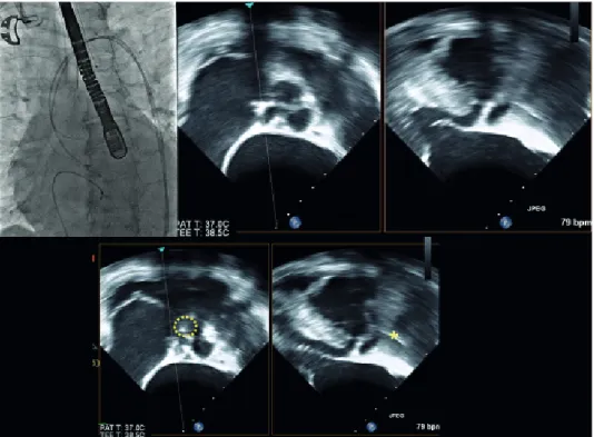
Under general anesthesia, temporary pacemaker was inserted through right femoral vein. 7 Fr sheath and 6 Fr pig-tail catheter were inserted through right femoral artery under sono-guided puncture technique. 8 Fr sheath was inserted through left femoral artery and replaced with 18 Fr Sentrant sheath. Straight coil wire under back-up with an AL 1 diagnostic catheter was crossed the stenotic aortic valve. Then, straight coil wire was changed to the round-shaped Amplatz stiff wire. Under TEE guidance, a 29-mm Evolut R prosthesis was placed at the optimal position and was deployed successfully (Fig. 3). After valve implantation, follow-up TEE showed mild para- valvular leak and mild to moderate AR. AR index was 24. All procedure was finished.
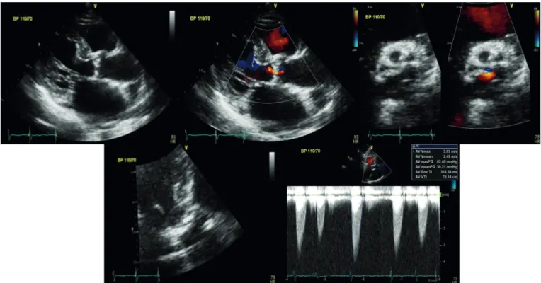
Fig. 1. Transthoracic echocardiogram showed severe AS (AVA: 0.96 cm2 by continuity equation) with peak/mean pressure gradient (64/33 mmHg).
AS; aortic stenosis, AVA; aortic valve area, BP; blood pressure.
After procedure, her renal function had not been aggravated until she was discharged (directly after: BUN, 46.1 mg/dL; creatinine, 3.33 mg/dL; eGFR, 11.7 mL/min/1.73 m2 and when she was discharged: BUN, 46.1 mg/dL; creatinine, 3.33 mg/dL; eGFR, 13 mL/min/1.73 m2).
DISCUSSION
We have reported a case of zero-contrast TAVR for severe AS in patient with CKD. This case is different from previous TAVR cases in terms of usage of contrast, 3D-echo in pre-evaluation, TEE during intervention, peripheral US in vascular access. TAVR may be an option for people who are considered at intermediate or high risk of complications from surgical aortic
Annulus mininum diameter
Annulus maxinum diameter 20.6 mm 24.6 mm
Fig. 2. Annulus dimension and perimeter derived diameter by 3-dimentional echocardiogram.
Fig. 3. Location of catheter was identified by using biplane TEE (dotted circle indicated tip of catheter and asterisk indicated catheter).
TEE; transesophageal echocardiography, PAT; patient, T; temperature.
restore renal perfusion promptly improves renal function. However, prolonged or profound prerenal azotemia can result in ischemic damage leading to ischemic AKI, particularly in combination with the presence of exogenous toxic compounds (e.g., aminoglycosides, contrast media).
So, AKI after TAVR can be considered as the common final path resulting from prerenal azotemia due to pre-, intra- and post-operative factors and additional nephrotoxic influences resulting in ATN.4-6
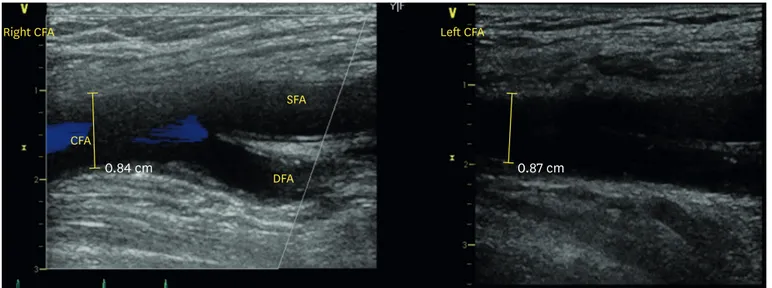
We particularly performed a femoral puncture by vascular ultrasound (Fig. 4). Furthermore, we used a 3D-echo in pre-evaluation and TEE during TAVR differently from the other cases (Fig. 5).7 Kidney function is a powerful long-term predictor of mortality in patients undergoing TAVR.
Patients with severe impaired renal function had the highest mortality after TAVR. Thus, the beneficial effects of TAVR in patients with severe impaired renal function seem to be limited and should be implemented in a decision-making process before TAVR.8-10
All the while, it has been important to do pre-procedural assessments using multimodality imaging including CT angiography and conventional angiography. So, CT angiography and conventional angiography has been thought essential. But, we could overcome this aspect by using 3D-echo in pre-evaluation.
CFA
0.84 cm 0.87 cm
SFA
DFA
Left CFA Right CFA
Fig. 4. Vascular ultrasound showed acceptable access site of left external iliac artery.
CFA; common femoral artery, SFA; superficial femoral artery, DFA; deep femoral artery.
And we have to pay the particular attention to do pre-procedural assessments by using 3D-echo to avoid the increment of complication or failure.
Furthermore, we used TEE during the procedure, TEE-TAVR was associated with similar and midterm results as angiography guiding TAVR and significantly reduced contrast media use during the procedures.11
In conclusion, this is the case report of zero contrast with TEE guidance. The zero contrast TAVR is more effective method in CKD patients in avoidance of AKI.
REFERENCES
1. Otto CM, Kumbhani DJ, Alexander KP, Calhoon JH, Desai MY, Kaul S, et al. 2017 ACC expert consensus decision pathway for transcatheter aortic valve replacement in the management of adults with aortic stenosis: a report of the American College of Cardiology Task Force on clinical expert consensus documents. J Am Coll Cardiol 2017;69:1313-1346.
PUBMED | CROSSREF Fig. 5. 3D-echo in pre-evaluation and TEE during TAVR.
TEE; transesophageal echocardiography, TAVR; transcatheter aortic valve replacement, Comm; commissure.
2015;7:1527-1535.
PUBMED
6. Thongprayoon C, Cheungpasitporn W, Srivali N, Ungprasert P, Kittanamongkolchai W, Greason KL, et al.
Acute kidney injury after transcatheter aortic valve replacement: a systematic review and meta-analysis.
Am J Nephrol 2015;41:372-382.
PUBMED | CROSSREF
7. Varga-Szemes A, Cannao PM, Muscogiuri G, De Cecco CN, Giri S, Piccini D, et al. Non-contrast 3D radial and QISS MRA for transcatheter aortic valve replacement planning. J Cardiovasc Magn Reson 2015;17:O71.
CROSSREF
8. Voigtländer L, Schewel J, Martin J, Schewel D, Frerker C, Wohlmuth P, et al. Impact of kidney function on mortality after transcatheter valve implantation in patients with severe aortic valvular stenosis. Int J Cardiol 2015;178:275-281.
PUBMED | CROSSREF
9. Bagur R, Webb JG, Nietlispach F, Dumont E, De Larochellière R, Doyle D, et al. Acute kidney injury following transcatheter aortic valve implantation: predictive factors, prognostic value, and comparison with surgical aortic valve replacement. Eur Heart J 2010;31:865-874.
PUBMED | CROSSREF
10. Barbash IM, Ben-Dor I, Dvir D, Maluenda G, Xue Z, Torguson R, et al. Incidence and predictors of acute kidney injury after transcatheter aortic valve replacement. Am Heart J 2012;163:1031-1036.
PUBMED | CROSSREF
11. Bagur R, Rodés-Cabau J, Doyle D, De Larochellière R, Villeneuve J, Lemieux J, et al. Usefulness of TEE as the primary imaging technique to guide transcatheter transapical aortic valve implantation. JACC Cardiovasc Imaging 2011;4:115-124.
PUBMED | CROSSREF