• 검색 결과가 없습니다.

Fatal Delayed Coronary Artery Perforation After Coronary Stent Implantation

N/A
N/A
Protected

Academic year: 2022

Share "Fatal Delayed Coronary Artery Perforation After Coronary Stent Implantation"

Copied!
3
0
0

로드 중.... (전체 텍스트 보기)

전체 글

(1)

352 Copyright © 2012 The Korean Society of Cardiology Korean Circulation Journal

Introduction

Coronary artery perforation (CP) during percutaneous coronary intervention (PCI) is a rare complication. The reported incidence of CPs ranges from 0.1% to 0.6%.

1-7)

Most CPs occur during the proce- dure, immediately after stenting or adjunctive ballooning. However, it is also well recognized that the hydrophilic wires and glycopro- tein IIb/IIIa antagonist during PCI increases the risk of delayed CP.

2)

However, those CP seemed to be minor and do not require pericar- dial drainage. However, we herein describe an unusual delayed CP (type III) at the site of stent implantation after PCI without any evid- ence of immediate perforations. To the best of our knowledge, this is the first case report of angiographic documentation and treat- ment of delayed CP at the site of stent, presented as cardiac arrest.

Case Report

http://dx.doi.org/10.4070/kcj.2012.42.5.352 Print ISSN 1738-5520 • On-line ISSN 1738-5555

Fatal Delayed Coronary Artery Perforation After Coronary Stent Implantation

Sang-Hoon Kim, MD, Jae-Youn Moon, MD, Jung-Hoon Sung, MD, In Jai Kim, MD, Sang-Wook Lim, MD, Dong-Hun Cha, MD, and Seung-Yun Cho, MD

Department of Cardiology, CHA Bundang Medical Center, CHA University, Seongnam, Korea

Most type I and II perforations are predominately caused by hydrophilic and stiff wires, often presented in the delayed form, and do not require pericardial drainage or surgical interventions. However, we report a type III delayed coronary artery perforation at the site of stent implantation after intervention without any evidence of immediate perforations. To the best of our knowledge, this is the first case report of angiographic documentation and treatment of delayed coronary perforation at the site of stent, presented as a cardiac arrest. (Korean Circ J 2012;42:352-354)

KEY WORDS: Cardiac tamponade; Complications; Percutaneous transluminal coronary angioplasty; Drug-eluting stent.

Received: August 31, 2011 Revision Received: October 4, 2011 Accepted: October 8, 2011

Correspondence: Jae-Youn Moon, MD, Department of Cardiology, CHA Bundang Medical Center, CHA University, 59 Yatap-ro, Bundang-gu, Seong- nam 463-712, Korea

Tel: 82-31-780-5585, Fax: 82-31-780-5857 E-mail: answodus@medimail.co.kr

• The authors have no financial conflicts of interest.

This is an Open Access article distributed under the terms of the Creative Commons Attribution Non-Commercial License (http://creativecommons.

org/licenses/by-nc/3.0) which permits unrestricted non-commercial use, distribution, and reproduction in any medium, provided the original work is properly cited.

Case

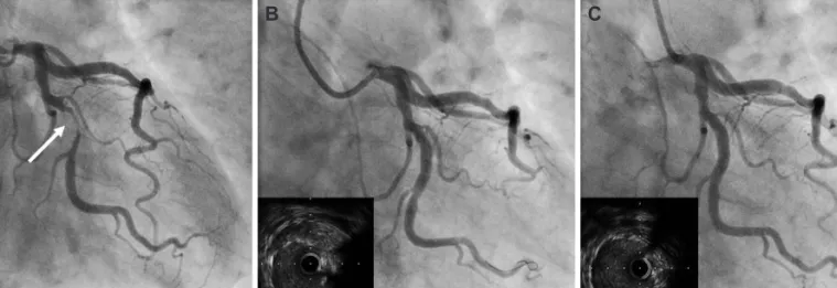
A 64 year-old male patient was admitted for chest pain. He had no specific cardiovascular risk factors except for a history of heavy smoking. Coronary angiogram (CAG) showed critical stenosis of the left circumflex artery (Fig. 1A).

Percutaneous coronary intervention was performed with the use of 6 Fr guiding catheter and hydrophilic guide-wire via trans-radial sheath. After wiring, pre-dilatation was done with a 2.0×15 mm co- ronary balloon. Next, an Endeavor splinter

TM

(3.5×18 mm) was im- planted with 10 atmosphere. After stenting, intravascular ultrasound (IVUS) showed suboptimal expansions (Fig. 1B). Therefore, adjunc- tive ballooning was performed with a 3.75×10 mm noncompliant balloon catheter. Final the CAG showed no residual stenosis with- out any complications and final IVUS images revealed good apposi- tion of the stent without any abnormal findings (Fig. 1C). Finally, we finished the procedure and the patient was transferred to the gen- eral ward. Till then, it was a routine PCI and no problems developed.

However, the patient complained of severe chest pain immedia-

tely after being moved to a general ward. Cardiac arrest developed

just at the moment of chest pain. The initial monitoring showed flat

electrocardiography. Therefore, cardiovascular resuscitation (CPR)

was performed for 15 minutes, but there was no response and the

pulse rate was not recovered. Retrospective analysis of the final an-

giogram, however, led to the suspicion that acute stent thrombosis

of drug-eluting stent or a delayed dissection of the major coronary

artery may be the cause of cardiac arrest at that time. Therefore, we

(2)

353 Sang-Hoon Kim, et al.

http://dx.doi.org/10.4070/kcj.2012.42.5.352 www.e-kcj.org

decided to perform a CAG again during CPR.

Before angiography, percutaneous cardiopulmonary support was immediately applied via the femoral artery during CPR. Next, the CAG was checked again. Surprisingly, CAG revealed coronary per- foration with active bleeding (type III) at the previous stent impl- antation site (Fig. 2A). Pericardiocentesis was performed success- fully and Jo graft stent (3.0×19 mm) was implanted for the active bleeding site (Fig. 2B). However, the leakage of blood still remained.

Therefore, we considered another graft stent. However, while pre- paring for another graft stent, the cardiac arrest redeveloped. The CPR was restarted and we were able to determine what the second problem was. Tension pneumothorax developed during ambu bag- ging, so we decided to insert the chest tube. Therefore, after inser- tion of another graft stent (3.5×19 mm), the chest tube was insert- ed to the pleural cavity by a thoracic surgeon simultaneously. After

that, the vital signs recovered and bleeding was controlled (Fig. 2C).

Finally, we were able to finish the procedures. The pneumothorax was resolved after 3 days and cardiac function totally recovered wi- thin 3 days. However, the brain of the patient was damaged by hy- poxic brain injury and he remained in a vegetative state.

Discussion

Coronary artery perforation was defined as evidence of extrava- sation of contrast dye or blood from the coronary artery during or following a coronary interventional procedure. Recently, the incid- ence of CP has increased slightly. Possibly due to various PCI devic- es being developed and more complex lesions or smaller vessels being included as the target lesions for PCI. The development of debulking devices has enabled the treatment of calcified and bifurca-

A   B   C  

Fig. 1. Coronary angiogram for pecutaneous coronary intervention for critical stenosis of left. Circumflex artery. A: coronary angiogram showed critical steno- sis of left circumflex artery (arrow). B: after stenting, angiogram showed mild residual stenosis in proximal stented lesion and intravascular ultrasound showed incomplete expansions. C: final angiogram showed no residual lesion and final intravascular ultrasound images revealed improved expansion and good apposition of the stent.

A   B   C  

Fig. 2. Coronary angiogram after cardiovascular resuscitation due to sudden cardiac arrest in general ward. A: angiogram showed type III coronary perfora-

tion with bleeding at the previous stent implantation site (arrow). B: before inflation, first Jo graft stent was placed for the active bleeding site. C: after stent-

ing of two graft stents, the final angiogram during cardiovascular resuscitation showed no additional bleeding.

(3)

354 Delayed Coronary Artery Perforation

http://dx.doi.org/10.4070/kcj.2012.42.5.352 www.e-kcj.org

tion lesions. The perforation type was determined based on the Ellis classification.

1)

A classification was based on angiographic appear- ance of the perforation (I, extraluminal crater without extravasa- tion; II, pericardial or myocardial blushing without contrast jet ex- travasation; III, extravasation though frank perforation with ≥1-mm diameter). The outcomes of CP varied considerably with the grade of perforation. Patients who sustained type I or II CP showed a tend- ency of good prognoses but, in contrast, patients with type III per- forations had a higher mortality rate.

According to a previous report,

2)

pericardial effusion or tampon- ade rarely became clinically manifested after several hours follow- ing the coronary procedure even though vessel perforation was not clinically or angiographically obvious at the end of the proce- dure. Also, the mechanism of vessel perforation was believed to be a distal branch puncture with the guidewire tip such as a hydrophilic wire and the administration of glycoprotein IIb/IIIa antagonist. How- ever, most of the type I and II perforations are predominately caused by hydrophilic and stiff wires, and do not require pericardial drain- age or surgical intervention.

8)

Type III perforations are more often associated with stent and device use, and manifested immediately.

A majority of type III perforations can be initially managed by per- cutaneous methods.

In this case, we have shown a very rare possibility which could have been a neglected cause of a sudden cardiac arrest after PCI.

In most cases of sudden arrest within 24 hours after PCI, and espe- cially when the time of death, the causes of death are almost always assumed to be cardiac arrest by a stent thrombosis or by malignant arrhythmia (ex. ventricular tachycardia) unless the patient has an autopsy. For the diagnosis of CP after PCI, echocardiography was the preferred method, and pericardial fluid collection on echocar- diography could be a clue for assuming CP after PCI. Definite diag- nosis was made by confirming the extravasation of contrast dye or blood in coronary angiography. However, in many cases, even CP st- rongly assumed by echocardiography, coronary angiography could not find the exact focus of perforation.

An interesting point of the present case is that the type III perfor- ations occurred late after the procedures and presented as a cardiac arrest. If we did not confirm CP angiographically, we could not have

known the cause of cardiac arrest and it was reasonable to assume that the cause may be a probable acute stent thrombosis even when there was no evidence of it.

The mechanism of delayed type III perforations was unclear. We could not detect any evidence of coronary artery rupture in the final angiogram and IVUS study. However, we may postulate heavy coro- nary calcification and use a larger adjunctive coronary balloon.

In conclusion, this case highlights that delayed CP exists in clinical practice and could be misdiagnosed as other events such as an acu- te stent thrombosis or malignant ventricular arrhythmia. Sudden deterioration of a patient after PCI with a high risk of perforation such as tortuous calcified lesions, CP should be considered as one of the causes of deterioration.

References

1. Ellis SG, Ajluni S, Arnold AZ, et al. Increased coronary perforation in the new device era: incidence, classification, management, and outcome.

Circulation 1994;90:2725-30.

2. Gunning MG, Williams IL, Jewitt DE, Shah AM, Wainwright RJ, Thomas MR. Coronary artery perforation during percutaneous intervention:

incidence and outcome. Heart 2002;88:495-8.

3. Gruberg L, Pinnow E, Flood R, et al. Incidence, management, and out- come of coronary artery perforation during percutaneous coronary in- tervention. Am J Cardiol 2000;86:680-2.

4. Shimony A, Zahger D, Van Straten M, et al. Incidence, risk factors, management and outcomes of coronary artery perforation during per- cutaneous coronary intervention. Am J Cardiol 2009;104:1674-7.

5. Von Sohsten R, Kopistansky C, Cohen M, Kussmaul WG 3rd. Cardiac tamponade in the “new device” era: evaluation of 6999 consecutive percutaneous coronary interventions. Am Heart J 2000;140:279-83.

6. Fasseas P, Orford JL, Panetta CJ, et al. Incidence, correlates, manage- ment, and clinical outcome of coronary perforation: analysis of 16,298 procedures. Am Heart J 2004;147:140-5.

7. Fukutomi T, Suzuki T, Popma JJ, et al. Early and late clinical outcomes fol- lowing coronary perforation in patients undergoing percutaneous co- ronary intervention. Circ J 2002;66:349-56.

8. Ramana RK, Arab D, Joyal D, et al. Coronary artery perforation during

percutaneous coronary intervention: incidence and outcomes in the

new interventional era. J Invasive Cardiol 2005;17:603-5.

수치

Fig. 1. Coronary angiogram for pecutaneous coronary intervention for critical stenosis of left

참조

관련 문서

Incident major cardiovascular events (coronary artery disease, ischemic stroke, hemorrhagic stroke and cardiovascular mortality) were set as primary end points.

Prevention of coronary and stroke events with atorvastatin in hypertensive patients who have average or lower-than-average cholesterol concentrations, in

Changes in functional physical fitness, after the experiment compared to before the experiment in the group of exercise, showed a significant difference in

First, after the Group Art Therapy, the scale of hopelessness depression in the test group decreased significantly in comparison with that before the group

success rates of dental implants placed at the time of or after alveolar ridge augmentation with an autogenous mandibular bone graft and titanium mesh: a 3-to

Therefore, cTnT is a recommended biomarker for use in the detection of myocardial infarction (MI) and in acute coronary syndromes.[8] Indeed, several authors

Diagnosis and Treatment of Oral admidstrated Bisphosphonate-related osteonecrosis of the jaw.. and implantation

The defect area was filled with new-bone formation around the graft material hydroxy-apatite (open stars) at the periphery and chronic inflammation with